Loading
PDBj
MenuPDBj@FacebookPDBj@TwitterPDBj@YouTubewwPDB FoundationwwPDB
RCSB PDBPDBeBMRBAdv. SearchSearch help
PDB: 74 results

6KOD
DownloadVisualize
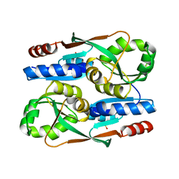
BU of 6kod by Molmil
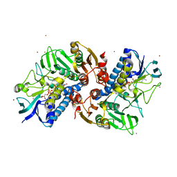
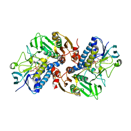
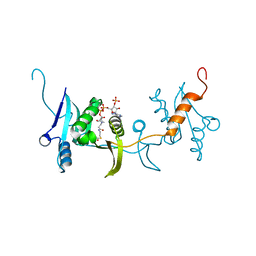
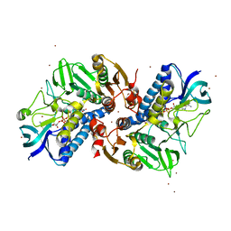
Cu(II) complex of HOCl-induced flavoprotein disulfide reductase RclA C43S mutant from Escherichia coli
Descriptor: CHLORIDE ION, COPPER (II) ION, FLAVIN-ADENINE DINUCLEOTIDE, ...
Authors:Baek, Y, Ha, N.-C.
Deposit date:2019-08-09
Release date:2020-02-05
Last modified:2023-11-22
Method:X-RAY DIFFRACTION (3 Å)
Cite:Structure and function of the hypochlorous acid-induced flavoprotein RclA fromEscherichia coli.
J.Biol.Chem., 295, 2020
7FB9
DownloadVisualize
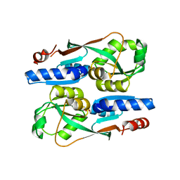
BU of 7fb9 by Molmil
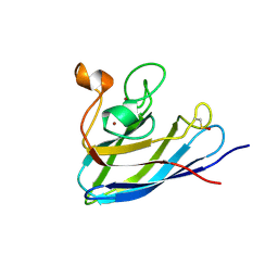
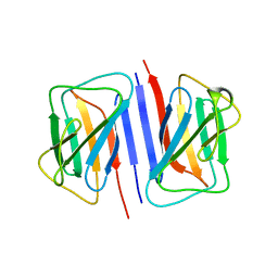
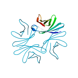
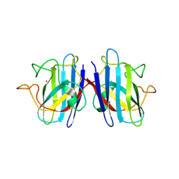
Crystal Structure of Human Cu, Zn Superoxide Dismutase (SOD1)
Descriptor: COPPER (II) ION, Superoxide dismutase [Cu-Zn], ZINC ION
Authors:Baek, Y, Ha, N.-C.
Deposit date:2021-07-08
Release date:2022-10-12
Last modified:2024-10-09
Method:X-RAY DIFFRACTION (2.7 Å)
Cite:Structural analysis of the overoxidized Cu/Zn-superoxide dismutase in ROS-induced ALS filament formation.
Commun Biol, 5, 2022
6KYY
DownloadVisualize
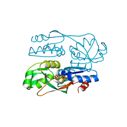
BU of 6kyy by Molmil
Cu(II) complex of HOCl-induced flavoprotein disulfide reductase RclA from Escherichia coli
Descriptor: CHLORIDE ION, COPPER (II) ION, FLAVIN-ADENINE DINUCLEOTIDE, ...
Authors:Baek, Y, Ha, N.-C.
Deposit date:2019-09-20
Release date:2020-02-05
Last modified:2023-11-22
Method:X-RAY DIFFRACTION (2.8 Å)
Cite:Structure and function of the hypochlorous acid-induced flavoprotein RclA fromEscherichia coli.
J.Biol.Chem., 295, 2020
6IY0
DownloadVisualize
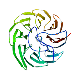
BU of 6iy0 by Molmil
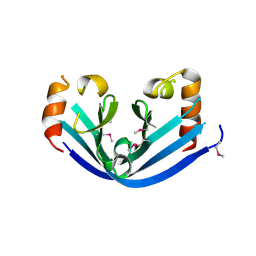
Crystal structure of conserved hypothetical protein SAV0927 from Staphylococcus aureus subsp. aureus Mu50
Descriptor: CHLORIDE ION, SAV0927
Authors:Jeong, S, Ha, N.-C.
Deposit date:2018-12-12
Release date:2019-12-18
Last modified:2020-07-01
Method:X-RAY DIFFRACTION (2.5 Å)
Cite:Crystal Structure of SAV0927 and Its Functional Implications.
J Microbiol Biotechnol., 29, 2019
6K25
DownloadVisualize
BU of 6k25 by Molmil
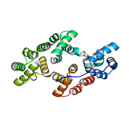
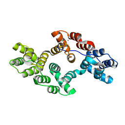
Crystal structure of Ca-unbound human Annexin A5 in low salt condition
Descriptor: Annexin A5
Authors:Hong, S, Ha, N.-C.
Deposit date:2019-05-13
Release date:2020-03-25
Last modified:2023-11-22
Method:X-RAY DIFFRACTION (2.401 Å)
Cite:High-resolution structures of annexin A5 in a two-dimensional array.
J.Struct.Biol., 209, 2020
7FB6
DownloadVisualize
BU of 7fb6 by Molmil
C57D/C146D mutant of Human Cu, Zn Superoxide Dismutase (SOD1)
Descriptor: CHLORIDE ION, COPPER (II) ION, Superoxide dismutase [Cu-Zn], ...
Authors:Baek, Y, Ha, N.-C.
Deposit date:2021-07-08
Release date:2022-10-12
Last modified:2023-11-29
Method:X-RAY DIFFRACTION (1.8 Å)
Cite:Structural analysis of the overoxidized Cu/Zn-superoxide dismutase in ROS-induced ALS filament formation.
Commun Biol, 5, 2022
6K22
DownloadVisualize
BU of 6k22 by Molmil
Crystal structure of Ca-bound human Annexin A5 in low salt condition
Descriptor: Annexin A5, CALCIUM ION
Authors:Hong, S, Ha, N.-C.
Deposit date:2019-05-13
Release date:2020-03-25
Last modified:2023-11-22
Method:X-RAY DIFFRACTION (2.747 Å)
Cite:High-resolution structures of annexin A5 in a two-dimensional array.
J.Struct.Biol., 209, 2020
6KGY
DownloadVisualize
BU of 6kgy by Molmil
HOCl-induced flavoprotein disulfide reductase RclA from Escherichia coli
Descriptor: CHLORIDE ION, FLAVIN-ADENINE DINUCLEOTIDE, Pyridine nucleotide-disulphide oxidoreductase dimerisation region
Authors:Baek, Y, Ha, N.-C.
Deposit date:2019-07-12
Release date:2020-02-05
Last modified:2024-04-03
Method:X-RAY DIFFRACTION (2.9 Å)
Cite:Structure and function of the hypochlorous acid-induced flavoprotein RclA fromEscherichia coli.
J.Biol.Chem., 295, 2020
7DNP
DownloadVisualize
BU of 7dnp by Molmil
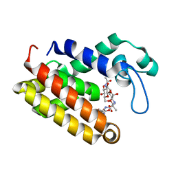
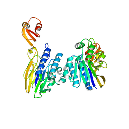
Structure of Brucella abortus SagA
Descriptor: (2R)-2-[[(2S)-2-[[(2R)-2-[(2R,3S,4R,5R,6S)-5-acetamido-3-[(2S,3R,4R,5S,6R)-3-acetamido-6-(hydroxymethyl)-4,5-bis(oxidanyl)oxan-2-yl]oxy-2-(hydroxymethyl)-6-oxidanyl-oxan-4-yl]oxypropanoyl]amino]propanoyl]amino]pentanedioic acid, Secretion activator protein, hypothetical
Authors:Hyun, Y, Ha, N.-C.
Deposit date:2020-12-10
Release date:2021-06-23
Last modified:2024-05-29
Method:X-RAY DIFFRACTION (2 Å)
Cite:Structure and Function of the Autolysin SagA in the Type IV Secretion System of Brucella abortus .
Mol.Cells, 44, 2021
7DPY
DownloadVisualize
BU of 7dpy by Molmil
Structure of Brucella abortus PhiA
Descriptor: Brucella Abortus PhiA
Authors:Hyun, Y, Ha, N.-C.
Deposit date:2020-12-22
Release date:2021-06-23
Last modified:2023-11-29
Method:X-RAY DIFFRACTION (1.8 Å)
Cite:Structure and Function of the Autolysin SagA in the Type IV Secretion System of Brucella abortus .
Mol.Cells, 44, 2021
7DTG
DownloadVisualize
BU of 7dtg by Molmil
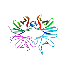
Crystal structure of lamin B1 Ig-like domain from human
Descriptor: Lamin-B1
Authors:Ahn, J, Lee, J, Ha, N.-C.
Deposit date:2021-01-05
Release date:2021-11-03
Last modified:2023-11-29
Method:X-RAY DIFFRACTION (3.6 Å)
Cite:Beta-strand-mediated dimeric formation of the Ig-like domains of human lamin A/C and B1.
Biochem.Biophys.Res.Commun., 550, 2021
7DXN
DownloadVisualize
BU of 7dxn by Molmil
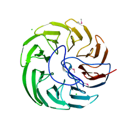
Plant growth-promoting factor YxaL from Bacillus velezensis
Descriptor: CHLORIDE ION, Membrane associated protein kinase with beta-propeller domain, pyrrolo-quinoline quinone beta-propeller repeat
Authors:Baek, Y, Ha, N.-C.
Deposit date:2021-01-19
Release date:2022-01-19
Method:X-RAY DIFFRACTION (1.8 Å)
Cite:Structure of the plant growth-promoting factor YxaL from the rhizobacterium Bacillus velezensis and its application to protein engineering.
Acta Crystallogr D Struct Biol, 78, 2022
7EQ5
DownloadVisualize
BU of 7eq5 by Molmil
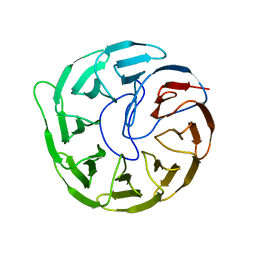
Plant growth-promoting factor YxaL mutant from Bacillus velezensis - T175W/W215G
Descriptor: Membrane associated protein kinase with beta-propeller domain, pyrrolo-quinoline quinone beta-propeller repeat
Authors:Kim, J, Ha, N.-C.
Deposit date:2021-04-30
Release date:2022-01-12
Last modified:2023-11-29
Method:X-RAY DIFFRACTION (2.6 Å)
Cite:Structure of the plant growth-promoting factor YxaL from the rhizobacterium Bacillus velezensis and its application to protein engineering.
Acta Crystallogr D Struct Biol, 78, 2022
7DOG
DownloadVisualize
BU of 7dog by Molmil
Crystal structure of a nuclease and capping domain of SbcD from Staphylococcus aureus
Descriptor: MANGANESE (II) ION, Nuclease SbcCD subunit D
Authors:Lee, J, Ha, N.-C.
Deposit date:2020-12-14
Release date:2021-05-05
Last modified:2023-11-29
Method:X-RAY DIFFRACTION (2.91 Å)
Cite:Crystal structure of the nuclease and capping domain of SbcD from Staphylococcus aureus.
J.Microbiol, 59, 2021
7ERQ
DownloadVisualize
BU of 7erq by Molmil
The regulatory domain of YeiE, a sulfite sensing LysR-type transcriptional regulator from Cronobacter sakazakii (ligand-free form)
Descriptor: LysR family transcriptional regulator
Authors:Hong, S, Ha, N.-C.
Deposit date:2021-05-06
Release date:2022-03-23
Last modified:2024-10-09
Method:X-RAY DIFFRACTION (1.95 Å)
Cite:Crystal structures of YeiE from Cronobacter sakazakii and the role of sulfite tolerance in gram-negative bacteria.
Proc.Natl.Acad.Sci.USA, 119, 2022
7ERP
DownloadVisualize
BU of 7erp by Molmil
The regulatory domain of YeiE, a sulfite sensing LysR-type transcriptional regulator from Cronobacter sakazakii (sulfite-bound form)
Descriptor: LysR family transcriptional regulator, SULFITE ION
Authors:Hong, S, Ha, N.-C.
Deposit date:2021-05-06
Release date:2022-03-23
Last modified:2024-04-03
Method:X-RAY DIFFRACTION (2.03 Å)
Cite:Crystal structures of YeiE from Cronobacter sakazakii and the role of sulfite tolerance in gram-negative bacteria.
Proc.Natl.Acad.Sci.USA, 119, 2022
7FDF
DownloadVisualize
BU of 7fdf by Molmil
The E145S mutant of the regulatory domain of YeiE, a sulfite sensing LysR-type transcriptional regulator from Cronobacter sakazakii (sulfate-bound form)
Descriptor: LysR family transcriptional regulator, SULFATE ION
Authors:Hong, S, Ha, N.-C.
Deposit date:2021-07-16
Release date:2022-03-23
Last modified:2023-11-29
Method:X-RAY DIFFRACTION (2.05 Å)
Cite:Crystal structures of YeiE from Cronobacter sakazakii and the role of sulfite tolerance in gram-negative bacteria.
Proc.Natl.Acad.Sci.USA, 119, 2022
7EVF
DownloadVisualize
BU of 7evf by Molmil
Plant growth-promoting factor YxaL mutant from Bacillus velezensis - T175W/S213G/W215A
Descriptor: PQQ_3 domain-containing protein
Authors:Kim, J, Ha, N.-C.
Deposit date:2021-05-21
Release date:2022-01-12
Last modified:2023-11-29
Method:X-RAY DIFFRACTION (1.5 Å)
Cite:Structure of the plant growth-promoting factor YxaL from the rhizobacterium Bacillus velezensis and its application to protein engineering.
Acta Crystallogr D Struct Biol, 78, 2022
7DQG
DownloadVisualize
BU of 7dqg by Molmil
Crystal structure of Salmonella phage acetyltransferase
Descriptor: ACETYL COENZYME *A, Putative acetyltransferase
Authors:Hyun, Y, Ha, N.-C.
Deposit date:2020-12-23
Release date:2021-12-29
Last modified:2023-11-29
Method:X-RAY DIFFRACTION (2.21 Å)
Cite:Crystal structure of the phage-encoded N-acetyltransferase in complex with acetyl-CoA
To Be Published
7DSG
DownloadVisualize
BU of 7dsg by Molmil
Crystal structure of Brucella abortus PhiA
Descriptor: Brucella Abortus PhiA
Authors:Hyun, Y, Ha, N.-C.
Deposit date:2020-12-31
Release date:2022-01-12
Last modified:2023-11-29
Method:X-RAY DIFFRACTION (1.9 Å)
Cite:Crystal structure of Brucella abortus PhiA
To Be Published
1OCL
DownloadVisualize
BU of 1ocl by Molmil
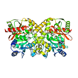
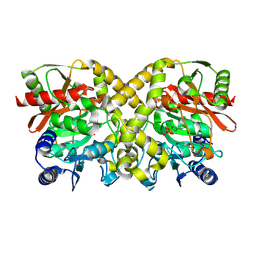
THE CRYSTAL STRUCTURE OF MALONAMIDASE E2 COMPLEXED WITH MALONATE FROM BRADYRHIZOBIUM JAPONICUM
Descriptor: MALONAMIDASE E2, MALONIC ACID
Authors:Shin, S, Ha, N.-C, Lee, T.-H, Oh, B.-H.
Deposit date:2003-02-08
Release date:2003-02-25
Last modified:2023-12-13
Method:X-RAY DIFFRACTION (2 Å)
Cite:Characterization of a Novel Ser-Cisser-Lys Catalytic Triad in Comparison with the Classical Ser-His-Asp Triad.
J.Biol.Chem., 278, 2003
1OCK
DownloadVisualize
BU of 1ock by Molmil
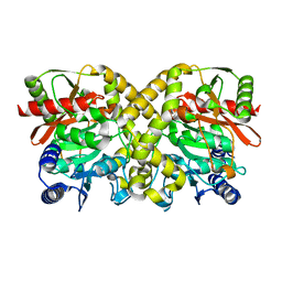
THE CRYSTAL STRUCTURE OF MALONAMIDASE E2 FROM BRADYRHIZOBIUM JAPONICUM
Descriptor: MALONAMIDASE E2
Authors:Shin, S, Ha, N.-C, Lee, T.-H, Oh, B.-H.
Deposit date:2003-02-08
Release date:2003-03-03
Last modified:2024-05-08
Method:X-RAY DIFFRACTION (1.8 Å)
Cite:Characterization of a Novel Ser-Cisser-Lys Catalytic Triad in Comparison with the Classical Ser-His-Asp Triad.
J.Biol.Chem., 278, 2003
1OCM
DownloadVisualize
BU of 1ocm by Molmil
THE CRYSTAL STRUCTURE OF MALONAMIDASE E2 COVALENTLY COMPLEXED WITH PYROPHOSPHATE FROM BRADYRHIZOBIUM JAPONICUM
Descriptor: MALONAMIDASE E2, PYROPHOSPHATE 2-
Authors:Shin, S, Ha, N.-C, Lee, T.-H, Oh, B.-H.
Deposit date:2003-02-08
Release date:2003-02-25
Last modified:2024-10-09
Method:X-RAY DIFFRACTION (1.9 Å)
Cite:Characterization of a Novel Ser-Cisser-Lys Catalytic Triad in Comparison with the Classical Ser-His-Asp Triad.
J.Biol.Chem., 278, 2003
1GS3
DownloadVisualize
BU of 1gs3 by Molmil
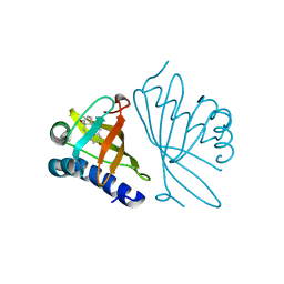
High resolution crystal structure of PI delta-5-3-Ketosteroid Isomerase mutants Y30F/Y55F/Y115F/D38N (Y32F/Y57F/Y119F/D40N, PI numbering)complexed with equilenin at 2.1 A resolution
Descriptor: EQUILENIN, STEROID DELTA-ISOMERASE
Authors:Shin, S, Ha, N.-C, Oh, B.-H.
Deposit date:2001-12-27
Release date:2003-01-23
Last modified:2023-12-13
Method:X-RAY DIFFRACTION (2.1 Å)
Cite:Contribution of a Low-Barrier Hydrogen Bond to Catalysis by Delta-5-3-Ketosteroid Isomerase is not Extremely High Compared to that of an Ordinary Hydrogen Bond. Low-Barrier Hydrogen Bond of Pi Ksi
To be Published
<123


226707

数据于2024-10-30公开中

PDB statisticsPDBj update infoContact PDBjnumon