Loading
PDBj
MenuPDBj@FacebookPDBj@TwitterPDBj@YouTubewwPDB FoundationwwPDB
RCSB PDBPDBeBMRBAdv. SearchSearch help
PDB: 66 results

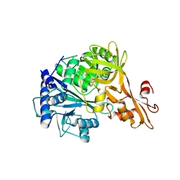
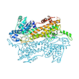
3UNV
DownloadVisualize
BU of 3unv by Molmil
Pantoea agglomerans Phenylalanine Aminomutase
Descriptor: (3S)-3-amino-3-phenylpropanoic acid, 1,2-ETHANEDIOL, AdmH, ...
Authors:Geiger, J, Strom, S.
Deposit date:2011-11-16
Release date:2012-02-22
Last modified:2023-11-15
Method:X-RAY DIFFRACTION (1.54 Å)
Cite:Insights into the mechanistic pathway of the Pantoea agglomerans phenylalanine aminomutase.
Angew.Chem.Int.Ed.Engl., 51, 2012
4EAT
DownloadVisualize
BU of 4eat by Molmil
Crystal structure of a benzoate coenzyme A ligase
Descriptor: 1,2-ETHANEDIOL, BENZOIC ACID, Benzoate-coenzyme A ligase, ...
Authors:Geiger, J, Strom, S.
Deposit date:2012-03-22
Release date:2013-03-27
Last modified:2024-02-28
Method:X-RAY DIFFRACTION (1.798 Å)
Cite:Kinetically and Crystallographically Guided Mutations of a Benzoate CoA Ligase (BadA) Elucidate Mechanism and Expand Substrate Permissivity.
Biochemistry, 54, 2015
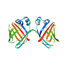
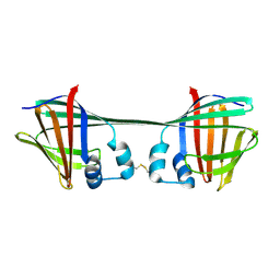
6MCU
DownloadVisualize
BU of 6mcu by Molmil
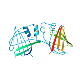
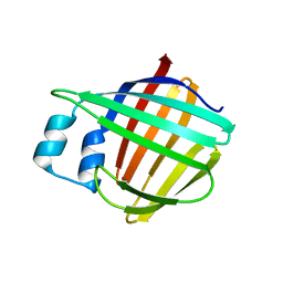
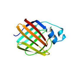
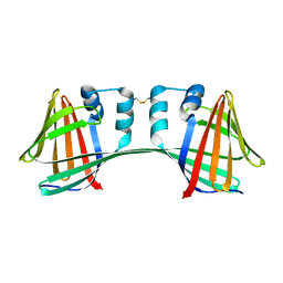
Crystal structure of the holo retinal-bound domain-swapped dimer Q108K:T51D:A28H mutant of human Cellular Retinol Binding Protein II
Descriptor: GLYCEROL, RETINAL, Retinol-binding protein 2
Authors:Ghanbarpour, A, Geiger, J.
Deposit date:2018-09-02
Release date:2019-10-16
Last modified:2023-10-11
Method:X-RAY DIFFRACTION (2.572 Å)
Cite:Engineering the hCRBPII Domain-Swapped Dimer into a New Class of Protein Switches.
J.Am.Chem.Soc., 141, 2019
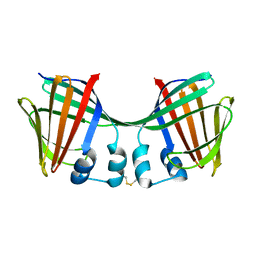
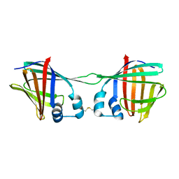
6ON7
DownloadVisualize
BU of 6on7 by Molmil
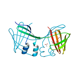
Crystal Structure of Apo Domain-Swapped Dimer Q108K:T51D:A28C:L36C Mutant of Human Cellular Retinol Binding Protein II
Descriptor: Retinol-binding protein 2
Authors:Ghanbarpour, A, Geiger, J.
Deposit date:2019-04-20
Release date:2019-10-16
Last modified:2023-10-11
Method:X-RAY DIFFRACTION (1.981 Å)
Cite:Engineering the hCRBPII Domain-Swapped Dimer into a New Class of Protein Switches.
J.Am.Chem.Soc., 141, 2019
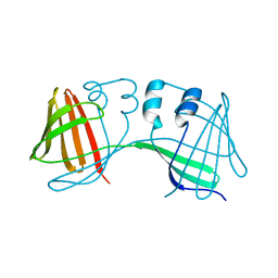
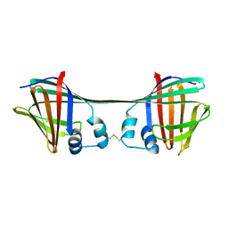
6E51
DownloadVisualize
BU of 6e51 by Molmil
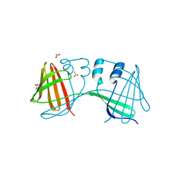
Crystal structure of the apo domain-swapped dimer Q108K:K40L:T51W mutant of human cellular retinol binding protein II
Descriptor: Retinol-binding protein 2
Authors:Ghanbarpour, A, Geiger, J.
Deposit date:2018-07-19
Release date:2019-10-16
Last modified:2023-10-11
Method:X-RAY DIFFRACTION (2.256 Å)
Cite:Engineering the hCRBPII Domain-Swapped Dimer into a New Class of Protein Switches.
J.Am.Chem.Soc., 141, 2019
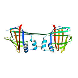
6E50
DownloadVisualize
BU of 6e50 by Molmil
Crystal structure of the apo domain-swapped dimer Q108K:K40L:T51F mutant of human Cellular Retinol Binding Protein II
Descriptor: ACETATE ION, Retinol-binding protein 2
Authors:Ghanbarpour, A, Geiger, J.
Deposit date:2018-07-18
Release date:2019-10-16
Last modified:2023-10-11
Method:X-RAY DIFFRACTION (1.965 Å)
Cite:Engineering the hCRBPII Domain-Swapped Dimer into a New Class of Protein Switches.
J.Am.Chem.Soc., 141, 2019
6E5E
DownloadVisualize
BU of 6e5e by Molmil
Crystal structure of the apo domain-swapped dimer Q108K:T51D mutant of human cellular retinol binding protein II
Descriptor: ACETATE ION, Retinol-binding protein 2
Authors:Ghanbarpour, A, Geiger, J.
Deposit date:2018-07-20
Release date:2019-10-16
Last modified:2023-10-11
Method:X-RAY DIFFRACTION (1.696 Å)
Cite:Engineering the hCRBPII Domain-Swapped Dimer into a New Class of Protein Switches.
J.Am.Chem.Soc., 141, 2019
6E5Q
DownloadVisualize
BU of 6e5q by Molmil
Crystal structure of the apo domain-swapped dimer Q108K:T51D:A28H mutant of human Cellular Retinol Binding Protein II
Descriptor: ACETATE ION, Retinol-binding protein 2
Authors:Ghanbarpour, A, Geiger, J.
Deposit date:2018-07-21
Release date:2019-10-16
Last modified:2023-10-11
Method:X-RAY DIFFRACTION (1.99 Å)
Cite:Engineering the hCRBPII Domain-Swapped Dimer into a New Class of Protein Switches.
J.Am.Chem.Soc., 141, 2019
6E7M
DownloadVisualize
BU of 6e7m by Molmil
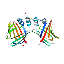
Crystal structure of the holo retinal-bound domain-swapped dimer Q108K:T51D:A28C mutant of human Cellular Retinol Binding Protein II
Descriptor: ACETATE ION, GLYCEROL, RETINAL, ...
Authors:Ghanbarpour, A, Geiger, J.
Deposit date:2018-07-26
Release date:2019-10-16
Last modified:2023-10-11
Method:X-RAY DIFFRACTION (2.7 Å)
Cite:Engineering the hCRBPII Domain-Swapped Dimer into a New Class of Protein Switches.
J.Am.Chem.Soc., 141, 2019
6DCM
DownloadVisualize
BU of 6dcm by Molmil
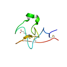
The atomic resolution crystal structure of Kringle 2 variant bound with EACA
Descriptor: 6-AMINOHEXANOIC ACID, Plasminogen
Authors:Ghanbarpour, A, Geiger, J.
Deposit date:2018-05-07
Release date:2019-05-15
Last modified:2023-11-15
Method:X-RAY DIFFRACTION (1.031 Å)
Cite:The atomic resolution crystal structure of Kringle 2 variant bound with EACA
To Be Published
5UVI
DownloadVisualize
BU of 5uvi by Molmil
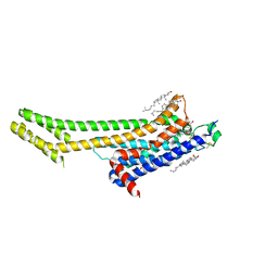
Serial Millisecond Crystallography of Membrane and Soluble Protein Micro-crystals using Synchrotron Radiation
Descriptor: (2R)-2,3-dihydroxypropyl (9Z)-octadec-9-enoate, 4-{2-[(7-amino-2-furan-2-yl[1,2,4]triazolo[1,5-a][1,3,5]triazin-5-yl)amino]ethyl}phenol, Adenosine receptor A2a,Soluble cytochrome b562,Adenosine receptor A2a, ...
Authors:Martin-Garcia, J.M, Conrad, C.E, Nelson, G, Stander, N, Zatsepin, N.A, Zook, J, Zhu, L, Geiger, J, Chun, E, Kissick, D, Hilgart, M.C, Ogata, C, Ishchenko, A, Nagaratnam, N, Roy-Chowdhury, S, Coe, J, Subramanian, G, Schaffer, A, James, D, Ketawala, G, Venugopalan, N, Xu, S, Corcoran, S, Ferguson, D, Weierstall, U, Spence, J.C.H, Cherezov, V, Fromme, P, Fischetti, R.F, Liu, W.
Deposit date:2017-02-20
Release date:2017-05-24
Last modified:2023-10-04
Method:X-RAY DIFFRACTION (3.2 Å)
Cite:Serial millisecond crystallography of membrane and soluble protein microcrystals using synchrotron radiation.
IUCrJ, 4, 2017
5UVJ
DownloadVisualize
BU of 5uvj by Molmil
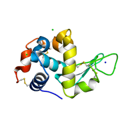
Serial Millisecond Crystallography of Membrane and Soluble Protein Micro-crystals using Synchrotron Radiation
Descriptor: CHLORIDE ION, Lysozyme C, SODIUM ION
Authors:Martin-Garcia, J.M, Conrad, C.E, Nelson, G, Stander, N, Zatsepin, N.A, Zook, J, Zhu, L, Geiger, J, Chun, E, Kissick, D, Hilgart, M.C, Ogata, C, Ishchenko, A, Nagaratnam, N, Roy-Chowdhury, S, Coe, J, Subramanian, G, Schaffer, A, James, D, Ketawala, G, Venugopalan, N, Xu, S, Corcoran, S, Ferguson, D, Weierstall, U, Spence, J.C.H, Cherezov, V, Fromme, P, Fischetti, R.F, Liu, W.
Deposit date:2017-02-20
Release date:2017-05-24
Last modified:2023-10-04
Method:X-RAY DIFFRACTION (2.05 Å)
Cite:Serial millisecond crystallography of membrane and soluble protein microcrystals using synchrotron radiation.
IUCrJ, 4, 2017
5UVK
DownloadVisualize
BU of 5uvk by Molmil
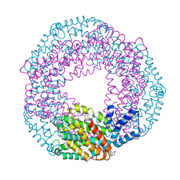
Serial Millisecond Crystallography of Membrane and Soluble Protein Micro-crystals using Synchrotron Radiation
Descriptor: C-phycocyanin alpha chain, C-phycocyanin beta chain, PHYCOCYANOBILIN
Authors:Martin-Garcia, J.M, Conrad, C.E, Nelson, G, Stander, N, Zatsepin, N.A, Zook, J, Zhu, L, Geiger, J, Chun, E, Kissick, D, Hilgart, M.C, Ogata, C, Ishchenko, A, Nagaratnam, N, Roy-Chowdhury, S, Coe, J, Subramanian, G, Schaffer, A, James, D, Ketawala, G, Venugopalan, N, Xu, S, Corcoran, S, Ferguson, D, Weierstall, U, Spence, J.C.H, Cherezov, V, Fromme, P, Fischetti, R.F, Liu, W.
Deposit date:2017-02-20
Release date:2017-05-24
Last modified:2023-10-04
Method:X-RAY DIFFRACTION (3.1 Å)
Cite:Serial millisecond crystallography of membrane and soluble protein microcrystals using synchrotron radiation.
Iucrj, 4, 2017
5UVL
DownloadVisualize
BU of 5uvl by Molmil
Serial Millisecond Crystallography of Membrane and Soluble Protein Micro-crystals using Synchrotron Radiation
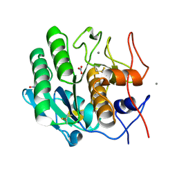
Descriptor: CALCIUM ION, NITRATE ION, Proteinase K
Authors:Martin-Garcia, J.M, Conrad, C.E, Nelson, G, Stander, N, Zatsepin, N.A, Zook, J, Zhu, L, Geiger, J, Chun, E, Kissick, D, Hilgart, M.C, Ogata, C, Ishchenko, A, Nagaratnam, N, Roy-Chowdhury, S, Coe, J, Subramanian, G, Schaffer, A, James, D, Ketawala, G, Venugopalan, N, Xu, S, Corcoran, S, Ferguson, D, Weierstall, U, Spence, J.C.H, Cherezov, V, Fromme, P, Fischetti, R.F, Liu, W.
Deposit date:2017-02-20
Release date:2017-05-24
Last modified:2023-10-04
Method:X-RAY DIFFRACTION (2.65 Å)
Cite:Serial millisecond crystallography of membrane and soluble protein microcrystals using synchrotron radiation.
IUCrJ, 4, 2017
2QZS
DownloadVisualize
BU of 2qzs by Molmil
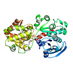
Crystal Structure of Wild-type E.coli GS in complex with ADP and Glucose(wtGSb)
Descriptor: (2R)-2-hydroxy-3-[4-(2-hydroxyethyl)piperazin-1-yl]propane-1-sulfonic acid, ADENOSINE-5'-DIPHOSPHATE, alpha-D-glucopyranose, ...
Authors:Sheng, F, Geiger, J.
Deposit date:2007-08-17
Release date:2008-09-09
Last modified:2023-08-30
Method:X-RAY DIFFRACTION (2.2 Å)
Cite:The Crystal Structures of the Open and Catalytically Competent Closed Conformation of Escherichia coli Glycogen Synthase.
J.Biol.Chem., 284, 2009
2R4U
DownloadVisualize
BU of 2r4u by Molmil
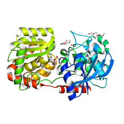
Crystal Structure of Wild-type E.coli GS in complex with ADP and Glucose(wtGSd)
Descriptor: (2R)-2-hydroxy-3-[4-(2-hydroxyethyl)piperazin-1-yl]propane-1-sulfonic acid, 3,6,9,12,15,18,21,24,27,30,33,36,39-TRIDECAOXAHENTETRACONTANE-1,41-DIOL, ADENOSINE-5'-DIPHOSPHATE, ...
Authors:Sheng, F, Geiger, J.
Deposit date:2007-09-01
Release date:2008-09-09
Last modified:2023-08-30
Method:X-RAY DIFFRACTION (2.367 Å)
Cite:The crystal structures of the open and catalytically competent closed conformation of Escherichia coli glycogen synthase.
J.Biol.Chem., 284, 2009
2R4T
DownloadVisualize
BU of 2r4t by Molmil
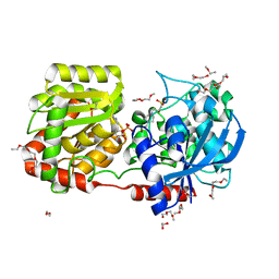
Crystal Structure of Wild-type E.coli GS in Complex with ADP and Glucose(wtGSc)
Descriptor: (2R)-2-hydroxy-3-[4-(2-hydroxyethyl)piperazin-1-yl]propane-1-sulfonic acid, 3,6,9,12,15,18,21,24,27,30,33,36,39-TRIDECAOXAHENTETRACONTANE-1,41-DIOL, ADENOSINE-5'-DIPHOSPHATE, ...
Authors:Sheng, F, Geiger, J.
Deposit date:2007-09-01
Release date:2008-09-09
Last modified:2023-08-30
Method:X-RAY DIFFRACTION (2.258 Å)
Cite:The crystal structures of the open and catalytically competent closed conformation of Escherichia coli glycogen synthase.
J.Biol.Chem., 284, 2009
7UCS
DownloadVisualize
BU of 7ucs by Molmil
The Crystal Structure of Domain-Swapped Dimer Q108K:T51D:A28C:L36C:F57:H:H:H:R58 Mutant of hCRBPII with Histidine Insertion in the Hinge Loop Region at 1.92 Angstrom Resolution
Descriptor: Retinol-binding protein 2
Authors:Ghanbarpour, A, Geiger, J.
Deposit date:2022-03-17
Release date:2023-10-18
Method:X-RAY DIFFRACTION (1.92 Å)
Cite:The Crystal Structure of Domain-Sawpped Dimer Q108K:T51D:A28C:L36C:F57:H:R58 Mutant of hCRBPII with a Histidine Insertion in the Hinge Loop Region at 1.96 Angstrom Resolution
To Be Published
7UCT
DownloadVisualize
BU of 7uct by Molmil
The Crystal Structure of Apo Domain-Swapped Dimer F57:H:H:H:H:H:H:R58 Mutant of HCRBPII with Histidine Insertion in the Hinge Loop Region at 2.5 Angstrom Resolution
Descriptor: Retinol-binding protein 2
Authors:Ghanbarpour, A, Geiger, J.
Deposit date:2022-03-17
Release date:2023-10-18
Method:X-RAY DIFFRACTION (2.52 Å)
Cite:The Crystal Structure of Domain-Sawpped Dimer Q108K:T51D:A28C:L36C:F57:H:R58 Mutant of hCRBPII with a Histidine Insertion in the Hinge Loop Region at 1.96 Angstrom Resolution
To Be Published
7UD3
DownloadVisualize
BU of 7ud3 by Molmil
The Crystal Structure of Domain-Swapped Dimer Q108K:T51D:A28C:L36C:F57:W:W:W:R58 Mutant of hCRBPII with a Histidine Insertion in the Hinge Loop Region at 2.36 Angstrom Resolution
Descriptor: Retinol-binding protein 2
Authors:Ghanbarpour, A, Geiger, J.
Deposit date:2022-03-17
Release date:2023-10-18
Method:X-RAY DIFFRACTION (2.37 Å)
Cite:The Crystal Structure of Domain-Sawpped Dimer Q108K:T51D:A28C:L36C:F57:H:R58 Mutant of hCRBPII with a Histidine Insertion in the Hinge Loop Region at 1.96 Angstrom Resolution
To Be Published
7UCV
DownloadVisualize
BU of 7ucv by Molmil
The Crystal Structure of Apo Domain-Swapped Dimer Q108K:T51D:A28CL36C R58:H:H:H:N59 HCRBPII with Histidine Insertion in the Hinge Loop Region at 2.19 Angstrom Resolution
Descriptor: Retinol-binding protein 2
Authors:Ghanbarpour, A, Geiger, J.
Deposit date:2022-03-17
Release date:2023-10-18
Method:X-RAY DIFFRACTION (2.19 Å)
Cite:The Crystal Structure of Domain-Sawpped Dimer Q108K:T51D:A28C:L36C:F57:H:R58 Mutant of hCRBPII with a Histidine Insertion in the Hinge Loop Region at 1.96 Angstrom Resolution
To Be Published
7UCZ
DownloadVisualize
BU of 7ucz by Molmil
The Crystal Structure of Apo Monomer F57:H:H:H:H:H:H:R58 Mutant of HCRBPII with Histidine Insertion in the Hinge Loop Region at 1.1 Angstrom Resolution
Descriptor: Retinol-binding protein 2
Authors:Ghanbarpour, A, Geiger, J.
Deposit date:2022-03-17
Release date:2023-10-18
Method:X-RAY DIFFRACTION (1.08 Å)
Cite:The Crystal Structure of Domain-Sawpped Dimer Q108K:T51D:A28C:L36C:F57:H:R58 Mutant of hCRBPII with a Histidine Insertion in the Hinge Loop Region at 1.96 Angstrom Resolution
To Be Published
7UD1
DownloadVisualize
BU of 7ud1 by Molmil
The Crystal Structure of Apo Monomer F57:H:H:H:R58 HCRBPII with Histidine Insertion in the Hinge Loop Region at 1.3 Angstrom Resolution
Descriptor: Retinol-binding protein 2
Authors:Ghanbarpour, A, Geiger, J.
Deposit date:2022-03-17
Release date:2023-10-18
Method:X-RAY DIFFRACTION (1.32 Å)
Cite:The Crystal Structure of Apo Monomer F57:H:H:H:R58 HCRBPII with Histidine Insertion in the Hinge Loop Region at 1.3 Angstrom Resolution
To Be Published
7UCN
DownloadVisualize
BU of 7ucn by Molmil
The Crystal Structure of Domain-Swapped Dimer Q108K:T51D:A28C:L36C:F57:H:R58 Mutant of hCRBPII with a Histidine Insertion in the Hinge Loop Region at 1.96 Angstrom Resolution
Descriptor: Retinol-binding protein 2
Authors:Ghanbarpour, A, Geiger, J.
Deposit date:2022-03-16
Release date:2023-10-18
Method:X-RAY DIFFRACTION (1.96 Å)
Cite:The Crystal Structure of Domain-Sawpped Dimer Q108K:T51D:A28C:L36C:F57:H:R58 Mutant of hCRBPII with a Histidine Insertion in the Hinge Loop Region at 1.96 Angstrom Resolution
To Be Published
6WP0
DownloadVisualize
BU of 6wp0 by Molmil
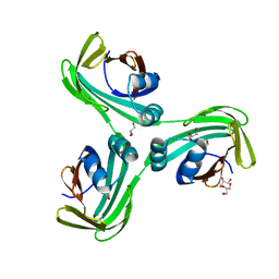
The Crystal Structure of Domain-Swapped Trimer Q108K:T51D variant of HCRBPII
Descriptor: ACETATE ION, GLYCEROL, Retinol-binding protein 2
Authors:Ghanbarpour, A, Geiger, J.
Deposit date:2020-04-26
Release date:2020-08-19
Last modified:2023-10-18
Method:X-RAY DIFFRACTION (2.78 Å)
Cite:Human Cellular Retinol Binding Protein II Forms a Domain-Swapped Trimer Representing a Novel Fold and a New Template for Protein Engineering.
Chembiochem, 21, 2020


123>

222624

PDB entries from 2024-07-17

PDB statisticsPDBj update infoContact PDBjnumon