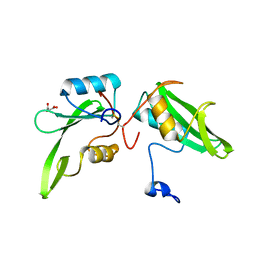
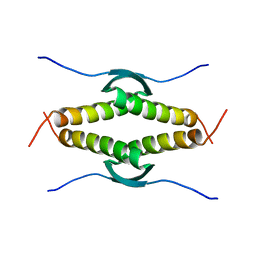
6D8L
 
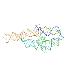 | |
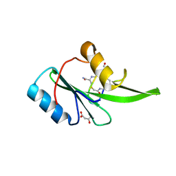
6DKU
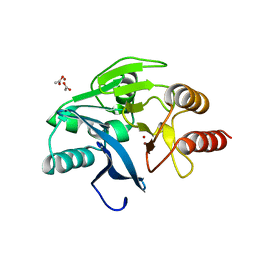
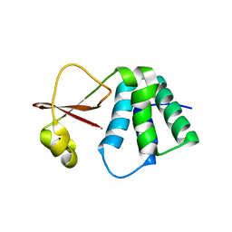 | | Crystal structure of Myotis VP35 mutant of interferon inhibitory domain | | 分子名称: | VP35 | | 著者 | Liu, H, Ginell, G.M, Keefe, L.J, Leung, D.W, Amarasinghe, G.K. | | 登録日 | 2018-05-30 | | 公開日 | 2018-07-18 | | 最終更新日 | 2023-10-11 | | 実験手法 | X-RAY DIFFRACTION (2.6 Å) | | 主引用文献 | Conservation of Structure and Immune Antagonist Functions of Filoviral VP35 Homologs Present in Microbat Genomes.
Cell Rep, 24, 2018
|
|
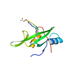
3EE6
 | | Crystal Structure Analysis of Tripeptidyl peptidase -I | | 分子名称: | 2-acetamido-2-deoxy-beta-D-glucopyranose, CALCIUM ION, CHLORIDE ION, ... | | 著者 | Pal, A, Kraetzner, R, Grapp, M, Gruene, T, Schreiber, K, Granborg, M, Urlaub, H, Asif, A.R, Becker, S, Gartner, J, Sheldrick, G.M, Steinfeld, R. | | 登録日 | 2008-09-04 | | 公開日 | 2008-11-25 | | 最終更新日 | 2020-07-29 | | 実験手法 | X-RAY DIFFRACTION (2.35 Å) | | 主引用文献 | Structure of tripeptidyl-peptidase I provides insight into the molecular basis of late infantile neuronal ceroid lipofuscinosis
J.Biol.Chem., 284, 2009
|
|
4CYE
 | | Crystal structure of avian FAK FERM domain FAK31-405 at 3.2A | | 分子名称: | FOCAL ADHESION KINASE 1 | | 著者 | Goni, G.M, Epifano, C, Boskovic, J, Camacho-Artacho, M, Zhou, J, Martin, M.T, Eck, M.J, Kremer, L, Graeter, F, Gervasio, F.L, Perez-Moreno, M, Lietha, D. | | 登録日 | 2014-04-10 | | 公開日 | 2014-04-23 | | 最終更新日 | 2023-12-20 | | 実験手法 | X-RAY DIFFRACTION (3.2 Å) | | 主引用文献 | Phosphatidylinositol 4,5-Bisphosphate Triggers Activation of Focal Adhesion Kinase by Inducing Clustering and Conformational Changes.
Proc.Natl.Acad.Sci.USA, 111, 2014
|
|
3F8L
 | | Crystal Structure of the Effector Domain of PhnF from Mycobacterium smegmatis | | 分子名称: | GLYCEROL, HTH-type transcriptional repressor phnF, SULFATE ION | | 著者 | Busby, J.N, Gebhard, S, Cook, G.M, Baker, E.N, Lott, S.J, Money, V.A. | | 登録日 | 2008-11-12 | | 公開日 | 2009-11-17 | | 最終更新日 | 2023-11-01 | | 実験手法 | X-RAY DIFFRACTION (1.9 Å) | | 主引用文献 | Crystal structure of PhnF, a GntR-family transcription regulator in Mycobacterium smegmatis
To be Published
|
|
4D18
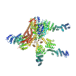 | | Crystal structure of the COP9 signalosome | | 分子名称: | COP9 SIGNALOSOME COMPLEX SUBUNIT 1, COP9 SIGNALOSOME COMPLEX SUBUNIT 2, COP9 SIGNALOSOME COMPLEX SUBUNIT 3, ... | | 著者 | Bunker, R.D, Lingaraju, G.M, Thoma, N.H. | | 登録日 | 2014-05-01 | | 公開日 | 2014-07-23 | | 最終更新日 | 2024-05-08 | | 実験手法 | X-RAY DIFFRACTION (4.08 Å) | | 主引用文献 | Crystal Structure of the Human Cop9 Signalosome
Nature, 512, 2014
|
|
4D0P
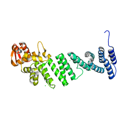 | | Crystal structure of human CSN4 | | 分子名称: | 1,2-ETHANEDIOL, CHLORIDE ION, COP9 SIGNALOSOME COMPLEX SUBUNIT 4, ... | | 著者 | Bunker, R.D, Lingaraju, G.M, Thoma, N.H. | | 登録日 | 2014-04-29 | | 公開日 | 2014-07-23 | | 最終更新日 | 2024-05-08 | | 実験手法 | X-RAY DIFFRACTION (1.6 Å) | | 主引用文献 | Crystal Structure of the Cop9 Signalosome
Nature, 512, 2014
|
|
4D10
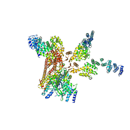 | | Crystal structure of the COP9 signalosome | | 分子名称: | COP9 SIGNALOSOME COMPLEX SUBUNIT 1, COP9 SIGNALOSOME COMPLEX SUBUNIT 2, COP9 SIGNALOSOME COMPLEX SUBUNIT 3, ... | | 著者 | Bunker, R.D, Lingaraju, G.M, Thoma, N.H. | | 登録日 | 2014-04-30 | | 公開日 | 2014-07-23 | | 最終更新日 | 2024-05-08 | | 実験手法 | X-RAY DIFFRACTION (3.8 Å) | | 主引用文献 | Crystal Structure of the Human Cop9 Signalosome
Nature, 512, 2014
|
|
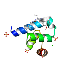
4TRX
 | |
5EEL
 | | Grb7 SH2 with bicyclic peptide inhibitor | | 分子名称: | Bicyclic Peptide Inhibitor, FORMIC ACID, Growth factor receptor-bound protein 7, ... | | 著者 | Watson, G.M, Gunzburg, M.J, Wilce, M.C.J, Wilce, J.A. | | 登録日 | 2015-10-23 | | 公開日 | 2016-06-15 | | 最終更新日 | 2018-12-26 | | 実験手法 | X-RAY DIFFRACTION (2.47 Å) | | 主引用文献 | Unexpected involvement of staple leads to redesign of selective bicyclic peptide inhibitor of Grb7.
Sci Rep, 6, 2016
|
|
5EG2
 | | SET7/9 N265A in complex with AdoHcy and TAF10 peptide | | 分子名称: | Histone-lysine N-methyltransferase SETD7, S-ADENOSYL-L-HOMOCYSTEINE, SODIUM ION, ... | | 著者 | Kroner, G.M, Fick, R.J, Trievel, R.C. | | 登録日 | 2015-10-26 | | 公開日 | 2016-01-13 | | 最終更新日 | 2023-09-27 | | 実験手法 | X-RAY DIFFRACTION (1.55 Å) | | 主引用文献 | Sulfur-Oxygen Chalcogen Bonding Mediates AdoMet Recognition in the Lysine Methyltransferase SET7/9.
Acs Chem.Biol., 11, 2016
|
|
7SX8
 | | T-Plastin-F-actin complex, parallel bundled state | | 分子名称: | ADENOSINE-5'-DIPHOSPHATE, Actin, alpha skeletal muscle, ... | | 著者 | Mei, L, Reynolds, M.J, Alushin, G.M. | | 登録日 | 2021-11-22 | | 公開日 | 2022-08-31 | | 最終更新日 | 2022-09-21 | | 実験手法 | ELECTRON MICROSCOPY (9 Å) | | 主引用文献 | Structural mechanism for bidirectional actin cross-linking by T-plastin.
Proc.Natl.Acad.Sci.USA, 119, 2022
|
|
7SX9
 | | T-Plastin-F-actin complex, anti-parallel bundled state | | 分子名称: | ADENOSINE-5'-DIPHOSPHATE, Actin, alpha skeletal muscle, ... | | 著者 | Mei, L, Reynolds, M.J, Alushin, G.M. | | 登録日 | 2021-11-22 | | 公開日 | 2022-08-31 | | 最終更新日 | 2022-09-21 | | 実験手法 | ELECTRON MICROSCOPY (10 Å) | | 主引用文献 | Structural mechanism for bidirectional actin cross-linking by T-plastin.
Proc.Natl.Acad.Sci.USA, 119, 2022
|
|
7SXA
 | | T-Plastin-F-actin complex, pre-bundling intermediate state | | 分子名称: | ADENOSINE-5'-DIPHOSPHATE, Actin, alpha skeletal muscle, ... | | 著者 | Mei, L, Reynolds, M.J, Alushin, G.M. | | 登録日 | 2021-11-22 | | 公開日 | 2022-08-31 | | 最終更新日 | 2022-09-21 | | 実験手法 | ELECTRON MICROSCOPY (6.87 Å) | | 主引用文献 | Structural mechanism for bidirectional actin cross-linking by T-plastin.
Proc.Natl.Acad.Sci.USA, 119, 2022
|
|
4WWQ
 | | Apo structure of the Grb7 SH2 domain | | 分子名称: | Growth factor receptor-bound protein 7, MALONIC ACID | | 著者 | Watson, G.M, Ambaye, N.D, Wilce, M.C, Wilce, J.A. | | 登録日 | 2014-11-12 | | 公開日 | 2015-09-23 | | 最終更新日 | 2023-09-27 | | 実験手法 | X-RAY DIFFRACTION (1.8 Å) | | 主引用文献 | Cyclic Peptides Incorporating Phosphotyrosine Mimetics as Potent and Specific Inhibitors of the Grb7 Breast Cancer Target.
J.Med.Chem., 58, 2015
|
|
4X6S
 | | Grb7 SH2 domain with phosphotyrosine mimetic inhibitor peptide | | 分子名称: | Growth factor receptor-bound protein 7, Phosphotyrosine mimetic inhibitor peptide G7-TEM1 | | 著者 | Watson, G.M, Panjikar, S, Wilce, M.C, Wilce, J.A. | | 登録日 | 2014-12-09 | | 公開日 | 2015-09-23 | | 最終更新日 | 2015-10-28 | | 実験手法 | X-RAY DIFFRACTION (2.55 Å) | | 主引用文献 | Cyclic Peptides Incorporating Phosphotyrosine Mimetics as Potent and Specific Inhibitors of the Grb7 Breast Cancer Target.
J.Med.Chem., 58, 2015
|
|
4WSN
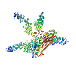 | | Crystal structure of the COP9 signalosome, a P1 crystal form | | 分子名称: | COP9 signalosome complex subunit 1, COP9 signalosome complex subunit 2, COP9 signalosome complex subunit 3, ... | | 著者 | Bunker, R.D, Lingaraju, G.M, Thoma, N.H. | | 登録日 | 2014-10-28 | | 公開日 | 2015-12-23 | | 最終更新日 | 2024-01-10 | | 実験手法 | X-RAY DIFFRACTION (5.5 Å) | | 主引用文献 | Cullin-RING ubiquitin E3 ligase regulation by the COP9 signalosome.
Nature, 531, 2016
|
|
1K3V
 | | Porcine Parvovirus Capsid | | 分子名称: | capsid protein VP2 | | 著者 | Simpson, A.A, Hebert, B, Sullivan, G.M, Parrish, C.R, Zadori, Z, Tijssen, P, Rossmann, M.G. | | 登録日 | 2001-10-04 | | 公開日 | 2001-10-24 | | 最終更新日 | 2024-04-03 | | 実験手法 | X-RAY DIFFRACTION (3.5 Å) | | 主引用文献 | The structure of porcine parvovirus: comparison with related viruses.
J.Mol.Biol., 315, 2002
|
|
1KO3
 | | VIM-2, a Zn-beta-lactamase from Pseudomonas aeruginosa with Cys221 reduced | | 分子名称: | ACETATE ION, CHLORIDE ION, HYDROXIDE ION, ... | | 著者 | Garcia-Saez, I, Docquier, J.-D, Rossolini, G.M, Dideberg, O. | | 登録日 | 2001-12-20 | | 公開日 | 2003-09-02 | | 最終更新日 | 2023-10-25 | | 実験手法 | X-RAY DIFFRACTION (1.91 Å) | | 主引用文献 | The three-dimensional structure of VIM-2, a Zn-beta-lactamase from Pseudomonas aeruginosa in its reduced and oxidised form
J.Mol.Biol., 375, 2008
|
|
2W7U
 | | SplA serine protease of Staphylococcus aureus (2.4A) | | 分子名称: | SERINE PROTEASE SPLA | | 著者 | Stec-Niemczyka, J, Pustelny, K, Kisielewska, M, Bista, M, Boulware, K.T, Stennicke, H.R, Thogersen, I.B, Daugherty, P.S, Enghild, J.J, Popowicz, G.M, Dubin, A, Potempa, J, Dubin, G. | | 登録日 | 2008-12-30 | | 公開日 | 2010-03-31 | | 最終更新日 | 2023-12-13 | | 実験手法 | X-RAY DIFFRACTION (2.43 Å) | | 主引用文献 | Structural and Functional Characterization of Spla, an Exclusively Specific Protease of Staphylococcus Aureus.
Biochem.J., 419, 2009
|
|
6T3C
 | | Crystal structure of PI3Kgamma in complex with DNA-PK inhibitor AZD7648 | | 分子名称: | 7-methyl-2-[(7-methyl-[1,2,4]triazolo[1,5-a]pyridin-6-yl)amino]-9-(oxan-4-yl)purin-8-one, Phosphatidylinositol 4,5-bisphosphate 3-kinase catalytic subunit gamma isoform | | 著者 | Schimpl, M, Goldberg, F.W, Finlay, M.R.V, Ting, A.K.T, Beattie, D, Lamont, G.M, Fallan, C, Wrigley, G.L, Howard, M.R, Williamson, B, Davies, B.R, Cadogan, E.B, Ramos-Montoya, A, Dean, E. | | 登録日 | 2019-10-10 | | 公開日 | 2020-01-01 | | 最終更新日 | 2024-01-24 | | 実験手法 | X-RAY DIFFRACTION (2.62 Å) | | 主引用文献 | The Discovery of 7-Methyl-2-[(7-methyl[1,2,4]triazolo[1,5-a]pyridin-6-yl)amino]-9-(tetrahydro-2H-pyran-4-yl)-7,9-dihydro-8H-purin-8-one (AZD7648), a Potent and Selective DNA-Dependent Protein Kinase (DNA-PK) Inhibitor.
J.Med.Chem., 63, 2020
|
|
6T2W
 | | Crystal structure of the CSF1R kinase domain with a dihydropurinone inhibitor (compound 4) | | 分子名称: | 2-[(4-methoxy-2-methyl-phenyl)amino]-7-methyl-9-(4-oxidanylcyclohexyl)purin-8-one, Macrophage colony-stimulating factor 1 receptor, SULFATE ION | | 著者 | Schimpl, M, Goldberg, F.W, Finlay, M.R.V, Ting, A.K.T, Beattie, D, Lamont, G.M, Fallan, C, Wrigley, G.L, Howard, M.R, Williamson, B, Davies, B.R, Cadogan, E.B, Ramos-Montoya, A, Dean, E. | | 登録日 | 2019-10-09 | | 公開日 | 2020-01-01 | | 最終更新日 | 2024-05-01 | | 実験手法 | X-RAY DIFFRACTION (1.7 Å) | | 主引用文献 | The Discovery of 7-Methyl-2-[(7-methyl[1,2,4]triazolo[1,5-a]pyridin-6-yl)amino]-9-(tetrahydro-2H-pyran-4-yl)-7,9-dihydro-8H-purin-8-one (AZD7648), a Potent and Selective DNA-Dependent Protein Kinase (DNA-PK) Inhibitor.
J.Med.Chem., 63, 2020
|
|
3SAK
 | |
5N8V
 | | Targeting the PEX14-PEX5 interaction by small molecules provides novel therapeutic routes to treat trypanosomiases. | | 分子名称: | 1-(2-azanylethyl)-5-[(4-methoxynaphthalen-1-yl)methyl]-~{N}-(naphthalen-1-ylmethyl)-6,7-dihydro-4~{H}-pyrazolo[4,3-c]pyridine-3-carboxamide, BETA-MERCAPTOETHANOL, CHLORIDE ION, ... | | 著者 | Dawidowski, M, Emmanouilidis, L, Sattler, M, Popowicz, G.M. | | 登録日 | 2017-02-24 | | 公開日 | 2017-03-15 | | 最終更新日 | 2024-05-08 | | 実験手法 | X-RAY DIFFRACTION (1.55 Å) | | 主引用文献 | Inhibitors of PEX14 disrupt protein import into glycosomes and kill Trypanosoma parasites.
Science, 355, 2017
|
|
4QQB
 | | Structural basis for the assembly of the SXL-UNR translation regulatory complex | | 分子名称: | Protein sex-lethal, Upstream of N-ras, isoform A, ... | | 著者 | Hennig, J, Popowicz, G.M, Sattler, M. | | 登録日 | 2014-06-27 | | 公開日 | 2014-09-03 | | 最終更新日 | 2023-09-20 | | 実験手法 | X-RAY DIFFRACTION (2.8 Å) | | 主引用文献 | Structural basis for the assembly of the Sxl-Unr translation regulatory complex.
Nature, 515, 2014
|
|


