Loading
PDBj
MenuPDBj@FacebookPDBj@TwitterPDBj@YouTubewwPDB FoundationwwPDB
RCSB PDBPDBeBMRBAdv. SearchSearch help
PDB: 158 results

1RFK
DownloadVisualize
BU of 1rfk by Molmil
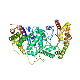
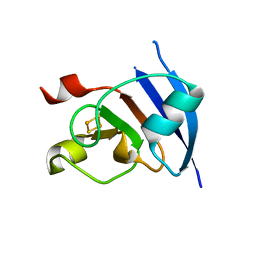
Crystal Structure of 2Fe2S Ferredoxin from Thermophilic Cyanobacterium Mastigocladus Laminosus
Descriptor: FE2/S2 (INORGANIC) CLUSTER, Ferredoxin
Authors:Fish, A, Nechushtai, R, Livnah, O.
Deposit date:2003-11-10
Release date:2005-04-12
Last modified:2024-02-14
Method:X-RAY DIFFRACTION (1.25 Å)
Cite:Structural basis for the thermostability of ferredoxin from the cyanobacterium Mastigocladus laminosus.
J.Mol.Biol., 350, 2005
7TNI
DownloadVisualize
BU of 7tni by Molmil
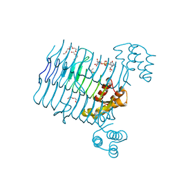
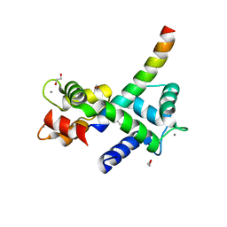
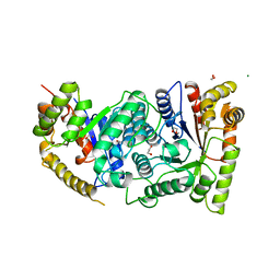
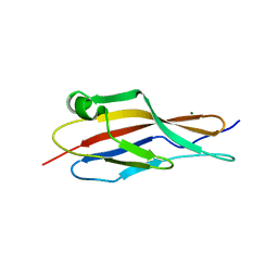
Structure of EC12 Y1392W variant of BT-R1 from Manduca sexta, a Cry1A toxin binding domain
Descriptor: Cadherin, MAGNESIUM ION
Authors:Fisher, A.J, Wilcox, X.E.
Deposit date:2022-01-21
Release date:2022-05-04
Last modified:2023-10-18
Method:X-RAY DIFFRACTION (1.9 Å)
Cite:Functional and Structural Analysis of the Toxin-Binding Site of the Cadherin G-Protein-Coupled Receptor, BT-R 1 , for Cry1A Toxins of Bacillus thuringiensis .
Biochemistry, 61, 2022
6VLB
DownloadVisualize
BU of 6vlb by Molmil
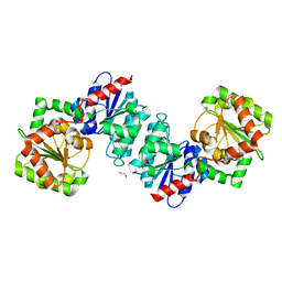
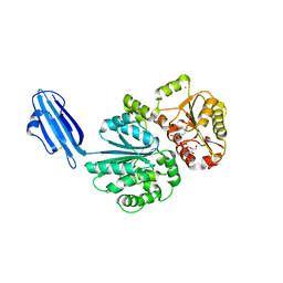
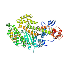
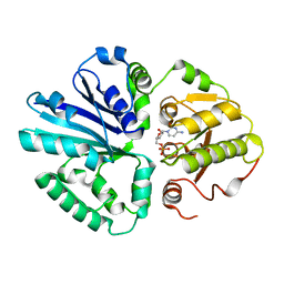
Crystal structure of ligand-free UDP-GlcNAc 2-epimerase from Neisseria meningitidis
Descriptor: 1,2-ETHANEDIOL, SODIUM ION, UDP-N-acetylglucosamine 2-epimerase
Authors:Fisher, A.J, Hurlburt, N.K.
Deposit date:2020-01-23
Release date:2020-11-11
Last modified:2023-10-11
Method:X-RAY DIFFRACTION (1.85 Å)
Cite:Structural characterization of a nonhydrolyzing UDP-GlcNAc 2-epimerase from Neisseria meningitidis serogroup A.
Acta Crystallogr.,Sect.F, 76, 2020
8E4X
DownloadVisualize
BU of 8e4x by Molmil
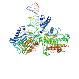
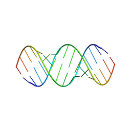
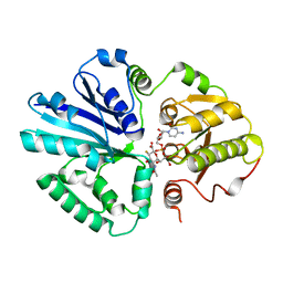
Human Adenosine Deaminase Acting on dsRNA (ADAR2-R2D) bound to dsRNA containing a G:3-deaza dA pair adjacent to the target site
Descriptor: Double-stranded RNA-specific editase 1, INOSITOL HEXAKISPHOSPHATE, RNA (5-R(*GP*CP*UP*CP*GP*CP*GP*AP*UP*GP*CP*GP*(8AZ)P*GP*AP*GP*GP*GP*CP* UP*CP*UP*GP*AP*UP*AP*GP*CP*UP*AP*CP*G)-3), ...
Authors:Fisher, A.J, Mendoza, H.G.
Deposit date:2022-08-19
Release date:2022-10-26
Last modified:2023-10-18
Method:X-RAY DIFFRACTION (2.8 Å)
Cite:ADAR activation by inducing a syn conformation at guanosine adjacent to an editing site.
Nucleic Acids Res., 50, 2022
6VLC
DownloadVisualize
BU of 6vlc by Molmil
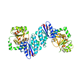
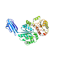
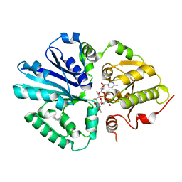
Crystal structure of UDP-GlcNAc 2-epimerase from Neisseria meningitidis bound to UDP-GlcNAc
Descriptor: UDP-N-acetylglucosamine 2-epimerase, URIDINE-DIPHOSPHATE-N-ACETYLGLUCOSAMINE
Authors:Fisher, A.J, Hurlburt, N.K.
Deposit date:2020-01-23
Release date:2020-11-11
Last modified:2023-10-11
Method:X-RAY DIFFRACTION (2.15 Å)
Cite:Structural characterization of a nonhydrolyzing UDP-GlcNAc 2-epimerase from Neisseria meningitidis serogroup A.
Acta Crystallogr.,Sect.F, 76, 2020
4R37
DownloadVisualize
BU of 4r37 by Molmil
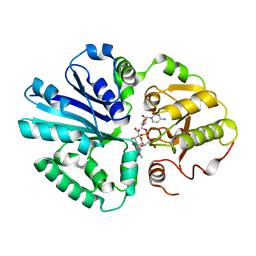
Crystal structure analysis of LpxA, a UDP-N-acetylglucosamine acyltransferase from Bacteroides fragilis 9343 with UDP-GlcNAc
Descriptor: 2-(2-METHOXYETHOXY)ETHANOL, 3,6,9,12,15,18,21,24-OCTAOXAHEXACOSAN-1-OL, ACETATE ION, ...
Authors:Fisher, A.J, Chen, X, Ngo, A.
Deposit date:2014-08-14
Release date:2015-05-06
Last modified:2023-09-20
Method:X-RAY DIFFRACTION (1.9 Å)
Cite:Structures of Bacteroides fragilis uridine 5'-diphosphate-N-acetylglucosamine (UDP-GlcNAc) acyltransferase (BfLpxA).
Acta Crystallogr.,Sect.D, 71, 2015
4R84
DownloadVisualize
BU of 4r84 by Molmil
Crystal structure of Sialyltransferase from Photobacterium damsela with CMP-3F(a)Neu5Ac bound
Descriptor: CALCIUM ION, CYTIDINE-5'-MONOPHOSPHATE-3-FLUORO-N-ACETYL-NEURAMINIC ACID, Sialyltransferase 0160
Authors:Fisher, A.J, Chen, X, Li, Y, Huynh, N.
Deposit date:2014-08-29
Release date:2014-12-03
Last modified:2023-09-20
Method:X-RAY DIFFRACTION (1.7 Å)
Cite:Crystal structures of sialyltransferase from Photobacterium damselae.
Febs Lett., 588, 2014
6BGB
DownloadVisualize
BU of 6bgb by Molmil
Crystal Structure of the 16mer GCAGNCUUAAGUCUGC containing BrPh 7-triazolyl-8-aza-7-deazaadenosine
Descriptor: (5'-R(*GP*CP*AP*GP*(A7C)P*CP*UP*UP*AP*AP*GP*UP*CP*UP*GP*C)-3')
Authors:Fisher, A.J, Beal, P.A, Mumbleau, M.M.
Deposit date:2017-10-27
Release date:2017-12-06
Last modified:2024-03-13
Method:X-RAY DIFFRACTION (1.65 Å)
Cite:Controlling miRNA-like off-target effects of an siRNA with nucleobase modifications.
Org. Biomol. Chem., 15, 2017
4R83
DownloadVisualize
BU of 4r83 by Molmil
Crystal structure of Sialyltransferase from Photobacterium damsela
Descriptor: CALCIUM ION, Sialyltransferase 0160
Authors:Fisher, A.J, Chen, X, Li, Y, Huynh, N.
Deposit date:2014-08-29
Release date:2014-12-03
Last modified:2023-09-20
Method:X-RAY DIFFRACTION (1.93 Å)
Cite:Crystal structures of sialyltransferase from Photobacterium damselae.
Febs Lett., 588, 2014
1P35
DownloadVisualize
BU of 1p35 by Molmil
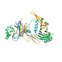
CRYSTAL STRUCTURE OF BACULOVIRUS P35
Descriptor: 1,2-ETHANEDIOL, P35, PHOSPHATE ION
Authors:Fisher, A.J, Delacruz, W.P, Zoog, S.J, Schneider, C.L, Friesen, P.D.
Deposit date:1998-09-18
Release date:1999-06-01
Last modified:2024-02-14
Method:X-RAY DIFFRACTION (2.2 Å)
Cite:Crystal structure of baculovirus P35: role of a novel reactive site loop in apoptotic caspase inhibition.
EMBO J., 18, 1999
7KL5
DownloadVisualize
BU of 7kl5 by Molmil
Structure of Calmodulin Bound to the Cardiac Ryanodine Receptor (RyR2) at Residues: Phe4246 to Val4271
Descriptor: 1,2-ETHANEDIOL, CALCIUM ION, Calmodulin-1, ...
Authors:Fisher, A.J, Ames, J.B, Yu, Q.
Deposit date:2020-10-29
Release date:2021-04-07
Last modified:2023-10-18
Method:X-RAY DIFFRACTION (1.65 Å)
Cite:The Crystal Structure of Calmodulin Bound to the Cardiac Ryanodine Receptor (RyR2) at Residues Phe4246-Val4271 Reveals a Fifth Calcium Binding Site.
Biochemistry, 60, 2021
4RAJ
DownloadVisualize
BU of 4raj by Molmil
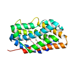
Crystal structure of heme oxygenase 2 from Chlamydomonas reinhardtii without heme.
Descriptor: 1-(2-METHOXY-ETHOXY)-2-{2-[2-(2-METHOXY-ETHOXY]-ETHOXY}-ETHANE, Heme oxygenase
Authors:Fisher, A.J, Lagarias, J.C.
Deposit date:2014-09-10
Release date:2015-09-16
Last modified:2023-09-20
Method:X-RAY DIFFRACTION (1.84 Å)
Cite:Crystal structure of heme oxygenase 2 from Chlamydomonas reinhardtii without heme.
To be Published
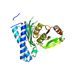
5DJI
DownloadVisualize
BU of 5dji by Molmil
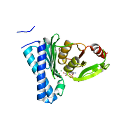
Structure of M. tuberculosis CysQ, a PAP phosphatase with AMP, PO4, and 2Mg bound
Descriptor: 3'-phosphoadenosine 5'-phosphate phosphatase, ADENOSINE MONOPHOSPHATE, MAGNESIUM ION, ...
Authors:Fisher, A.J, Erickson, A.I.
Deposit date:2015-09-02
Release date:2015-11-11
Last modified:2023-09-27
Method:X-RAY DIFFRACTION (1.659 Å)
Cite:Crystal Structures of Mycobacterium tuberculosis CysQ, with Substrate and Products Bound.
Biochemistry, 54, 2015
5DJH
DownloadVisualize
BU of 5djh by Molmil
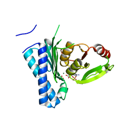
Structure of M. tuberculosis CysQ, a PAP phosphatase with AMP, PO4, and 3Mg bound
Descriptor: 3'-phosphoadenosine 5'-phosphate phosphatase, ADENOSINE MONOPHOSPHATE, MAGNESIUM ION, ...
Authors:Fisher, A.J, Erickson, A.I.
Deposit date:2015-09-02
Release date:2015-11-11
Last modified:2023-09-27
Method:X-RAY DIFFRACTION (1.451 Å)
Cite:Crystal Structures of Mycobacterium tuberculosis CysQ, with Substrate and Products Bound.
Biochemistry, 54, 2015
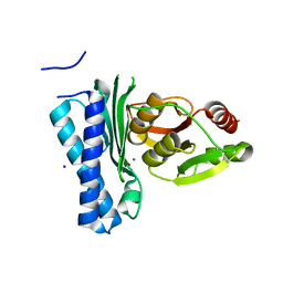
5DJJ
DownloadVisualize
BU of 5djj by Molmil
Structure of M. tuberculosis CysQ, a PAP phosphatase with PO4 and 2Mg bound
Descriptor: 3'-phosphoadenosine 5'-phosphate phosphatase, MAGNESIUM ION, PHOSPHATE ION, ...
Authors:Fisher, A.J, Erickson, A.I.
Deposit date:2015-09-02
Release date:2015-11-11
Last modified:2023-09-27
Method:X-RAY DIFFRACTION (1.501 Å)
Cite:Crystal Structures of Mycobacterium tuberculosis CysQ, with Substrate and Products Bound.
Biochemistry, 54, 2015
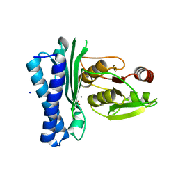
5DJK
DownloadVisualize
BU of 5djk by Molmil
Structure of M. tuberculosis CysQ, a PAP phosphatase with PO4 and 2Ca bound
Descriptor: 3'-phosphoadenosine 5'-phosphate phosphatase, CALCIUM ION, PHOSPHATE ION, ...
Authors:Fisher, A.J, Erickson, A.I.
Deposit date:2015-09-02
Release date:2015-11-11
Last modified:2023-09-27
Method:X-RAY DIFFRACTION (1.799 Å)
Cite:Crystal Structures of Mycobacterium tuberculosis CysQ, with Substrate and Products Bound.
Biochemistry, 54, 2015
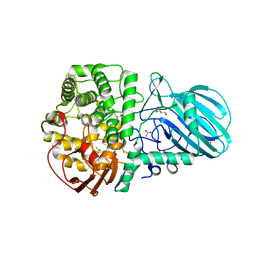
6DWO
DownloadVisualize
BU of 6dwo by Molmil
Crystal structure of alpha-1-2-mannosidase from Enterococcus faecalis V583
Descriptor: ACETATE ION, Alpha-1,2-mannosidase, CALCIUM ION, ...
Authors:Fisher, A.J, Li, Y.
Deposit date:2018-06-26
Release date:2019-10-30
Last modified:2023-10-11
Method:X-RAY DIFFRACTION (2.15 Å)
Cite:Enterococcus faecalis alpha 1-2-mannosidase (EfMan-I): an efficient catalyst for glycoprotein N-glycan modification.
Febs Lett., 594, 2020
5DJG
DownloadVisualize
BU of 5djg by Molmil
Structure of M. tuberculosis CysQ, a PAP phosphatase with PAP, Mg, and Li bound
Descriptor: 3'-phosphoadenosine 5'-phosphate phosphatase, ADENOSINE-3'-5'-DIPHOSPHATE, LITHIUM ION, ...
Authors:Fisher, A.J, Erickson, A.I.
Deposit date:2015-09-02
Release date:2015-11-11
Last modified:2023-09-27
Method:X-RAY DIFFRACTION (1.951 Å)
Cite:Crystal Structures of Mycobacterium tuberculosis CysQ, with Substrate and Products Bound.
Biochemistry, 54, 2015
1BRL
DownloadVisualize
BU of 1brl by Molmil
THREE-DIMENSIONAL STRUCTURE OF BACTERIAL LUCIFERASE FROM VIBRIO HARVEYI AT 2.4 ANGSTROMS RESOLUTION
Descriptor: BACTERIAL LUCIFERASE, PHOSPHATE ION
Authors:Fisher, A.J, Rayment, I.
Deposit date:1995-03-20
Release date:1996-04-01
Last modified:2024-02-07
Method:X-RAY DIFFRACTION (2.4 Å)
Cite:Three-dimensional structure of bacterial luciferase from Vibrio harveyi at 2.4 A resolution.
Biochemistry, 34, 1995
1LUC
DownloadVisualize
BU of 1luc by Molmil
BACTERIAL LUCIFERASE
Descriptor: 1,2-ETHANEDIOL, BACTERIAL LUCIFERASE, MAGNESIUM ION
Authors:Fisher, A.J, Rayment, I.
Deposit date:1996-05-10
Release date:1996-12-07
Last modified:2024-02-14
Method:X-RAY DIFFRACTION (1.5 Å)
Cite:The 1.5-A resolution crystal structure of bacterial luciferase in low salt conditions.
J.Biol.Chem., 271, 1996
1MMD
DownloadVisualize
BU of 1mmd by Molmil
TRUNCATED HEAD OF MYOSIN FROM DICTYOSTELIUM DISCOIDEUM COMPLEXED WITH MGADP-BEF3
Descriptor: ADENOSINE-5'-DIPHOSPHATE, BERYLLIUM TRIFLUORIDE ION, MAGNESIUM ION, ...
Authors:Fisher, A.J, Holden, H.M, Rayment, I.
Deposit date:1995-03-21
Release date:1996-08-17
Last modified:2024-02-14
Method:X-RAY DIFFRACTION (2 Å)
Cite:X-ray structures of the myosin motor domain of Dictyostelium discoideum complexed with MgADP.BeFx and MgADP.AlF4-.
Biochemistry, 34, 1995
2IHK
DownloadVisualize
BU of 2ihk by Molmil
crystal structure of multifunctional sialyltransferase from pasteurella multocida with CMP-3F(equatorial)-Neu5Ac bound
Descriptor: Alpha-2,3/2,6-sialyltransferase/sialidase, CYTIDINE-5'-MONOPHOSPHATE-3-FLUORO-N-ACETYL-NEURAMINIC ACID
Authors:Fisher, A.J, Ni, L.
Deposit date:2006-09-26
Release date:2007-05-22
Last modified:2023-08-30
Method:X-RAY DIFFRACTION (1.9 Å)
Cite:Crystal Structures of Pasteurella multocida Sialyltransferase Complexes with Acceptor and Donor Analogues Reveal Substrate Binding Sites and Catalytic Mechanism.
Biochemistry, 46, 2007
2IHJ
DownloadVisualize
BU of 2ihj by Molmil
crystal structure of multifunctional sialyltransferase from pasteurella multocida with CMP-3F-Neu5Ac bound
Descriptor: Alpha-2,3/2,6-sialyltransferase/sialidase, CYTIDINE-5'-MONOPHOSPHATE-3-FLUORO-N-ACETYL-NEURAMINIC ACID
Authors:Fisher, A.J, Ni, L.
Deposit date:2006-09-26
Release date:2007-05-22
Last modified:2023-08-30
Method:X-RAY DIFFRACTION (2 Å)
Cite:Crystal Structures of Pasteurella multocida Sialyltransferase Complexes with Acceptor and Donor Analogues Reveal Substrate Binding Sites and Catalytic Mechanism.
Biochemistry, 46, 2007
2IHZ
DownloadVisualize
BU of 2ihz by Molmil
Crystal structure of multifunctional sialyltransferase from pasteurella multocida with CMP-3F-Neu5Ac and alpha-lactose bound
Descriptor: Alpha-2,3/2,6-sialyltransferase/sialidase, CYTIDINE-5'-MONOPHOSPHATE-3-FLUORO-N-ACETYL-NEURAMINIC ACID, beta-D-galactopyranose-(1-4)-alpha-D-glucopyranose
Authors:Fisher, A.J, Ni, L.
Deposit date:2006-09-27
Release date:2007-05-22
Last modified:2023-08-30
Method:X-RAY DIFFRACTION (2 Å)
Cite:Crystal Structures of Pasteurella multocida Sialyltransferase Complexes with Acceptor and Donor Analogues Reveal Substrate Binding Sites and Catalytic Mechanism.
Biochemistry, 46, 2007
2ILV
DownloadVisualize
BU of 2ilv by Molmil
crystal structure of multifunctional sialyltransferase from Pasteurella multocida with CMP and alpha-lactose bound
Descriptor: Alpha-2,3/2,6-sialyltransferase/sialidase, CYTIDINE-5'-MONOPHOSPHATE, beta-D-galactopyranose-(1-4)-alpha-D-glucopyranose
Authors:Fisher, A.J, Ni, L.
Deposit date:2006-10-03
Release date:2007-05-22
Last modified:2023-08-30
Method:X-RAY DIFFRACTION (2.27 Å)
Cite:Crystal Structures of Pasteurella multocida Sialyltransferase Complexes with Acceptor and Donor Analogues Reveal Substrate Binding Sites and Catalytic Mechanism.
Biochemistry, 46, 2007

222624

PDB entries from 2024-07-17

PDB statisticsPDBj update infoContact PDBjnumon