Loading
PDBj
MenuPDBj@FacebookPDBj@TwitterPDBj@YouTubewwPDB FoundationwwPDB
RCSB PDBPDBeBMRBAdv. SearchSearch help
PDB: 197 results

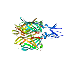
6RCP
DownloadVisualize
BU of 6rcp by Molmil
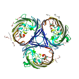
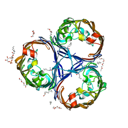
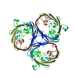
Crystal structure of the OmpK36 clinical isolate ST258 from Klebsiella pneumonia
Descriptor: OmpK36
Authors:Beis, K, Romano, M, Kwong, J.
Deposit date:2019-04-11
Release date:2019-09-11
Last modified:2024-01-24
Method:X-RAY DIFFRACTION (3.231 Å)
Cite:OmpK36-mediated Carbapenem resistance attenuates ST258 Klebsiella pneumoniae in vivo.
Nat Commun, 10, 2019
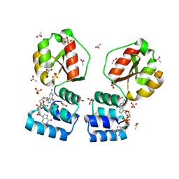
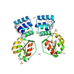
1WAM
DownloadVisualize
BU of 1wam by Molmil
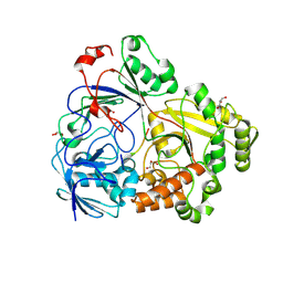
Structure of UDP-galactopyranose mutase from Klebsiella Pneumoniae with FADH-
Descriptor: FLAVIN-ADENINE DINUCLEOTIDE, UDP-GALACTOPYRANOSE MUTASE
Authors:Beis, K, Srikannathasan, V, Naismith, J.H.
Deposit date:2004-10-27
Release date:2006-05-24
Last modified:2023-12-13
Method:X-RAY DIFFRACTION (2.35 Å)
Cite:Crystal Structures of Mycobacteria Tuberculosis and Klebsiella Pneumoniae Udp-Galactopyranose Mutase in the Oxidised State and Klebsiella Pneumoniae Udp-Galactopyranose Mutase in the (Active) Reduced State.
J.Mol.Biol., 348, 2005
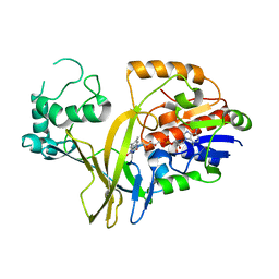
6RD3
DownloadVisualize
BU of 6rd3 by Molmil
Crystal structure of the wild type OmpK36 from Klebsiella pneumonia
Descriptor: (HYDROXYETHYLOXY)TRI(ETHYLOXY)OCTANE, MAGNESIUM ION, OmpK36
Authors:Beis, K, Romano, M, Kwong, J.
Deposit date:2019-04-12
Release date:2019-09-11
Last modified:2024-01-24
Method:X-RAY DIFFRACTION (1.98 Å)
Cite:OmpK36-mediated Carbapenem resistance attenuates ST258 Klebsiella pneumoniae in vivo.
Nat Commun, 10, 2019
7Z6F
DownloadVisualize
BU of 7z6f by Molmil
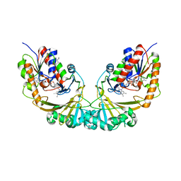
Crystal structure of the substrate-binding protein YejA in complex with peptide fragment
Descriptor: CHLORIDE ION, GLYCEROL, MAGNESIUM ION, ...
Authors:Beis, K, Qu, F.
Deposit date:2022-03-11
Release date:2023-09-20
Method:X-RAY DIFFRACTION (1.65 Å)
Cite:Crystal structure of the substrate-binding protein YejA in complex with peptide fragment
To Be Published
6STL
DownloadVisualize
BU of 6stl by Molmil
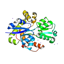
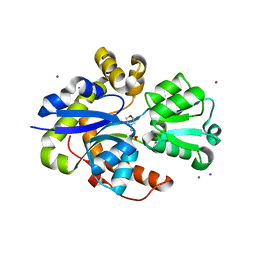
Taurine ABC transporter substrate binding protein TauA from E. coli in complex with taurine
Descriptor: 2-AMINOETHANESULFONIC ACID, IODIDE ION, Taurine-binding periplasmic protein
Authors:Beis, K, Qu, F, Wagner, A, ElOmari, K.
Deposit date:2019-09-10
Release date:2019-12-18
Last modified:2024-05-15
Method:X-RAY DIFFRACTION (1.3 Å)
Cite:Desolvation of the substrate-binding protein TauA dictates ligand specificity for the alkanesulfonate ABC importer TauABC.
Biochem.J., 476, 2019
6SSY
DownloadVisualize
BU of 6ssy by Molmil
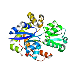
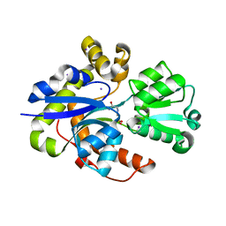
Taurine ABC transporter substrate binding protein TauA from E. coli in complex with 2-Aminoethylphosphonic acid
Descriptor: (2-aminoethyl)phosphonic acid, GLYCEROL, IODIDE ION, ...
Authors:Beis, K, Qu, F.
Deposit date:2019-09-09
Release date:2019-12-18
Last modified:2024-01-24
Method:X-RAY DIFFRACTION (1.62 Å)
Cite:Desolvation of the substrate-binding protein TauA dictates ligand specificity for the alkanesulfonate ABC importer TauABC.
Biochem.J., 476, 2019
6ST1
DownloadVisualize
BU of 6st1 by Molmil
Taurine ABC transporter substrate binding protein TauA from E. coli in complex with 2-(N-Morpholino)ethanesulfonic acid (MES)
Descriptor: 2-(N-MORPHOLINO)-ETHANESULFONIC ACID, GLYCEROL, IODIDE ION, ...
Authors:Beis, K, Qu, F.
Deposit date:2019-09-09
Release date:2019-12-18
Last modified:2024-01-24
Method:X-RAY DIFFRACTION (1.55 Å)
Cite:Desolvation of the substrate-binding protein TauA dictates ligand specificity for the alkanesulfonate ABC importer TauABC.
Biochem.J., 476, 2019
6ST0
DownloadVisualize
BU of 6st0 by Molmil
Taurine ABC transporter substrate binding protein TauA from E. coli in complex with N-(2-Acetamido)-2-aminoethanesulfonic acid
Descriptor: IODIDE ION, N-(2-acetamido)-2-aminoethanesulfonic acid, Taurine-binding periplasmic protein
Authors:Beis, K, Qu, F.
Deposit date:2019-09-09
Release date:2019-12-18
Last modified:2024-01-24
Method:X-RAY DIFFRACTION (1.5 Å)
Cite:Desolvation of the substrate-binding protein TauA dictates ligand specificity for the alkanesulfonate ABC importer TauABC.
Biochem.J., 476, 2019
8PFA
DownloadVisualize
BU of 8pfa by Molmil
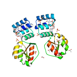
X-ray structure of the Thermus thermophilus K167R mutant of the PilF-GSPIIB domain in the c-di-GMP bound state
Descriptor: 9,9'-[(2R,3R,3aS,5S,7aR,9R,10R,10aS,12S,14aR)-3,5,10,12-tetrahydroxy-5,12-dioxidooctahydro-2H,7H-difuro[3,2-d:3',2'-j][1,3,7,9,2,8]tetraoxadiphosphacyclododecine-2,9-diyl]bis(2-amino-1,9-dihydro-6H-purin-6-one), ACETATE ION, SULFATE ION, ...
Authors:Neissner, K, Woehnert, J.
Deposit date:2023-06-15
Release date:2024-06-26
Method:X-RAY DIFFRACTION (1.8 Å)
Cite:X-ray structure of the Thermus thermophilus K167R mutant of the PilF-GSPIIB domain in the c-di-GMP bound state
To Be Published
6RCK
DownloadVisualize
BU of 6rck by Molmil
Crystal structure of the OmpK36 GD insertion chimera from Klebsiella pneumonia
Descriptor: (2S)-3-{[{[(2S)-2,3-DIHYDROXYPROPYL]OXY}(HYDROXY)PHOSPHORYL]OXY}-2-[(6E)-HEXADEC-6-ENOYLOXY]PROPYL (8E)-OCTADEC-8-ENOATE, (HYDROXYETHYLOXY)TRI(ETHYLOXY)OCTANE, LAURYL DIMETHYLAMINE-N-OXIDE, ...
Authors:Beis, K, Romano, M, Kwong, J.
Deposit date:2019-04-11
Release date:2019-09-11
Last modified:2024-01-24
Method:X-RAY DIFFRACTION (2.029 Å)
Cite:OmpK36-mediated Carbapenem resistance attenuates ST258 Klebsiella pneumoniae in vivo.
Nat Commun, 10, 2019
8PDK
DownloadVisualize
BU of 8pdk by Molmil
X-ray structure of the Thermus thermophilus PilF-GSPIIB domain in the c-di-GMP bound state
Descriptor: 9,9'-[(2R,3R,3aS,5S,7aR,9R,10R,10aS,12S,14aR)-3,5,10,12-tetrahydroxy-5,12-dioxidooctahydro-2H,7H-difuro[3,2-d:3',2'-j][1,3,7,9,2,8]tetraoxadiphosphacyclododecine-2,9-diyl]bis(2-amino-1,9-dihydro-6H-purin-6-one), ACETATE ION, SULFATE ION, ...
Authors:Neissner, K, Woehnert, J.
Deposit date:2023-06-12
Release date:2024-06-26
Method:X-RAY DIFFRACTION (2 Å)
Cite:X-ray structure of the PilF-GSPIIB domain in the c-di-GMP bound state
To Be Published
8PE0
DownloadVisualize
BU of 8pe0 by Molmil
X-ray structure of the Thermus thermophilus K167L mutant of the PilF-GSPIIB domain in the c-di-GMP bound state
Descriptor: 9,9'-[(2R,3R,3aS,5S,7aR,9R,10R,10aS,12S,14aR)-3,5,10,12-tetrahydroxy-5,12-dioxidooctahydro-2H,7H-difuro[3,2-d:3',2'-j][1,3,7,9,2,8]tetraoxadiphosphacyclododecine-2,9-diyl]bis(2-amino-1,9-dihydro-6H-purin-6-one), ACETATE ION, SULFATE ION, ...
Authors:Neissner, K, Woehnert, J.
Deposit date:2023-06-13
Release date:2024-06-26
Method:X-RAY DIFFRACTION (1.9 Å)
Cite:X-ray structure of the K167L mutant of PilF-GSPIIB domain in the c-di-GMP bound state
To Be Published
4XC5
DownloadVisualize
BU of 4xc5 by Molmil
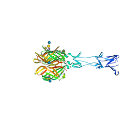
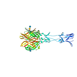
CRYSTAL STRUCTURE OF THE T1L REOVIRUS ATTACHMENT PROTEIN SIGMA1
Descriptor: ACETATE ION, CHLORIDE ION, GLYCEROL, ...
Authors:Reiss, K, Stehle, T.
Deposit date:2014-12-17
Release date:2015-04-01
Last modified:2024-01-10
Method:X-RAY DIFFRACTION (2.2 Å)
Cite:Structure of Serotype 1 Reovirus Attachment Protein sigma 1 in Complex with Junctional Adhesion Molecule A Reveals a Conserved Serotype-Independent Binding Epitope.
J.Virol., 89, 2015
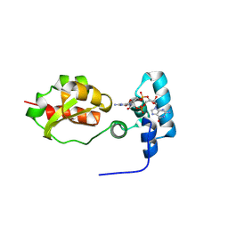
8PKZ
DownloadVisualize
BU of 8pkz by Molmil
NMR solution structure of PilF-GSPIIB in the c-di-GMP bound state
Descriptor: ATP-binding motif-containing protein pilF, GUANOSINE-5'-MONOPHOSPHATE
Authors:Neissner, K, Woehnert, J.
Deposit date:2023-06-27
Release date:2024-07-10
Method:SOLUTION NMR
Cite:NMR solution structure of GSPIIB of Thermus thermophilus in the c-di-GMP bound state
To Be Published
3U9C
DownloadVisualize
BU of 3u9c by Molmil
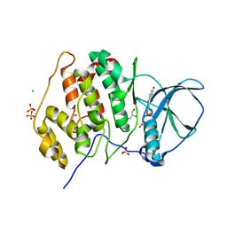
Structure of a C-terminal deletion mutant of human protein kinase CK2 catalytic subunit with the ATP-competitive inhibitor resorufin
Descriptor: 7-hydroxy-3H-phenoxazin-3-one, CHLORIDE ION, Casein kinase II subunit alpha, ...
Authors:Klopffleisch, K, Issinger, O.-G, Niefind, K.
Deposit date:2011-10-18
Release date:2012-05-30
Last modified:2023-09-13
Method:X-RAY DIFFRACTION (3.2 Å)
Cite:Low-density crystal packing of human protein kinase CK2 catalytic subunit in complex with resorufin or other ligands: a tool to study the unique hinge-region plasticity of the enzyme without packing bias.
Acta Crystallogr.,Sect.D, 68, 2012
7ZVS
DownloadVisualize
BU of 7zvs by Molmil
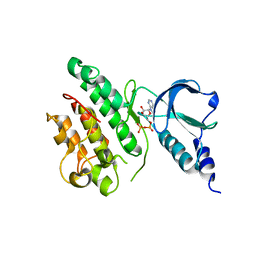
Crystal structure of the Schistosoma mansoni VKR2 kinase domain in an active-like state (ADP-bound)
Descriptor: ADENOSINE-5'-DIPHOSPHATE, Venus kinase receptor 2
Authors:Beis, K, Mathavan, I.
Deposit date:2022-05-17
Release date:2022-10-12
Last modified:2024-01-31
Method:X-RAY DIFFRACTION (3.07 Å)
Cite:Identification of Inhibitors of the Schistosoma mansoni VKR2 Kinase Domain.
Acs Med.Chem.Lett., 13, 2022
6YCV
DownloadVisualize
BU of 6ycv by Molmil
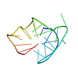
2'-F-riboguanosine and LNA modified hybrid type G-quadruplex with V-loop
Descriptor: DNA (5'-D(*GP*GP*GP*AP*TP*GP*GP*GP*AP*CP*AP*CP*AP*(GF2))-R(P*(LCG))-D(P*GP*GP*AP*CP*GP*GP*G)-3')
Authors:Weisz, K, Haase, L.
Deposit date:2020-03-19
Release date:2020-09-02
Last modified:2024-05-15
Method:SOLUTION NMR
Cite:Locked nucleic acid building blocks as versatile tools for advanced G-quadruplex design.
Nucleic Acids Res., 48, 2020
6YEP
DownloadVisualize
BU of 6yep by Molmil
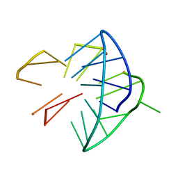
LNA modified G-quadruplex with flipped G-tract and central tetrad
Descriptor: DNA (5'-D(*GP*GP*GP*AP*TP*GP*GP*GP*AP*CP*AP*CP*AP*G)-R(P*(LCG))-D(P*GP*GP*AP*CP*GP*GP*G)-3')
Authors:Weisz, K, Haase, L.
Deposit date:2020-03-25
Release date:2020-09-02
Last modified:2024-05-15
Method:SOLUTION NMR
Cite:Locked nucleic acid building blocks as versatile tools for advanced G-quadruplex design.
Nucleic Acids Res., 48, 2020
3S96
DownloadVisualize
BU of 3s96 by Molmil
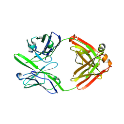
Crystal structure of 3B5H10
Descriptor: 3B5H10 FAB heavy chain, 3B5H10 FAB light chain
Authors:Weisgraber, K, Peters-Libeu, C, Rutenber, E, Newhouse, Y, Finkbeiner, S.
Deposit date:2011-05-31
Release date:2012-02-15
Last modified:2024-04-03
Method:X-RAY DIFFRACTION (1.9 Å)
Cite:Disease-associated polyglutamine stretches in monomeric huntingtin adopt a compact structure.
J.Mol.Biol., 421, 2012
1OC2
DownloadVisualize
BU of 1oc2 by Molmil
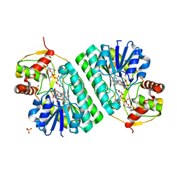
The structure of NADH in the dTDP-D-glucose dehydratase (RmlB) enzyme
Descriptor: DTDP-GLUCOSE 4,6-DEHYDRATASE, NICOTINAMIDE-ADENINE-DINUCLEOTIDE, SULFATE ION, ...
Authors:Beis, K, Naismith, J.H.
Deposit date:2003-02-04
Release date:2003-10-02
Last modified:2023-12-13
Method:X-RAY DIFFRACTION (1.5 Å)
Cite:The Structure of Nadh in the Enzyme Dtdp-D-Glucose Dehydratase (Rmlb)
J.Am.Chem.Soc., 125, 2003
1V0J
DownloadVisualize
BU of 1v0j by Molmil
Udp-galactopyranose mutase from Mycobacterium tuberculosis
Descriptor: BICINE, FLAVIN-ADENINE DINUCLEOTIDE, UDP-GALACTOPYRANOSE MUTASE
Authors:Beis, K, Naismith, J.H.
Deposit date:2004-03-30
Release date:2005-01-26
Last modified:2023-12-13
Method:X-RAY DIFFRACTION (2.25 Å)
Cite:Crystal structures of Mycobacteria tuberculosis and Klebsiella pneumoniae UDP-galactopyranose mutase in the oxidised state and Klebsiella pneumoniae UDP-galactopyranose mutase in the (active) reduced state.
J. Mol. Biol., 348, 2005
4GU3
DownloadVisualize
BU of 4gu3 by Molmil
Crystal structure of the T1L reovirus attachment protein sigma1 in complex with the GM2 glycan
Descriptor: N-acetyl-alpha-neuraminic acid-(2-3)-[2-acetamido-2-deoxy-beta-D-galactopyranose-(1-4)]beta-D-galactopyranose-(1-4)-beta-D-glucopyranose, Outer capsid protein sigma-1
Authors:Reiss, K, Stehle, T.
Deposit date:2012-08-29
Release date:2012-12-05
Last modified:2024-02-28
Method:X-RAY DIFFRACTION (3.6 Å)
Cite:The GM2 Glycan Serves as a Functional Coreceptor for Serotype 1 Reovirus.
Plos Pathog., 8, 2012
4GU4
DownloadVisualize
BU of 4gu4 by Molmil
Crystal structure of the T1L reovirus attachment protein sigma1 in complex with alpha-2,3-sialyllactose
Descriptor: N-acetyl-alpha-neuraminic acid-(2-3)-beta-D-galactopyranose, N-acetyl-alpha-neuraminic acid-(2-3)-beta-D-galactopyranose-(1-4)-beta-D-glucopyranose, Outer capsid protein sigma-1
Authors:Reiss, K, Stehle, T.
Deposit date:2012-08-29
Release date:2012-12-05
Last modified:2024-02-28
Method:X-RAY DIFFRACTION (3.5 Å)
Cite:The GM2 Glycan Serves as a Functional Coreceptor for Serotype 1 Reovirus.
Plos Pathog., 8, 2012
5OFR
DownloadVisualize
BU of 5ofr by Molmil
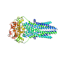
Structure of the antibacterial peptide ABC transporter McjD in a high energy outward occluded intermediate state
Descriptor: ADENOSINE-5'-DIPHOSPHATE, MAGNESIUM ION, Microcin-J25 export ATP-binding/permease protein McjD, ...
Authors:Beis, K, Bountra, K.
Deposit date:2017-07-11
Release date:2017-09-06
Last modified:2024-01-17
Method:X-RAY DIFFRACTION (3.4 Å)
Cite:Structural basis for antibacterial peptide self-immunity by the bacterial ABC transporter McjD.
EMBO J., 36, 2017
5OFP
DownloadVisualize
BU of 5ofp by Molmil
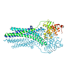
Structure of the antibacterial peptide ABC transporter McjD in an apo inward occluded conformation
Descriptor: Microcin-J25 export ATP-binding/permease protein McjD
Authors:Beis, K, Choudhury, H.G.
Deposit date:2017-07-11
Release date:2017-09-06
Last modified:2024-01-17
Method:X-RAY DIFFRACTION (4.71 Å)
Cite:Structural basis for antibacterial peptide self-immunity by the bacterial ABC transporter McjD.
EMBO J., 36, 2017

222624

PDB entries from 2024-07-17

PDB statisticsPDBj update infoContact PDBjnumon