Loading
PDBj
MenuPDBj@FacebookPDBj@TwitterPDBj@YouTubewwPDB FoundationwwPDB
RCSB PDBPDBeBMRBAdv. SearchSearch help
PDB: 58 results

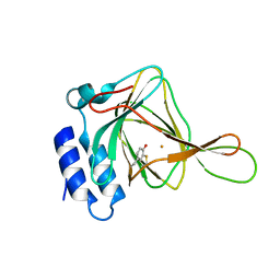
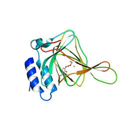
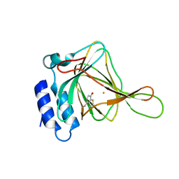
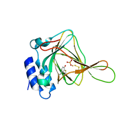
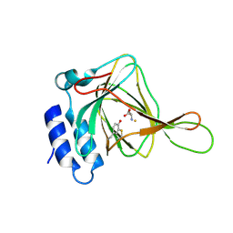
4XFF
DownloadVisualize
BU of 4xff by Molmil
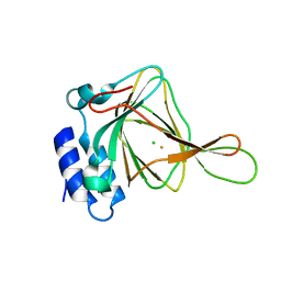
cysteine dioxygenase variant - Y157F at pH 6.2 with dithionite
Descriptor: CHLORIDE ION, Cysteine dioxygenase type 1, FE (III) ION, ...
Authors:Driggers, C.M, Karplus, P.A.
Deposit date:2014-12-26
Release date:2016-02-24
Last modified:2023-09-27
Method:X-RAY DIFFRACTION (1.25 Å)
Cite:Structure-Based Insights into the Role of the Cys-Tyr Crosslink and Inhibitor Recognition by Mammalian Cysteine Dioxygenase.
J. Mol. Biol., 428, 2016
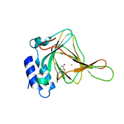
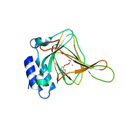
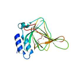
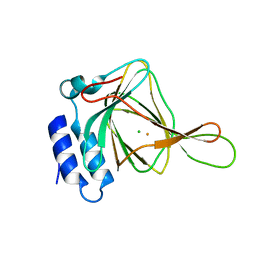
4JTO
DownloadVisualize
BU of 4jto by Molmil
Cysteine bound Cysteine Dioxygenase at pH 8.0 in the presence of Cys and dithionite
Descriptor: CYSTEINE, Cysteine dioxygenase type 1, FE (II) ION
Authors:Driggers, C.M, Karplus, P.A.
Deposit date:2013-03-23
Release date:2013-06-26
Last modified:2013-09-11
Method:X-RAY DIFFRACTION (2 Å)
Cite:Cysteine Dioxygenase Structures from pH4 to 9: Consistent Cys-Persulfenate Formation at Intermediate pH and a Cys-Bound Enzyme at Higher pH.
J.Mol.Biol., 425, 2013
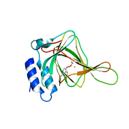
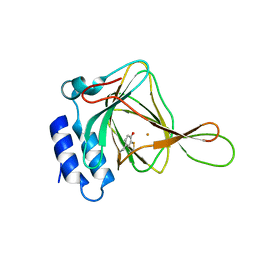
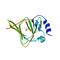
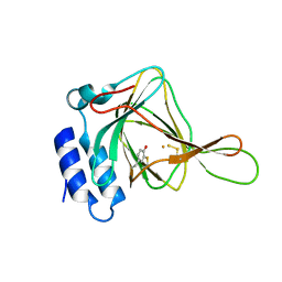
4JTN
DownloadVisualize
BU of 4jtn by Molmil
Cysteine Dioxygenase at pH 8.0 in the presence of dithionite
Descriptor: Cysteine dioxygenase type 1, FE (II) ION
Authors:Driggers, C.M, Karplus, P.A.
Deposit date:2013-03-23
Release date:2013-06-26
Last modified:2013-09-11
Method:X-RAY DIFFRACTION (1.593 Å)
Cite:Cysteine Dioxygenase Structures from pH4 to 9: Consistent Cys-Persulfenate Formation at Intermediate pH and a Cys-Bound Enzyme at Higher pH.
J.Mol.Biol., 425, 2013
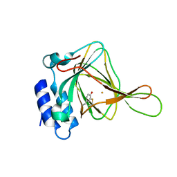
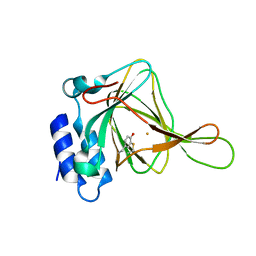
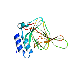
4IEQ
DownloadVisualize
BU of 4ieq by Molmil
unliganded Cysteine Dioxygenase at pH 5.0 in the presence of Cys
Descriptor: Cysteine dioxygenase type 1, FE (II) ION
Authors:Driggers, C.M, Cooley, R.B, Karplus, P.A.
Deposit date:2012-12-13
Release date:2013-06-26
Last modified:2013-09-11
Method:X-RAY DIFFRACTION (1.396 Å)
Cite:Cysteine Dioxygenase Structures from pH4 to 9: Consistent Cys-Persulfenate Formation at Intermediate pH and a Cys-Bound Enzyme at Higher pH.
J.Mol.Biol., 425, 2013
4IEY
DownloadVisualize
BU of 4iey by Molmil
Cys-persulfenate bound Cysteine Dioxygenase at pH 7.0 in the presence of Cys, home-source structure
Descriptor: Cysteine dioxygenase type 1, FE (II) ION, S-HYDROPEROXYCYSTEINE
Authors:Driggers, C.M, Cooley, R.B, Karplus, P.A.
Deposit date:2012-12-13
Release date:2013-06-26
Last modified:2013-09-11
Method:X-RAY DIFFRACTION (1.63 Å)
Cite:Cysteine Dioxygenase Structures from pH4 to 9: Consistent Cys-Persulfenate Formation at Intermediate pH and a Cys-Bound Enzyme at Higher pH.
J.Mol.Biol., 425, 2013
4IEP
DownloadVisualize
BU of 4iep by Molmil
unliganded Cysteine Dioxygenase at pH 4.5 in the presence of Cys
Descriptor: Cysteine dioxygenase type 1, FE (II) ION
Authors:Driggers, C.M, Cooley, R.B, Karplus, P.A.
Deposit date:2012-12-13
Release date:2013-06-26
Last modified:2013-09-11
Method:X-RAY DIFFRACTION (1.45 Å)
Cite:Cysteine Dioxygenase Structures from pH4 to 9: Consistent Cys-Persulfenate Formation at Intermediate pH and a Cys-Bound Enzyme at Higher pH.
J.Mol.Biol., 425, 2013
4IEU
DownloadVisualize
BU of 4ieu by Molmil
Cys-persulfenate bound Cysteine Dioxygenase at pH 7.0 in the presence of Cys
Descriptor: Cysteine dioxygenase type 1, FE (II) ION, S-HYDROPEROXYCYSTEINE
Authors:Driggers, C.M, Cooley, R.B, Karplus, P.A.
Deposit date:2012-12-13
Release date:2013-06-26
Last modified:2013-09-11
Method:X-RAY DIFFRACTION (1.25 Å)
Cite:Cysteine Dioxygenase Structures from pH4 to 9: Consistent Cys-Persulfenate Formation at Intermediate pH and a Cys-Bound Enzyme at Higher pH.
J.Mol.Biol., 425, 2013
4IER
DownloadVisualize
BU of 4ier by Molmil
Cys-persulfenate bound Cysteine Dioxygenase at pH 5.5 in the presence of Cys
Descriptor: Cysteine dioxygenase type 1, FE (II) ION, S-HYDROPEROXYCYSTEINE
Authors:Driggers, C.M, Cooley, R.B, Karplus, P.A.
Deposit date:2012-12-13
Release date:2013-06-26
Last modified:2013-09-11
Method:X-RAY DIFFRACTION (1.45 Å)
Cite:Cysteine Dioxygenase Structures from pH4 to 9: Consistent Cys-Persulfenate Formation at Intermediate pH and a Cys-Bound Enzyme at Higher pH.
J.Mol.Biol., 425, 2013
4IEZ
DownloadVisualize
BU of 4iez by Molmil
unliganded Cysteine Dioxygenase at pH 8.0
Descriptor: Cysteine dioxygenase type 1, FE (II) ION
Authors:Driggers, C.M, Cooley, R.B, Karplus, P.A.
Deposit date:2012-12-13
Release date:2013-06-26
Last modified:2013-09-11
Method:X-RAY DIFFRACTION (1.39 Å)
Cite:Cysteine Dioxygenase Structures from pH4 to 9: Consistent Cys-Persulfenate Formation at Intermediate pH and a Cys-Bound Enzyme at Higher pH.
J.Mol.Biol., 425, 2013
4IEO
DownloadVisualize
BU of 4ieo by Molmil
unliganded Cysteine Dioxygenase at pH 4.0 in the presence of Cys
Descriptor: Cysteine dioxygenase type 1, FE (II) ION
Authors:Driggers, C.M, Cooley, R.B, Karplus, P.A.
Deposit date:2012-12-13
Release date:2013-06-26
Last modified:2013-09-11
Method:X-RAY DIFFRACTION (1.55 Å)
Cite:Cysteine Dioxygenase Structures from pH4 to 9: Consistent Cys-Persulfenate Formation at Intermediate pH and a Cys-Bound Enzyme at Higher pH.
J.Mol.Biol., 425, 2013
4IEV
DownloadVisualize
BU of 4iev by Molmil
Cys-only bound Cysteine Dioxygenase at pH 8.0 in the presence of Cys
Descriptor: CYSTEINE, Cysteine dioxygenase type 1, FE (II) ION
Authors:Driggers, C.M, Cooley, R.B, Karplus, P.A.
Deposit date:2012-12-13
Release date:2013-06-26
Last modified:2013-09-11
Method:X-RAY DIFFRACTION (1.6 Å)
Cite:Cysteine Dioxygenase Structures from pH4 to 9: Consistent Cys-Persulfenate Formation at Intermediate pH and a Cys-Bound Enzyme at Higher pH.
J.Mol.Biol., 425, 2013
4IET
DownloadVisualize
BU of 4iet by Molmil
Cys-persulfenate bound Cysteine Dioxygenase at pH 6.8 in the presence of Cys
Descriptor: Cysteine dioxygenase type 1, FE (II) ION, S-HYDROPEROXYCYSTEINE
Authors:Driggers, C.M, Cooley, R.B, Karplus, P.A.
Deposit date:2012-12-13
Release date:2013-06-26
Last modified:2015-06-24
Method:X-RAY DIFFRACTION (1.4 Å)
Cite:Cysteine dioxygenase structures from pH4 to 9: consistent cys-persulfenate formation at intermediate pH and a Cys-bound enzyme at higher pH.
J.Mol.Biol., 425, 2013
4QM9
DownloadVisualize
BU of 4qm9 by Molmil
Crystal Structure of a Putative Cysteine Dioxygenase From Bacillus subtilis with Cys-bound
Descriptor: CYSTEINE, Cysteine dioxygenase, FE (III) ION
Authors:Hartman, S.H, Driggers, C.M, Karplus, P.A.
Deposit date:2014-06-15
Release date:2014-11-19
Last modified:2023-09-20
Method:X-RAY DIFFRACTION (2.3 Å)
Cite:Structures of Arg- and Gln-type bacterial cysteine dioxygenase homologs.
Protein Sci., 24, 2015
5I0T
DownloadVisualize
BU of 5i0t by Molmil
Thiosulfate bound Cysteine Dioxygenase at pH 6.8
Descriptor: Cysteine dioxygenase type 1, FE (III) ION, THIOSULFATE
Authors:Kean, K.M, Driggers, C.M, Karplus, P.A.
Deposit date:2016-02-04
Release date:2016-12-14
Last modified:2023-11-15
Method:X-RAY DIFFRACTION (1.37 Å)
Cite:Structure-Based Insights into the Role of the Cys-Tyr Crosslink and Inhibitor Recognition by Mammalian Cysteine Dioxygenase.
J. Mol. Biol., 428, 2016
5I0S
DownloadVisualize
BU of 5i0s by Molmil
Thiosulfate bound Cysteine Dioxygenase at pH 6.2
Descriptor: Cysteine dioxygenase type 1, FE (III) ION, THIOSULFATE
Authors:Kean, K.M, Driggers, C.M, Karplus, P.A.
Deposit date:2016-02-04
Release date:2016-12-14
Last modified:2023-11-15
Method:X-RAY DIFFRACTION (1.3 Å)
Cite:Structure-Based Insights into the Role of the Cys-Tyr Crosslink and Inhibitor Recognition by Mammalian Cysteine Dioxygenase.
J. Mol. Biol., 428, 2016
5I0R
DownloadVisualize
BU of 5i0r by Molmil
D-cysteine bound C93A mutant of Cysteine Dioxygenase at pH 8
Descriptor: CHLORIDE ION, Cysteine dioxygenase type 1, D-CYSTEINE, ...
Authors:Kean, K.M, Driggers, C.M, Karplus, P.A.
Deposit date:2016-02-04
Release date:2016-12-14
Last modified:2023-09-27
Method:X-RAY DIFFRACTION (1.35 Å)
Cite:Structure-Based Insights into the Role of the Cys-Tyr Crosslink and Inhibitor Recognition by Mammalian Cysteine Dioxygenase.
J. Mol. Biol., 428, 2016
5I0U
DownloadVisualize
BU of 5i0u by Molmil
Incompletely interpreted D-cysteine soak of Cysteine Dioxygenase at pH 7.0
Descriptor: Cysteine dioxygenase type 1, D-CYSTEINE, FE (III) ION
Authors:Kean, K.M, Driggers, C.M, Karplus, P.A.
Deposit date:2016-02-04
Release date:2017-02-22
Last modified:2023-11-15
Method:X-RAY DIFFRACTION (1.25 Å)
Cite:Structure-Based Insights into the Role of the Cys-Tyr Crosslink and Inhibitor Recognition by Mammalian Cysteine Dioxygenase.
J. Mol. Biol., 428, 2016
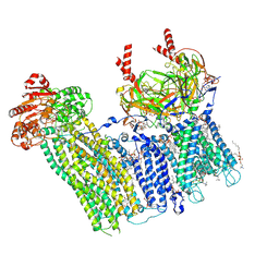
7TYS
DownloadVisualize
BU of 7tys by Molmil
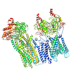
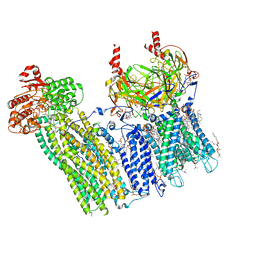
Cryo-EM structure of the pancreatic ATP-sensitive potassium channel bound to ATP and repaglinide with Kir6.2-CTD in the up conformation
Descriptor: (2S)-3-(hexadecanoyloxy)-2-[(9Z)-octadec-9-enoyloxy]propyl 2-(trimethylammonio)ethyl phosphate, (9R,12R)-15-amino-12-hydroxy-6,12-dioxo-7,11,13-trioxa-12lambda~5~-phosphapentadecan-9-yl undecanoate, 2-acetamido-2-deoxy-beta-D-glucopyranose-(1-4)-2-acetamido-2-deoxy-beta-D-glucopyranose, ...
Authors:Shyng, S.L, Sung, M.W, Driggers, C.M.
Deposit date:2022-02-14
Release date:2022-08-31
Last modified:2022-09-14
Method:ELECTRON MICROSCOPY (3.41 Å)
Cite:Ligand-mediated Structural Dynamics of a Mammalian Pancreatic K ATP Channel.
J.Mol.Biol., 434, 2022
7U1E
DownloadVisualize
BU of 7u1e by Molmil
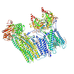
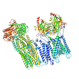
CryoEM structure of the pancreatic ATP-sensitive potassium channel bound to ATP with Kir6.2-CTD in the down conformation
Descriptor: 2-acetamido-2-deoxy-beta-D-glucopyranose, ADENOSINE-5'-TRIPHOSPHATE, ATP-binding cassette sub-family C member 8, ...
Authors:Shyng, S.L, Sung, M.W, Driggers, C.M.
Deposit date:2022-02-21
Release date:2022-08-31
Last modified:2022-09-14
Method:ELECTRON MICROSCOPY (4.52 Å)
Cite:Ligand-mediated Structural Dynamics of a Mammalian Pancreatic K ATP Channel.
J.Mol.Biol., 434, 2022
7TYT
DownloadVisualize
BU of 7tyt by Molmil
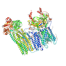
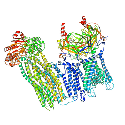
Cryo-EM structure of the pancreatic ATP-sensitive potassium channel bound to ATP and repaglinide with Kir6.2-CTD in the down conformation
Descriptor: (2S)-3-(hexadecanoyloxy)-2-[(9Z)-octadec-9-enoyloxy]propyl 2-(trimethylammonio)ethyl phosphate, 2-acetamido-2-deoxy-beta-D-glucopyranose-(1-4)-2-acetamido-2-deoxy-beta-D-glucopyranose, ADENOSINE-5'-TRIPHOSPHATE, ...
Authors:Shyng, S.L, Sung, M.W, Driggers, C.M.
Deposit date:2022-02-14
Release date:2022-08-31
Last modified:2022-09-14
Method:ELECTRON MICROSCOPY (3.6 Å)
Cite:Ligand-mediated Structural Dynamics of a Mammalian Pancreatic K ATP Channel.
J.Mol.Biol., 434, 2022
7U1S
DownloadVisualize
BU of 7u1s by Molmil
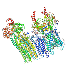
Cryo-EM structure of the pancreatic ATP-sensitive potassium channel bound to ATP and repaglinide with SUR1-out conformation
Descriptor: (2S)-3-(hexadecanoyloxy)-2-[(9Z)-octadec-9-enoyloxy]propyl 2-(trimethylammonio)ethyl phosphate, 2-acetamido-2-deoxy-beta-D-glucopyranose-(1-4)-2-acetamido-2-deoxy-beta-D-glucopyranose, ADENOSINE-5'-TRIPHOSPHATE, ...
Authors:Shyng, S.L, Sung, M.W, Driggers, C.M.
Deposit date:2022-02-22
Release date:2022-08-31
Last modified:2022-09-14
Method:ELECTRON MICROSCOPY (3.8 Å)
Cite:Ligand-mediated Structural Dynamics of a Mammalian Pancreatic K ATP Channel.
J.Mol.Biol., 434, 2022
7U1Q
DownloadVisualize
BU of 7u1q by Molmil
Cryo-EM structure of the pancreatic ATP-sensitive potassium channel bound to ATP and repaglinide with SUR1-in conformation
Descriptor: (2S)-3-(hexadecanoyloxy)-2-[(9Z)-octadec-9-enoyloxy]propyl 2-(trimethylammonio)ethyl phosphate, 2-acetamido-2-deoxy-beta-D-glucopyranose-(1-4)-2-acetamido-2-deoxy-beta-D-glucopyranose, ADENOSINE-5'-TRIPHOSPHATE, ...
Authors:Shyng, S.L, Sung, M.W, Driggers, C.M.
Deposit date:2022-02-21
Release date:2022-08-31
Last modified:2022-09-14
Method:ELECTRON MICROSCOPY (3.9 Å)
Cite:Ligand-mediated Structural Dynamics of a Mammalian Pancreatic K ATP Channel.
J.Mol.Biol., 434, 2022
7U24
DownloadVisualize
BU of 7u24 by Molmil
Cryo-EM structure of the pancreatic ATP-sensitive potassium channel bound to ATP and glibenclamide with Kir6.2-CTD in the up conformation
Descriptor: (2S)-3-(hexadecanoyloxy)-2-[(9Z)-octadec-9-enoyloxy]propyl 2-(trimethylammonio)ethyl phosphate, 2-acetamido-2-deoxy-beta-D-glucopyranose, 5-chloro-N-(2-{4-[(cyclohexylcarbamoyl)sulfamoyl]phenyl}ethyl)-2-methoxybenzamide, ...
Authors:Shyng, S.L, Sung, M.W, Driggers, C.M.
Deposit date:2022-02-23
Release date:2022-08-31
Last modified:2022-09-14
Method:ELECTRON MICROSCOPY (3.58 Å)
Cite:Ligand-mediated Structural Dynamics of a Mammalian Pancreatic K ATP Channel.
J.Mol.Biol., 434, 2022
7U6Y
DownloadVisualize
BU of 7u6y by Molmil
Cryo-EM structure of the pancreatic ATP-sensitive potassium channel in the presence of glibenclamide and ATP with Kir6.2-CTD in the down conformation
Descriptor: ADENOSINE-5'-TRIPHOSPHATE, ATP-binding cassette sub-family C member 8, ATP-sensitive inward rectifier potassium channel 11
Authors:Shyng, S.L, Sung, M.W, Driggers, C.M.
Deposit date:2022-03-06
Release date:2022-08-31
Last modified:2024-06-12
Method:ELECTRON MICROSCOPY (7.4 Å)
Cite:Ligand-mediated Structural Dynamics of a Mammalian Pancreatic K ATP Channel.
J.Mol.Biol., 434, 2022
7U7M
DownloadVisualize
BU of 7u7m by Molmil
Cryo-EM structure of the pancreatic ATP-sensitive potassium channel in the presence of carbamazepine and ATP with Kir6.2-CTD in the up conformation
Descriptor: ADENOSINE-5'-TRIPHOSPHATE, ATP-binding cassette sub-family C member 8, ATP-sensitive inward rectifier potassium channel 11
Authors:Shyng, S.L, Sung, M.W, Driggers, C.M.
Deposit date:2022-03-07
Release date:2022-08-31
Last modified:2024-06-12
Method:ELECTRON MICROSCOPY (5.2 Å)
Cite:Ligand-mediated Structural Dynamics of a Mammalian Pancreatic K ATP Channel.
J.Mol.Biol., 434, 2022

224931

건을2024-09-11부터공개중

PDB statisticsPDBj update infoContact PDBjnumon