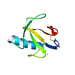
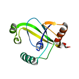
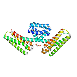
2UP1
 
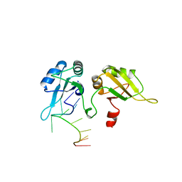 | | STRUCTURE OF UP1-TELOMERIC DNA COMPLEX | | 分子名称: | DNA (5'-D(P*TP*AP*GP*GP*GP*TP*TP*AP*GP*GP*G)-3'), PROTEIN (HETEROGENEOUS NUCLEAR RIBONUCLEOPROTEIN A1) | | 著者 | Ding, J, Hayashi, M.K, Krainer, A.R, Xu, R.-M. | | 登録日 | 1998-07-10 | | 公開日 | 1999-11-10 | | 最終更新日 | 2023-08-30 | | 実験手法 | X-RAY DIFFRACTION (2.1 Å) | | 主引用文献 | Crystal structure of the two-RRM domain of hnRNP A1 (UP1) complexed with single-stranded telomeric DNA.
Genes Dev., 13, 1999
|
|
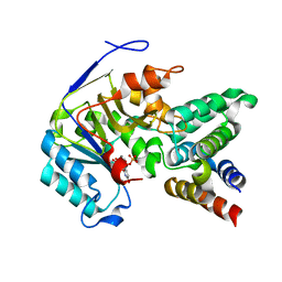
6RNT
 | | CRYSTAL STRUCTURE OF RIBONUCLEASE T1 COMPLEXED WITH ADENOSINE 2'-MONOPHOSPHATE AT 1.8-ANGSTROMS RESOLUTION | | 分子名称: | ADENOSINE-2'-MONOPHOSPHATE, CALCIUM ION, RIBONUCLEASE T1 | | 著者 | Ding, J, Koellner, G, Grunert, H.-P, Saenger, W. | | 登録日 | 1991-08-20 | | 公開日 | 1993-01-15 | | 最終更新日 | 2017-11-29 | | 実験手法 | X-RAY DIFFRACTION (1.8 Å) | | 主引用文献 | Crystal structure of ribonuclease T1 complexed with adenosine 2'-monophosphate at 1.8-A resolution.
J.Biol.Chem., 266, 1991
|
|
6ACI
 | |
6KN1
 | |
6KN0
 | |
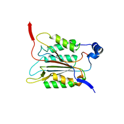
8RNT
 | | STRUCTURE OF RIBONUCLEASE T1 COMPLEXED WITH ZINC(II) AT 1.8 ANGSTROMS RESOLUTION: A ZN2+.6H2O.CARBOXYLATE CLATHRATE | | 分子名称: | RIBONUCLEASE T1, ZINC ION | | 著者 | Ding, J, Choe, H.-W, Granzin, J, Saenger, W. | | 登録日 | 1991-09-23 | | 公開日 | 1993-01-15 | | 最終更新日 | 2017-11-29 | | 実験手法 | X-RAY DIFFRACTION (1.8 Å) | | 主引用文献 | Structure of ribonuclease T1 complexed with zinc(II) at 1.8 A resolution: a Zn2+.6H2O.carboxylate clathrate.
Acta Crystallogr.,Sect.B, 48, 1992
|
|
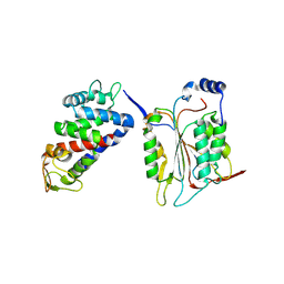
3WLD
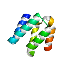
 | | Crystal structure of monomeric GCaMP6m | | 分子名称: | CALCIUM ION, Myosin light chain kinase, Green fluorescent protein, ... | | 著者 | Ding, J, Luo, A.F, Hu, L.Y, Wang, D.C, Shao, F. | | 登録日 | 2013-11-08 | | 公開日 | 2014-01-22 | | 最終更新日 | 2024-10-09 | | 実験手法 | X-RAY DIFFRACTION (2.7 Å) | | 主引用文献 | Structural basis of the ultrasensitive calcium indicator GCaMP6.
Sci China Life Sci, 57, 2014
|
|
3WLC
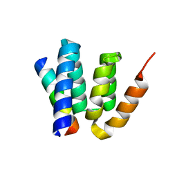
 | | Crystal structure of dimeric GCaMP6m | | 分子名称: | CALCIUM ION, Myosin light chain kinase, Green fluorescent protein, ... | | 著者 | Ding, J, Luo, A.F, Hu, L.Y, Wang, D.C, Shao, F. | | 登録日 | 2013-11-08 | | 公開日 | 2014-01-22 | | 最終更新日 | 2024-10-09 | | 実験手法 | X-RAY DIFFRACTION (2.49 Å) | | 主引用文献 | Structural basis of the ultrasensitive calcium indicator GCaMP6.
Sci China Life Sci, 57, 2014
|
|
8YEY
 | | TRIP4 ASCH domain in complex with ssDNA-1 | | 分子名称: | Activating signal cointegrator 1, DNA (5'-D(*GP*TP*TP*TP*C)-3') | | 著者 | Ding, J, Yang, H, Hu, C. | | 登録日 | 2024-02-23 | | 公開日 | 2024-06-26 | | 最終更新日 | 2024-08-28 | | 実験手法 | X-RAY DIFFRACTION (2 Å) | | 主引用文献 | Biochemical and structural characterization of the DNA-binding properties of human TRIP4 ASCH domain reveals insights into its functional role.
Structure, 32, 2024
|
|
8YFI
 | |
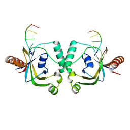
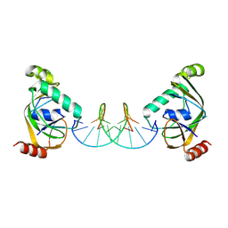
8YXX
 | | TRIP4 ASCH domain in complex with a 12bp dsDNA (5'-TGAGGTACCTCG-3') | | 分子名称: | Activating signal cointegrator 1, DNA (5'-D(*CP*GP*AP*GP*GP*TP*AP*CP*CP*TP*CP*A)-3'), DNA (5'-D(*TP*GP*AP*GP*GP*TP*AP*CP*CP*TP*CP*G)-3') | | 著者 | Ding, J, Yang, H, Hu, C. | | 登録日 | 2024-04-03 | | 公開日 | 2024-06-26 | | 最終更新日 | 2024-08-28 | | 実験手法 | X-RAY DIFFRACTION (2.65 Å) | | 主引用文献 | Biochemical and structural characterization of the DNA-binding properties of human TRIP4 ASCH domain reveals insights into its functional role.
Structure, 32, 2024
|
|
8YEW
 | | TRIP4 ASCH domain in unliganded form | | 分子名称: | Activating signal cointegrator 1 | | 著者 | Ding, J, Yang, H, Hu, C. | | 登録日 | 2024-02-23 | | 公開日 | 2024-06-26 | | 最終更新日 | 2024-08-28 | | 実験手法 | X-RAY DIFFRACTION (1.9 Å) | | 主引用文献 | Biochemical and structural characterization of the DNA-binding properties of human TRIP4 ASCH domain reveals insights into its functional role.
Structure, 32, 2024
|
|
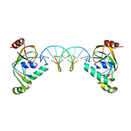
8YXW
 | | TRIP4 ASCH domain in complex with a 12bp dsDNA (5'-TGAGGTACCTCC-3') | | 分子名称: | Activating signal cointegrator 1, DNA (5'-D(*GP*GP*AP*GP*GP*TP*AP*CP*CP*TP*CP*A)-3'), DNA (5'-D(*TP*GP*AP*GP*GP*TP*AP*CP*CP*TP*CP*C)-3') | | 著者 | Ding, J, Yang, H, Hu, C. | | 登録日 | 2024-04-03 | | 公開日 | 2024-06-26 | | 最終更新日 | 2024-08-28 | | 実験手法 | X-RAY DIFFRACTION (2.1 Å) | | 主引用文献 | Biochemical and structural characterization of the DNA-binding properties of human TRIP4 ASCH domain reveals insights into its functional role.
Structure, 32, 2024
|
|
8YFJ
 | |
1K5M
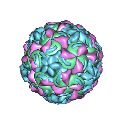 | | Crystal Structure of a Human Rhinovirus Type 14:Human Immunodeficiency Virus Type 1 V3 Loop Chimeric Virus MN-III-2 | | 分子名称: | CHIMERA OF HRV14 COAT PROTEIN VP2 (P1B) AND the V3 loop of HIV-1 gp120, COAT PROTEIN VP1 (P1D), COAT PROTEIN VP3 (P1C), ... | | 著者 | Ding, J, Smith, A.D, Geisler, S.C, Ma, X, Arnold, G.F, Arnold, E. | | 登録日 | 2001-10-11 | | 公開日 | 2002-07-17 | | 最終更新日 | 2023-08-16 | | 実験手法 | X-RAY DIFFRACTION (2.7 Å) | | 主引用文献 | Crystal Structure of a Human Rhinovirus
that Displays Part of the HIV-1 V3 Loop and
Induces Neutralizing Antibodies against
HIV-1
Structure, 10, 2002
|
|
4YKD
 | |
4YL6
 | |
4YKC
 | |
8SA2
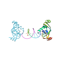
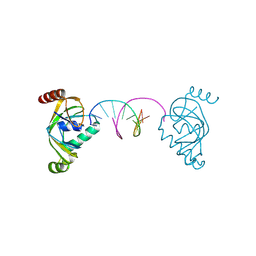
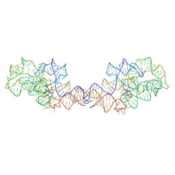
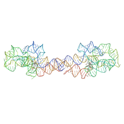 | | Adenosylcobalamin-bound riboswitch dimer, form 1 | | 分子名称: | Adenosylcobalamin, adenosylcobalamin riboswitch form 1 | | 著者 | Ding, J, Deme, J.C, Stagno, J.R, Yu, P, Lea, S.M, Wang, Y.X. | | 登録日 | 2023-03-31 | | 公開日 | 2023-07-26 | | 最終更新日 | 2023-10-25 | | 実験手法 | ELECTRON MICROSCOPY (3.1 Å) | | 主引用文献 | Capturing heterogeneous conformers of cobalamin riboswitch by cryo-EM.
Nucleic Acids Res., 51, 2023
|
|
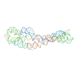
8SA3
 | | Adenosylcobalamin-bound riboswitch dimer, form 2 | | 分子名称: | Adenosylcobalamin, adenosylcobalamin riboswitch form 2 | | 著者 | Ding, J, Deme, J.C, Stagno, J.R, Yu, P, Lea, S.M, Wang, Y.X. | | 登録日 | 2023-03-31 | | 公開日 | 2023-07-26 | | 最終更新日 | 2023-10-25 | | 実験手法 | ELECTRON MICROSCOPY (3 Å) | | 主引用文献 | Capturing heterogeneous conformers of cobalamin riboswitch by cryo-EM.
Nucleic Acids Res., 51, 2023
|
|
8SA6
 | | apo form of adenosylcobalamin riboswitch dimer | | 分子名称: | apo form of adenosylcobalamin riboswitch dimer | | 著者 | Ding, J, Deme, J.C, Stagno, J.R, Yu, P, Lea, S.M, Wang, Y.X. | | 登録日 | 2023-03-31 | | 公開日 | 2023-07-26 | | 最終更新日 | 2023-10-25 | | 実験手法 | ELECTRON MICROSCOPY (5.3 Å) | | 主引用文献 | Capturing heterogeneous conformers of cobalamin riboswitch by cryo-EM.
Nucleic Acids Res., 51, 2023
|
|
8SA5
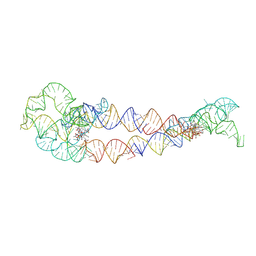 | | Adenosylcobalamin-bound riboswitch dimer, form 4 | | 分子名称: | Adenosylcobalamin, adenosylcobalamin riboswitch form 4 | | 著者 | Ding, J, Deme, J.C, Stagno, J.R, Yu, P, Lea, S.M, Wang, Y.X. | | 登録日 | 2023-03-31 | | 公開日 | 2023-07-26 | | 最終更新日 | 2023-10-25 | | 実験手法 | ELECTRON MICROSCOPY (3.5 Å) | | 主引用文献 | Capturing heterogeneous conformers of cobalamin riboswitch by cryo-EM.
Nucleic Acids Res., 51, 2023
|
|
8SA4
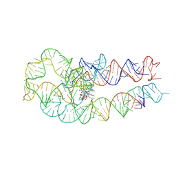 | | Adenosylcobalamin-bound riboswitch dimer, form 3 | | 分子名称: | Adenosylcobalamin, adenosylcobalamin riboswitch form 3 | | 著者 | Ding, J, Deme, J.C, Stagno, J.R, Yu, P, Lea, S.M, Wang, Y.X. | | 登録日 | 2023-03-31 | | 公開日 | 2023-07-26 | | 最終更新日 | 2023-10-25 | | 実験手法 | ELECTRON MICROSCOPY (3.1 Å) | | 主引用文献 | Capturing heterogeneous conformers of cobalamin riboswitch by cryo-EM.
Nucleic Acids Res., 51, 2023
|
|
3AJM
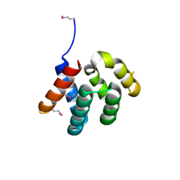
 | | Crystal structure of programmed cell death 10 in complex with inositol 1,3,4,5-tetrakisphosphate | | 分子名称: | INOSITOL-(1,3,4,5)-TETRAKISPHOSPHATE, Programmed cell death protein 10 | | 著者 | Ding, J, Wang, D.C. | | 登録日 | 2010-06-09 | | 公開日 | 2010-06-30 | | 最終更新日 | 2024-03-13 | | 実験手法 | X-RAY DIFFRACTION (2.3 Å) | | 主引用文献 | Crystal structure of human programmed cell death 10 complexed with inositol-(1,3,4,5)-tetrakisphosphate: a novel adaptor protein involved in human cerebral cavernous malformation.
Biochem.Biophys.Res.Commun., 399, 2010
|
|
6AC0
 | |


