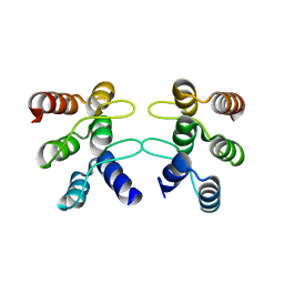
2GCO
 
 | |
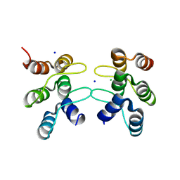
2GCN
 | |
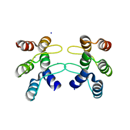
2GCP
 | |
3FEY
 | |
3FEX
 | |
4BQM
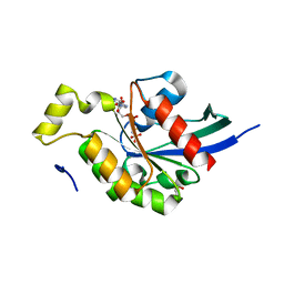
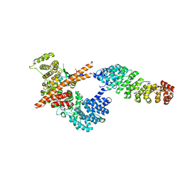
 | | Crystal structure of human liver-type glutaminase, catalytic domain | | 分子名称: | 1,2-ETHANEDIOL, CHLORIDE ION, GLUTAMINASE LIVER ISOFORM, ... | | 著者 | Ferreira, I.M, Vollmar, M, Krojer, T, Strain-Damerell, C, Froese, S, Coutandin, D, Williams, E, Burgess-Brown, N, von Delft, F, Arrowsmith, C.H, Bountra, C, Edwards, A, Dias, S.M.G, Ambrosio, A.L.B, Yue, W.W. | | 登録日 | 2013-05-31 | | 公開日 | 2013-07-10 | | 最終更新日 | 2023-12-20 | | 実験手法 | X-RAY DIFFRACTION (2.18 Å) | | 主引用文献 | Crystal Structure of Human Liver-Type Glutaminase, Catalytic Domain
To be Published
|
|
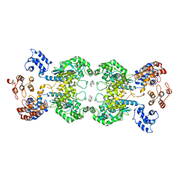
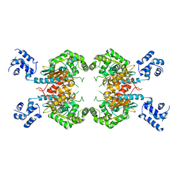
4JKT
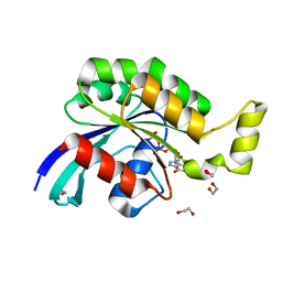
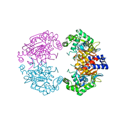
 | | Crystal structure of mouse Glutaminase C, BPTES-bound form | | 分子名称: | Glutaminase kidney isoform, mitochondrial, N,N'-[sulfanediylbis(ethane-2,1-diyl-1,3,4-thiadiazole-5,2-diyl)]bis(2-phenylacetamide) | | 著者 | Fornezari, C, Ferreira, A.P.S, Dias, S.M.G, Ambrosio, A.L.B. | | 登録日 | 2013-03-11 | | 公開日 | 2013-08-14 | | 最終更新日 | 2023-09-20 | | 実験手法 | X-RAY DIFFRACTION (2.77 Å) | | 主引用文献 | Active Glutaminase C Self-assembles into a Supratetrameric Oligomer That Can Be Disrupted by an Allosteric Inhibitor.
J.Biol.Chem., 288, 2013
|
|
3GWS
 | | Crystal Structure of T3-Bound Thyroid Hormone Receptor | | 分子名称: | 3,5,3'TRIIODOTHYRONINE, Thyroid hormone receptor beta | | 著者 | Nascimento, A.S, Dias, S.M.G, Nunes, F.M, Aparicio, R, Polikarpov, I, Baxter, J.D, Webb, P. | | 登録日 | 2009-04-01 | | 公開日 | 2009-04-28 | | 最終更新日 | 2023-11-15 | | 実験手法 | X-RAY DIFFRACTION (2.2 Å) | | 主引用文献 | Structural rearrangements in the thyroid hormone receptor hinge domain and their putative role in the receptor function
J.Mol.Biol., 360, 2006
|
|
2H77
 | | Crystal structure of human TR alpha bound T3 in monoclinic space group | | 分子名称: | 3,5,3'TRIIODOTHYRONINE, THRA protein | | 著者 | Nascimento, A.S, Dias, S.M.G, Nunes, F.M, Aparicio, R, Bleicher, L, Ambrosio, A.L.B, Figueira, A.C.M, Santos, M.A.M, Neto, M.O, Fischer, H, Togashi, H.F.M, Craievich, A.F, Garrat, R.C, Baxter, J.D, Webb, P, Polikarpov, I. | | 登録日 | 2006-06-01 | | 公開日 | 2006-07-25 | | 最終更新日 | 2023-11-15 | | 実験手法 | X-RAY DIFFRACTION (2.33 Å) | | 主引用文献 | Structural rearrangements in the thyroid hormone receptor hinge domain and their putative role in the receptor function.
J.Mol.Biol., 360, 2006
|
|
2H79
 | | Crystal Structure of human TR alpha bound T3 in orthorhombic space group | | 分子名称: | 3,5,3'TRIIODOTHYRONINE, THRA protein | | 著者 | Nascimento, A.S, Dias, S.M.G, Nunes, F.M, Aparicio, R, Bleicher, L, Ambrosio, A.L.B, Figueira, A.C.M, Santos, M.A.M, Neto, M.O, Fischer, H, Togashi, H.F.M, Craievich, A.F, Garrat, R.C, Baxter, J.D, Webb, P, Polikarpov, I. | | 登録日 | 2006-06-01 | | 公開日 | 2006-07-25 | | 最終更新日 | 2023-11-15 | | 実験手法 | X-RAY DIFFRACTION (1.87 Å) | | 主引用文献 | Structural rearrangements in the thyroid hormone receptor hinge domain and their putative role in the receptor function.
J.Mol.Biol., 360, 2006
|
|
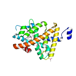
5U0K
 | | C-terminal ankyrin repeats from human liver-type glutaminase (GAB/LGA) | | 分子名称: | Glutaminase liver isoform, mitochondrial | | 著者 | Ferreira, I.M, Pasquali, C.C, Gonzalez, A, Dias, S.M.G, Ambrosio, A.L.B. | | 登録日 | 2016-11-24 | | 公開日 | 2017-05-24 | | 最終更新日 | 2023-10-04 | | 実験手法 | X-RAY DIFFRACTION (2.548 Å) | | 主引用文献 | The origin and evolution of human glutaminases and their atypical C-terminal ankyrin repeats.
J. Biol. Chem., 292, 2017
|
|
5U0I
 | | C-terminal ankyrin repeats from human kidney-type glutaminase (KGA) - tetragonal crystal form | | 分子名称: | CHLORIDE ION, Glutaminase kidney isoform, mitochondrial, ... | | 著者 | Pasquali, C.C, Gonzalez, A, Dias, S.M.G, Ambrosio, A.L.B. | | 登録日 | 2016-11-24 | | 公開日 | 2017-05-24 | | 最終更新日 | 2024-03-06 | | 実験手法 | X-RAY DIFFRACTION (1.423 Å) | | 主引用文献 | The origin and evolution of human glutaminases and their atypical C-terminal ankyrin repeats.
J. Biol. Chem., 292, 2017
|
|
5U0J
 | | C-terminal ankyrin repeats from human kidney-type glutaminase (KGA) - monoclinic crystal form | | 分子名称: | Glutaminase kidney isoform, mitochondrial, SODIUM ION | | 著者 | Pasquali, C.C, Gonzalez, A, Dias, S.M.G, Ambrosio, A.L.B. | | 登録日 | 2016-11-24 | | 公開日 | 2017-05-24 | | 最終更新日 | 2023-10-04 | | 実験手法 | X-RAY DIFFRACTION (1.72 Å) | | 主引用文献 | The origin and evolution of human glutaminases and their atypical C-terminal ankyrin repeats.
J. Biol. Chem., 292, 2017
|
|
3JZB
 | | Crystal Structure of TR-alfa bound to the selective thyromimetic TRIAC | | 分子名称: | THRA protein, [4-(4-HYDROXY-3-IODO-PHENOXY)-3,5-DIIODO-PHENYL]-ACETIC ACID | | 著者 | Nascimento, A.S, Dias, S.M.G, Nunes, F.M, Aparicio, R. | | 登録日 | 2009-09-23 | | 公開日 | 2009-12-08 | | 最終更新日 | 2023-09-06 | | 実験手法 | X-RAY DIFFRACTION (2.007 Å) | | 主引用文献 | Gaining ligand selectivity in thyroid hormone receptors via entropy.
Proc.Natl.Acad.Sci.USA, 106, 2009
|
|
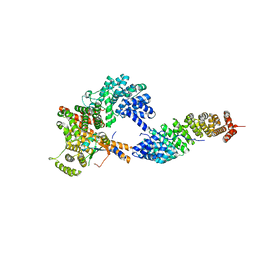
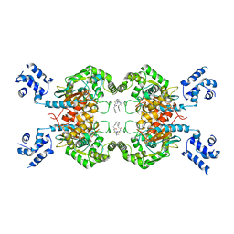
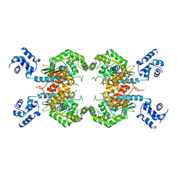
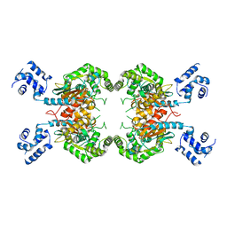
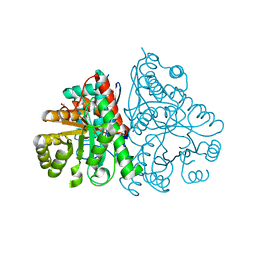
5UQE
 | | Multidomain structure of human kidney-type glutaminase(KGA/GLS) | | 分子名称: | Glutaminase kidney isoform, mitochondrial, N,N'-[sulfanediylbis(ethane-2,1-diyl-1,3,4-thiadiazole-5,2-diyl)]bis(2-phenylacetamide) | | 著者 | Pasquali, C.C, Dias, S.M.G, Ambrosio, A.L.B. | | 登録日 | 2017-02-08 | | 公開日 | 2017-05-24 | | 最終更新日 | 2024-03-06 | | 実験手法 | X-RAY DIFFRACTION (3.6 Å) | | 主引用文献 | The origin and evolution of human glutaminases and their atypical C-terminal ankyrin repeats.
J. Biol. Chem., 292, 2017
|
|
3SS3
 | |
3SS5
 | |
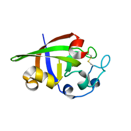
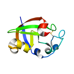
3SUJ
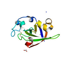 | | Crystal structure of cerato-platanin 1 from M. perniciosa (MpCP1) | | 分子名称: | ACETATE ION, CHLORIDE ION, Cerato-platanin 1, ... | | 著者 | Oliveira, J.F, Barsottini, M.R.O, Zaparoli, G, Machado, L.O, Dias, S.M.G, Pereira, G.A.G, Ambrosio, A.L.B. | | 登録日 | 2011-07-11 | | 公開日 | 2012-07-11 | | 最終更新日 | 2019-02-06 | | 実験手法 | X-RAY DIFFRACTION (1.34 Å) | | 主引用文献 | Functional diversification of cerato-platanins in Moniliophthora perniciosa as seen by differential expression and protein function specialization.
Mol. Plant Microbe Interact., 26, 2013
|
|
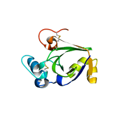
3SUK
 | | Crystal structure of cerato-platanin 2 from M. perniciosa (MpCP2) | | 分子名称: | Cerato-platanin-like protein | | 著者 | Oliveira, J.F, Barsottini, M.R.O, Zaparoli, G, Machado, L.O, Dias, S.M.G, Pereira, G.A.G, Ambrosio, A.L.B. | | 登録日 | 2011-07-11 | | 公開日 | 2012-07-11 | | 最終更新日 | 2023-09-13 | | 実験手法 | X-RAY DIFFRACTION (1.34 Å) | | 主引用文献 | Functional diversification of cerato-platanins in Moniliophthora perniciosa as seen by differential expression and protein function specialization.
Mol. Plant Microbe Interact., 26, 2013
|
|
3SUM
 | | Crystal structure of cerato-platanin 5 from M. perniciosa (MpCP5) | | 分子名称: | Cerato-platanin-like protein | | 著者 | Oliveira, J.F, Barsottini, M.R.O, Zaparoli, G, Machado, L.O, Dias, S.M.G, Pereira, G.A.G, Ambrosio, A.L.B. | | 登録日 | 2011-07-11 | | 公開日 | 2012-07-11 | | 最終更新日 | 2023-09-13 | | 実験手法 | X-RAY DIFFRACTION (1.87 Å) | | 主引用文献 | Functional diversification of cerato-platanins in Moniliophthora perniciosa as seen by differential expression and protein function specialization.
Mol. Plant Microbe Interact., 26, 2013
|
|
3ST1
 | | Crystal structure of Necrosis and Ethylene inducing Protein 2 from the causal agent of cocoa's Witches Broom disease | | 分子名称: | Necrosis-and ethylene-inducing protein, SODIUM ION, ZINC ION | | 著者 | Oliveira, J.F, Zaparoli, G, Barsottini, M.R.O, Ambrosio, A.L.B, Pereira, G.A.G, Dias, S.M.G. | | 登録日 | 2011-07-08 | | 公開日 | 2011-11-02 | | 最終更新日 | 2023-09-13 | | 実験手法 | X-RAY DIFFRACTION (1.8 Å) | | 主引用文献 | The Crystal Structure of Necrosis- and Ethylene-Inducing Protein 2 from the Causal Agent of Cacao's Witches' Broom Disease Reveals Key Elements for Its Activity.
Biochemistry, 50, 2011
|
|
3SS4
 | |
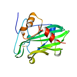
3SUL
 | | Crystal structure of cerato-platanin 3 from M. perniciosa (MpCP3) | | 分子名称: | Cerato-platanin-like protein | | 著者 | Oliveira, J.F, Barsottini, M.R.O, Zaparoli, G, Machado, L.O, Dias, S.M.G, Pereira, G.A.G, Ambrosio, A.L.B. | | 登録日 | 2011-07-11 | | 公開日 | 2012-07-11 | | 最終更新日 | 2023-09-13 | | 実験手法 | X-RAY DIFFRACTION (1.63 Å) | | 主引用文献 | Functional diversification of cerato-platanins in Moniliophthora perniciosa as seen by differential expression and protein function specialization.
Mol. Plant Microbe Interact., 26, 2013
|
|
2PTW
 | | Crystal Structure of the T. brucei enolase complexed with sulphate, identification of a metal binding site IV | | 分子名称: | 1,2-ETHANEDIOL, Enolase, SULFATE ION, ... | | 著者 | Navarro, M.V.A.S, Rigden, D.J, Garratt, R.C, Dias, S.M.G. | | 登録日 | 2007-05-08 | | 公開日 | 2007-11-20 | | 最終更新日 | 2023-08-30 | | 実験手法 | X-RAY DIFFRACTION (1.9 Å) | | 主引用文献 | Structural flexibility in Trypanosoma brucei enolase revealed by X-ray crystallography and molecular dynamics.
Febs J., 274, 2007
|
|
2PTZ
 | | Crystal Structure of the T. brucei enolase complexed with phosphonoacetohydroxamate (PAH), His156-out conformation | | 分子名称: | 1,2-ETHANEDIOL, Enolase, PHOSPHONOACETOHYDROXAMIC ACID, ... | | 著者 | Navarro, M.V.A.S, Rigden, D.J, Garratt, R.C, Dias, S.M.G. | | 登録日 | 2007-05-08 | | 公開日 | 2007-11-20 | | 最終更新日 | 2023-08-30 | | 実験手法 | X-RAY DIFFRACTION (1.65 Å) | | 主引用文献 | Structural flexibility in Trypanosoma brucei enolase revealed by X-ray crystallography and molecular dynamics.
Febs J., 274, 2007
|
|


