5HGC
 
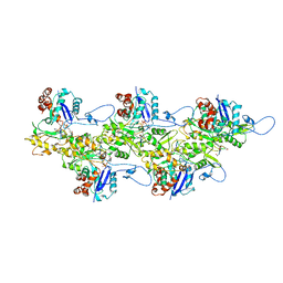
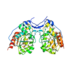
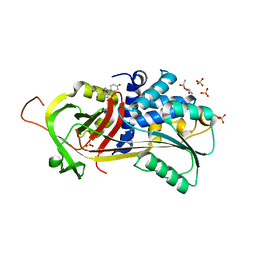 | | A Serpin structure | | 分子名称: | (11alpha,14beta)-11,17,21-trihydroxypregn-4-ene-3,20-dione, 2,6-diamino-2,6-dideoxy-alpha-D-glucopyranose, SULFATE ION, ... | | 著者 | Das, S, Vashchenko, G.V, Van Petegem, F. | | 登録日 | 2016-01-08 | | 公開日 | 2016-04-06 | | 最終更新日 | 2023-09-27 | | 実験手法 | X-RAY DIFFRACTION (2.43 Å) | | 主引用文献 | A Serpin structure
J.Biol.Chem., 2016
|
|
8GMX
 | |
6U3E
 | |
6U3G
 | |
6U96
 | | Actin phalloidin at BeFx state | | 分子名称: | ADENOSINE-5'-DIPHOSPHATE, Actin, alpha skeletal muscle, ... | | 著者 | Das, S, Ge, P, Durer, Z.A.O, Grintsevich, E.E, Zhou, Z.H, Reisler, E. | | 登録日 | 2019-09-06 | | 公開日 | 2020-05-13 | | 最終更新日 | 2020-05-20 | | 実験手法 | ELECTRON MICROSCOPY (3.8 Å) | | 主引用文献 | D-loop Dynamics and Near-Atomic-Resolution Cryo-EM Structure of Phalloidin-Bound F-Actin.
Structure, 28, 2020
|
|
6VSV
 | |
6VRR
 | |
5GO6
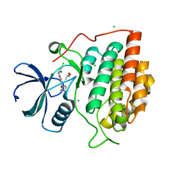
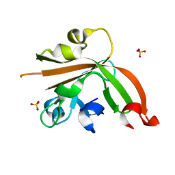 | | Structure of sortase E T196V mutant from Streptomyces avermitilis | | 分子名称: | Putative secreted protein, SULFATE ION | | 著者 | Das, S, Pawale, V.S, Dadireddy, V, Roy, R.P, Ramakumar, S. | | 登録日 | 2016-07-26 | | 公開日 | 2017-07-26 | | 最終更新日 | 2023-11-08 | | 実験手法 | X-RAY DIFFRACTION (1.7 Å) | | 主引用文献 | Structure of sortase E T196V mutant from Streptomyces avermitilis
To Be Published
|
|
5GO5
 | | Structure of sortase E from Streptomyces avermitilis | | 分子名称: | GLYCINE, SULFATE ION, sortase | | 著者 | Das, S, Pawale, V.S, Dadireddy, V, Roy, R.P, Ramakumar, S. | | 登録日 | 2016-07-26 | | 公開日 | 2017-03-15 | | 最終更新日 | 2023-11-08 | | 実験手法 | X-RAY DIFFRACTION (1.65 Å) | | 主引用文献 | Structure and specificity of a new class of Ca2+-independent housekeeping sortase from Streptomyces avermitilis provide insights into its non-canonical substrate preference.
J. Biol. Chem., 292, 2017
|
|
5FDY
 | |
5FEB
 | |
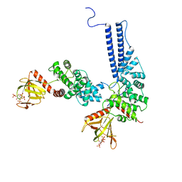
6BBP
 | | Model for compact volume of truncated monomeric Cytohesin-3 (Grp1; amino acids 63-399) E161A 6GS Arf6 Q67L fusion protein | | 分子名称: | Cytohesin-3,ADP-ribosylation factor 6, GUANOSINE-5'-TRIPHOSPHATE, INOSITOL-(1,3,4,5)-TETRAKISPHOSPHATE, ... | | 著者 | Das, S, Malaby, A.W, Lambright, D.G. | | 登録日 | 2017-10-19 | | 公開日 | 2018-01-10 | | 最終更新日 | 2023-11-29 | | 実験手法 | ELECTRON MICROSCOPY (35 Å) | | 主引用文献 | Structural Dynamics Control Allosteric Activation of Cytohesin Family Arf GTPase Exchange Factors.
Structure, 26, 2018
|
|
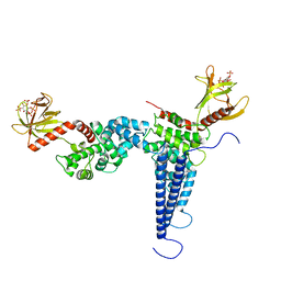
6BBQ
 | | Model for extended volume of truncated monomeric Cytohesin-3 (Grp1; amino acids 63-399) E161A Arf6 Q67L fusion protein | | 分子名称: | Cytohesin-3,ADP-ribosylation factor 6, GUANOSINE-5'-TRIPHOSPHATE, INOSITOL-(1,3,4,5)-TETRAKISPHOSPHATE, ... | | 著者 | Das, S, Malaby, A.W, Lambright, D.G. | | 登録日 | 2017-10-19 | | 公開日 | 2018-01-10 | | 最終更新日 | 2023-11-29 | | 実験手法 | ELECTRON MICROSCOPY (35 Å) | | 主引用文献 | Structural Dynamics Control Allosteric Activation of Cytohesin Family Arf GTPase Exchange Factors.
Structure, 26, 2018
|
|
2GES
 | | Pantothenate kinase from Mycobacterium tuberculosis (MtPanK) in complex with a coenzyme A derivative, Form-I (RT) | | 分子名称: | Pantothenate kinase, [(2R,3S,4R,5R)-5-(6-AMINO-9H-PURIN-9-YL)-4-HYDROXY-3-(PHOSPHONOOXY)TETRAHYDROFURAN-2-YL]METHYL (3R)-3-HYDROXY-4-{[3-({2-[(2-HYDROXYETHYL)DITHIO]ETHYL}AMINO)-3-OXOPROPYL]AMINO}-2,2-DIMETHYL-4-OXOBUTYL DIHYDROGEN DIPHOSPHATE | | 著者 | Das, S, Kumar, P, Bhor, V, Surolia, A, Vijayan, M. | | 登録日 | 2006-03-20 | | 公開日 | 2006-06-06 | | 最終更新日 | 2023-10-25 | | 実験手法 | X-RAY DIFFRACTION (2.4 Å) | | 主引用文献 | Invariance and variability in bacterial PanK: a study based on the crystal structure of Mycobacterium tuberculosis PanK.
Acta Crystallogr.,Sect.D, 62, 2006
|
|
2GEU
 | | Pantothenate kinase from Mycobacterium tuberculosis (MtPanK) in complex with a coenzyme A derivative, Form-II (RT) | | 分子名称: | Pantothenate kinase, [(2R,3S,4R,5R)-5-(6-AMINO-9H-PURIN-9-YL)-4-HYDROXY-3-(PHOSPHONOOXY)TETRAHYDROFURAN-2-YL]METHYL (3R)-3-HYDROXY-4-{[3-({2-[(2-HYDROXYETHYL)DITHIO]ETHYL}AMINO)-3-OXOPROPYL]AMINO}-2,2-DIMETHYL-4-OXOBUTYL DIHYDROGEN DIPHOSPHATE | | 著者 | Das, S, Kumar, P, Bhor, V, Surolia, A, Vijayan, M. | | 登録日 | 2006-03-20 | | 公開日 | 2006-06-06 | | 最終更新日 | 2023-10-25 | | 実験手法 | X-RAY DIFFRACTION (2.9 Å) | | 主引用文献 | Invariance and variability in bacterial PanK: a study based on the crystal structure of Mycobacterium tuberculosis PanK.
Acta Crystallogr.,Sect.D, 62, 2006
|
|
2GEV
 | | Pantothenate kinase from Mycobacterium tuberculosis (MtPanK) in complex with a coenzyme A derivative, Form-II (LT) | | 分子名称: | 2-AMINO-2-HYDROXYMETHYL-PROPANE-1,3-DIOL, GLYCEROL, Pantothenate kinase, ... | | 著者 | Das, S, Kumar, P, Bhor, V, Surolia, A, Vijayan, M. | | 登録日 | 2006-03-20 | | 公開日 | 2006-06-06 | | 最終更新日 | 2023-10-25 | | 実験手法 | X-RAY DIFFRACTION (2.35 Å) | | 主引用文献 | Invariance and variability in bacterial PanK: a study based on the crystal structure of Mycobacterium tuberculosis PanK.
Acta Crystallogr.,Sect.D, 62, 2006
|
|
2GET
 | | Pantothenate kinase from Mycobacterium tuberculosis (MtPanK) in complex with a coenzyme A derivative, Form-I (LT) | | 分子名称: | GLYCEROL, Pantothenate kinase, [(2R,3S,4R,5R)-5-(6-AMINO-9H-PURIN-9-YL)-4-HYDROXY-3-(PHOSPHONOOXY)TETRAHYDROFURAN-2-YL]METHYL (3R)-3-HYDROXY-4-{[3-({2-[(2-HYDROXYETHYL)DITHIO]ETHYL}AMINO)-3-OXOPROPYL]AMINO}-2,2-DIMETHYL-4-OXOBUTYL DIHYDROGEN DIPHOSPHATE | | 著者 | Das, S, Kumar, P, Bhor, V, Surolia, A, Vijayan, M. | | 登録日 | 2006-03-20 | | 公開日 | 2006-06-06 | | 最終更新日 | 2023-10-25 | | 実験手法 | X-RAY DIFFRACTION (2.35 Å) | | 主引用文献 | Invariance and variability in bacterial PanK: a study based on the crystal structure of Mycobacterium tuberculosis PanK.
Acta Crystallogr.,Sect.D, 62, 2006
|
|
4I2N
 | |
4MZ2
 | |
4MZ3
 | |
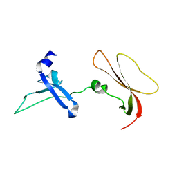
2LXD
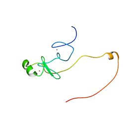 | | Backbone 1H, 13C, and 15N Chemical Shift Assignments for LMO2(LIM2)-Ldb1(LID) | | 分子名称: | Rhombotin-2,LIM domain-binding protein 1, ZINC ION | | 著者 | Dastmalchi, S, Wilkinson-White, L, Kwan, A.H, Gamsjaeger, R, Mackay, J.P, Matthews, J.M. | | 登録日 | 2012-08-20 | | 公開日 | 2012-09-12 | | 最終更新日 | 2024-05-15 | | 実験手法 | SOLUTION NMR | | 主引用文献 | Solution structure of a tethered Lmo2(LIM2) /Ldb1(LID) complex.
Protein Sci., 21, 2012
|
|
5V3I
 | |
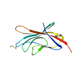
6B3K
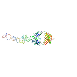 | | Crystal structure of mutant Spinach RNA aptamer in complex with Fab BL3-6 | | 分子名称: | Heavy chain of Fab BL3-6, Light chain of Fab BL3-6, MAGNESIUM ION, ... | | 著者 | DasGupta, S, Koirala, D, Shelke, S.A, Piccirilli, J.A. | | 登録日 | 2017-09-22 | | 公開日 | 2017-12-27 | | 最終更新日 | 2023-10-04 | | 実験手法 | X-RAY DIFFRACTION (2.09 Å) | | 主引用文献 | Affinity maturation of a portable Fab-RNA module for chaperone-assisted RNA crystallography.
Nucleic Acids Res., 46, 2018
|
|
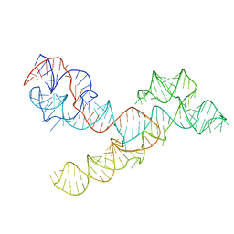
6B14
 | | Crystal structure of Spinach RNA aptamer in complex with Fab BL3-6S97N | | 分子名称: | Heavy chain of Fab BL3-6S97N, Light chain of Fab BL3-6S97N, MAGNESIUM ION, ... | | 著者 | DasGupta, S, Shelke, S.A, Piccirilli, J.A. | | 登録日 | 2017-09-16 | | 公開日 | 2017-12-27 | | 最終更新日 | 2023-10-04 | | 実験手法 | X-RAY DIFFRACTION (1.64 Å) | | 主引用文献 | Affinity maturation of a portable Fab-RNA module for chaperone-assisted RNA crystallography.
Nucleic Acids Res., 46, 2018
|
|
2IZS
 | | Structure of casein kinase gamma 3 in complex with inhibitor | | 分子名称: | CASEIN KINASE I ISOFORM GAMMA-3, CHLORIDE ION, MAGNESIUM ION, ... | | 著者 | Bunkoczi, G, Salah, E, Rellos, P, Das, S, Fedorov, O, Savitsky, P, Debreczeni, J.E, Gileadi, O, Sundstrom, M, Edwards, A, Arrowsmith, C, Weigelt, J, von Delft, F, Knapp, S. | | 登録日 | 2006-07-26 | | 公開日 | 2006-08-01 | | 最終更新日 | 2023-12-13 | | 実験手法 | X-RAY DIFFRACTION (1.95 Å) | | 主引用文献 | Inhibitor Binding by Casein Kinases
To be Published
|
|


