Loading
PDBj
MenuPDBj@FacebookPDBj@TwitterPDBj@YouTubewwPDB FoundationwwPDB
RCSB PDBPDBeBMRBAdv. SearchSearch help
PDB: 32 results

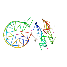
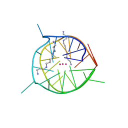
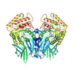
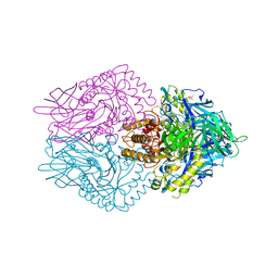
7D5D
DownloadVisualize
BU of 7d5d by Molmil
Left-handed G-quadruplex containing one bulge
Descriptor: 1xBulge-LHG4motif, POTASSIUM ION, SPERMINE
Authors:Das, P, Ngo, K.H, Winnerdy, F.R, Maity, A, Bakalar, B, Mechulam, Y, Schmitt, E, Phan, A.T.
Deposit date:2020-09-25
Release date:2021-02-10
Last modified:2023-11-29
Method:X-RAY DIFFRACTION (1.18 Å)
Cite:Bulges in left-handed G-quadruplexes.
Nucleic Acids Res., 49, 2021
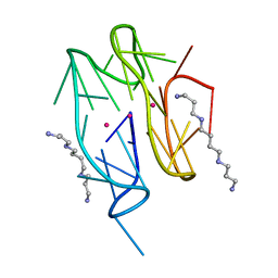
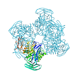
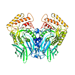
7D5E
DownloadVisualize
BU of 7d5e by Molmil
Left-handed G-quadruplex containing two bulges
Descriptor: 2xBulge-LHG4motif, POTASSIUM ION, SODIUM ION, ...
Authors:Das, P, Maity, A, Ngo, K.H, Winnerdy, F.R, Bakalar, B, Mechulam, Y, Schmitt, E, Phan, A.T.
Deposit date:2020-09-25
Release date:2021-02-10
Last modified:2023-11-29
Method:X-RAY DIFFRACTION (1.296 Å)
Cite:Bulges in left-handed G-quadruplexes.
Nucleic Acids Res., 49, 2021
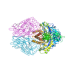
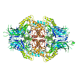
7DFY
DownloadVisualize
BU of 7dfy by Molmil
Novel motif for left-handed G-quadruplex formation
Descriptor: (4S)-2-METHYL-2,4-PENTANEDIOL, 2xMotif2, POTASSIUM ION, ...
Authors:Das, P, Winnerdy, F.R, Maity, A, Mechulam, Y, Phan, A.T.
Deposit date:2020-11-10
Release date:2021-09-01
Last modified:2023-11-29
Method:X-RAY DIFFRACTION (1.69 Å)
Cite:A novel minimal motif for left-handed G-quadruplex formation.
Chem.Commun.(Camb.), 57, 2021
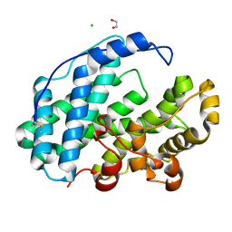
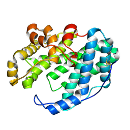
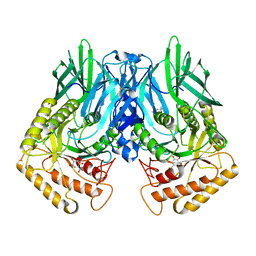
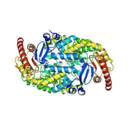
7WXM
DownloadVisualize
BU of 7wxm by Molmil
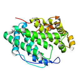
Crystal Structure of PL-5 family polysaccharide lyase PanPL from Pandoraea apista at pH6.5 in apo form
Descriptor: 1,2-ETHANEDIOL, CHLORIDE ION, poly(beta-D-mannuronate) lyase
Authors:Dash, P, Acharya, R.
Deposit date:2022-02-15
Release date:2022-08-10
Last modified:2024-10-30
Method:X-RAY DIFFRACTION (1.23 Å)
Cite:Distinct Modes of Hidden Structural Dynamics in the Functioning of an Allosteric Polysaccharide Lyase.
Acs Cent.Sci., 8, 2022
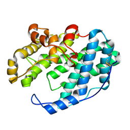
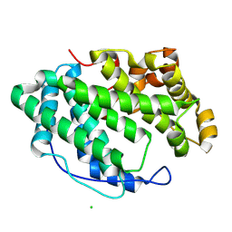
7WXN
DownloadVisualize
BU of 7wxn by Molmil
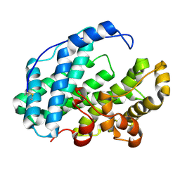
Crystal Structure of PL-5 family polysaccharide lyase PanPL from Pandoraea apista at pH7.5 in apo form
Descriptor: poly(beta-D-mannuronate) lyase
Authors:Dash, P, Acharya, R.
Deposit date:2022-02-15
Release date:2022-08-10
Last modified:2024-10-16
Method:X-RAY DIFFRACTION (2.4 Å)
Cite:Distinct Modes of Hidden Structural Dynamics in the Functioning of an Allosteric Polysaccharide Lyase.
Acs Cent.Sci., 8, 2022
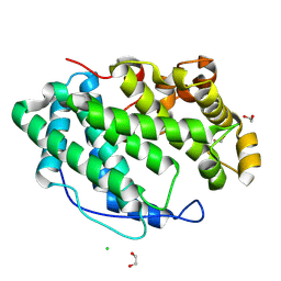
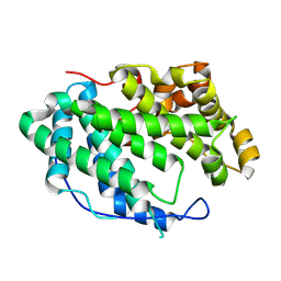
7WXP
DownloadVisualize
BU of 7wxp by Molmil
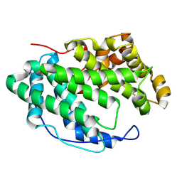
Crystal Structure of PL-5 family polysaccharide lyase PanPL-H172A mutant from Pandoraea apista in ManA bound form
Descriptor: beta-D-mannopyranuronic acid-(1-4)-beta-D-mannopyranuronic acid, poly(beta-D-mannuronate) lyase
Authors:Dash, P, Acharya, R.
Deposit date:2022-02-15
Release date:2022-08-10
Last modified:2023-11-29
Method:X-RAY DIFFRACTION (2.2 Å)
Cite:Distinct Modes of Hidden Structural Dynamics in the Functioning of an Allosteric Polysaccharide Lyase.
Acs Cent.Sci., 8, 2022
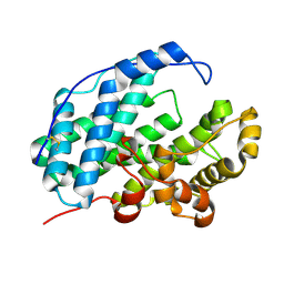
7WXL
DownloadVisualize
BU of 7wxl by Molmil
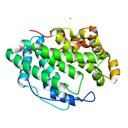
Crystal Structure of PL-5 family polysaccharide lyase PanPL from Pandoraea apista at pH5.5 in apo form
Descriptor: CHLORIDE ION, poly(beta-D-mannuronate) lyase
Authors:Dash, P, Acharya, R.
Deposit date:2022-02-15
Release date:2022-08-10
Last modified:2024-10-23
Method:X-RAY DIFFRACTION (1.45 Å)
Cite:Distinct Modes of Hidden Structural Dynamics in the Functioning of an Allosteric Polysaccharide Lyase.
Acs Cent.Sci., 8, 2022
7WXR
DownloadVisualize
BU of 7wxr by Molmil
Crystal Structure of PL-5 family polysaccharide lyase PanPL-N171L mutant from Pandoraea apista in apo form
Descriptor: poly(beta-D-mannuronate) lyase
Authors:Dash, P, Acharya, R.
Deposit date:2022-02-15
Release date:2022-08-10
Last modified:2024-10-09
Method:X-RAY DIFFRACTION (1.88 Å)
Cite:Distinct Modes of Hidden Structural Dynamics in the Functioning of an Allosteric Polysaccharide Lyase.
Acs Cent.Sci., 8, 2022
7WXO
DownloadVisualize
BU of 7wxo by Molmil
Crystal Structure of PL-5 family polysaccharide lyase PanPL from Pandoraea apista at pH8.5 in apo form
Descriptor: poly(beta-D-mannuronate) lyase
Authors:Dash, P, Acharya, R.
Deposit date:2022-02-15
Release date:2022-08-10
Last modified:2023-11-29
Method:X-RAY DIFFRACTION (1.96 Å)
Cite:Distinct Modes of Hidden Structural Dynamics in the Functioning of an Allosteric Polysaccharide Lyase.
Acs Cent.Sci., 8, 2022
7WXK
DownloadVisualize
BU of 7wxk by Molmil
Crystal Structure of PL-5 family polysaccharide lyase PanPL from Pandoraea apista at pH4.5 in apo form
Descriptor: poly(beta-D-mannuronate) lyase
Authors:Dash, P, Acharya, R.
Deposit date:2022-02-15
Release date:2022-08-10
Last modified:2023-11-29
Method:X-RAY DIFFRACTION (1.68 Å)
Cite:Distinct Modes of Hidden Structural Dynamics in the Functioning of an Allosteric Polysaccharide Lyase.
Acs Cent.Sci., 8, 2022
7WXQ
DownloadVisualize
BU of 7wxq by Molmil
Crystal Structure of PL-5 family polysaccharide lyase PanPL-H172A mutant from Pandoraea apista in apo form
Descriptor: CHLORIDE ION, poly(beta-D-mannuronate) lyase
Authors:Dash, P, Acharya, R.
Deposit date:2022-02-15
Release date:2022-08-10
Last modified:2024-10-16
Method:X-RAY DIFFRACTION (2.03 Å)
Cite:Distinct Modes of Hidden Structural Dynamics in the Functioning of an Allosteric Polysaccharide Lyase.
Acs Cent.Sci., 8, 2022
7XTF
DownloadVisualize
BU of 7xtf by Molmil
Crystal Structure of PL-5 family polysaccharide lyase PanPL-Y226F mutant from Pandoraea apista in apo form at pH5.5
Descriptor: 1,2-ETHANEDIOL, CHLORIDE ION, Poly(Beta-D-mannuronate) lyase
Authors:Dash, P, Acharya, R.
Deposit date:2022-05-17
Release date:2022-08-10
Last modified:2024-10-23
Method:X-RAY DIFFRACTION (1.73 Å)
Cite:Distinct Modes of Hidden Structural Dynamics in the Functioning of an Allosteric Polysaccharide Lyase.
Acs Cent.Sci., 8, 2022
7WXJ
DownloadVisualize
BU of 7wxj by Molmil
Crystal Structure of PL-5 family polysaccharide lyase PanPL from Pandoraea apista at pH3.5 in apo form
Descriptor: poly(beta-D-mannuronate) lyase
Authors:Dash, P, Acharya, R.
Deposit date:2022-02-15
Release date:2022-08-10
Last modified:2024-10-09
Method:X-RAY DIFFRACTION (2.1 Å)
Cite:Distinct Modes of Hidden Structural Dynamics in the Functioning of an Allosteric Polysaccharide Lyase.
Acs Cent.Sci., 8, 2022
7XTE
DownloadVisualize
BU of 7xte by Molmil
Crystal Structure of PL-5 family polysaccharide lyase PanPL-N171L mutant from Pandoraea apista in apo form at pH3.5
Descriptor: Poly(Beta-D-mannuronate) lyase
Authors:Dash, P, Acharya, R.
Deposit date:2022-05-17
Release date:2022-08-10
Last modified:2024-10-16
Method:X-RAY DIFFRACTION (2.3 Å)
Cite:Distinct Modes of Hidden Structural Dynamics in the Functioning of an Allosteric Polysaccharide Lyase.
Acs Cent.Sci., 8, 2022
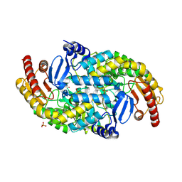
6JZ8
DownloadVisualize
BU of 6jz8 by Molmil
b-glucuronidase from Ruminococcus gnavus in complex with D-glucaro 1,5-lactone
Descriptor: (2S,3S,4S,5R)-3,4,5-trihydroxy-6-oxo-oxane-2-carboxylic acid, Beta-glucuronidase
Authors:Dashnyam, P, Lin, H.Y.
Deposit date:2019-04-30
Release date:2020-06-10
Last modified:2023-11-22
Method:X-RAY DIFFRACTION (1.583 Å)
Cite:Substituent Position of Iminocyclitols Determines the Potency and Selectivity for Gut Microbial Xenobiotic-Reactivating Enzymes.
J.Med.Chem., 63, 2020
6JZ5
DownloadVisualize
BU of 6jz5 by Molmil
b-glucuronidase from Ruminococcus gnavus in complex with D-glucuronic acid
Descriptor: Beta-glucuronidase, beta-D-glucopyranuronic acid
Authors:Dashnyam, P, Lin, H.Y.
Deposit date:2019-04-30
Release date:2020-06-10
Last modified:2023-11-22
Method:X-RAY DIFFRACTION (1.58 Å)
Cite:Substituent Position of Iminocyclitols Determines the Potency and Selectivity for Gut Microbial Xenobiotic-Reactivating Enzymes.
J.Med.Chem., 63, 2020
6JZ3
DownloadVisualize
BU of 6jz3 by Molmil
b-glucuronidase from Ruminococcus gnavus in complex with uronic deoxynojirimycin
Descriptor: (2~{S},3~{R},4~{R},5~{S})-3,4,5-tris(oxidanyl)piperidine-2-carboxylic acid, (4R)-2-METHYLPENTANE-2,4-DIOL, Beta-glucuronidase
Authors:Dashnyam, P, Lin, H.Y.
Deposit date:2019-04-30
Release date:2020-05-13
Last modified:2024-03-27
Method:X-RAY DIFFRACTION (1.502 Å)
Cite:Substituent Position of Iminocyclitols Determines the Potency and Selectivity for Gut Microbial Xenobiotic-Reactivating Enzymes.
J.Med.Chem., 63, 2020
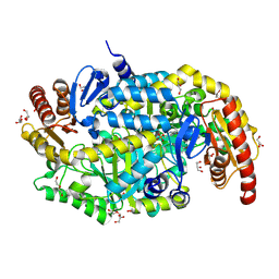
5Z19
DownloadVisualize
BU of 5z19 by Molmil
The crystal structure of Ruminococcus gnavus beta-glucuronidase in complex with uronic isofagomine
Descriptor: (3S,4R,5R)-4,5-dihydroxypiperidine-3-carboxylic acid, Beta-glucuronidase
Authors:Dashnyam, P, Lin, H.Y, Lin, C.H.
Deposit date:2017-12-25
Release date:2018-12-12
Last modified:2024-03-27
Method:X-RAY DIFFRACTION (2.503 Å)
Cite:Dissection of the substrate preference and structure of gut microbial beta-glucuronidases identifies the major bacteria causing xenobiotic toxicity
To Be Published
5Z1A
DownloadVisualize
BU of 5z1a by Molmil
The crystal structure of Bacteroides fragilis beta-glucuronidase in complex with uronic isofagomine
Descriptor: (3S,4R,5R)-4,5-dihydroxypiperidine-3-carboxylic acid, Putative beta-galactosidase
Authors:Dashnyam, P, Lin, H.Y, Lin, C.H.
Deposit date:2017-12-25
Release date:2018-12-12
Last modified:2024-03-27
Method:X-RAY DIFFRACTION (1.859 Å)
Cite:Dissection of the substrate preference and structure of gut microbial-glucuronidases identifies the major bacteria causing xenobiotic toxicity
To Be Published
6JZ2
DownloadVisualize
BU of 6jz2 by Molmil
b-glucuronidase from Ruminococcus gnavus in complex with uronic isofagomine at 1.3 Angstroms resolution
Descriptor: (3S,4R,5R)-4,5-dihydroxypiperidine-3-carboxylic acid, (4R)-2-METHYLPENTANE-2,4-DIOL, (4S)-2-METHYL-2,4-PENTANEDIOL, ...
Authors:Dashnyam, P, Lin, H.Y.
Deposit date:2019-04-30
Release date:2020-06-10
Last modified:2023-11-22
Method:X-RAY DIFFRACTION (1.29 Å)
Cite:Substituent Position of Iminocyclitols Determines the Potency and Selectivity for Gut Microbial Xenobiotic-Reactivating Enzymes.
J.Med.Chem., 63, 2020
6JZ4
DownloadVisualize
BU of 6jz4 by Molmil
b-glucuronidase from Ruminococcus gnavus in complex with D-glucaro-d-lactam
Descriptor: (2S,3R,4S,5R)-3,4,5-trihydroxy-6-oxopiperidine-2-carboxylic acid, (4R)-2-METHYLPENTANE-2,4-DIOL, Beta-glucuronidase
Authors:Dashnyam, P, Lin, H.Y.
Deposit date:2019-04-30
Release date:2020-06-03
Last modified:2024-03-27
Method:X-RAY DIFFRACTION (1.712 Å)
Cite:Substituent Position of Iminocyclitols Determines the Potency and Selectivity for Gut Microbial Xenobiotic-Reactivating Enzymes.
J.Med.Chem., 63, 2020
5Z1B
DownloadVisualize
BU of 5z1b by Molmil
Structure of Bifidobacterium dentium beta-glucuronidase complexed with coumarin-3-O-glucuronide
Descriptor: 3-HYDROXY-2H-CHROMEN-2-ONE, Glycosyl hydrolase family 2, TIM barrel domain protein, ...
Authors:Dashnyam, P, Lin, H.Y, Lin, C.H.
Deposit date:2017-12-25
Release date:2018-12-12
Last modified:2024-03-27
Method:X-RAY DIFFRACTION (1.45 Å)
Cite:Dissection of the substrate preference and structure of gut microbial-glucuronidases identifies the major bacteria causing xenobiotic toxicity
To Be Published
6JZ7
DownloadVisualize
BU of 6jz7 by Molmil
b-glucuronidase from Ruminococcus gnavus in complex with N1-substituted uronic isofagomine
Descriptor: (3~{S},4~{R},5~{R})-4,5-bis(oxidanyl)-1-propyl-piperidine-3-carboxylic acid, Beta-glucuronidase
Authors:Dashnyam, P, Lin, H.Y.
Deposit date:2019-04-30
Release date:2020-06-10
Last modified:2023-11-22
Method:X-RAY DIFFRACTION (1.45 Å)
Cite:Substituent Position of Iminocyclitols Determines the Potency and Selectivity for Gut Microbial Xenobiotic-Reactivating Enzymes.
J.Med.Chem., 63, 2020
6JZ6
DownloadVisualize
BU of 6jz6 by Molmil
b-glucuronidase from Ruminococcus gnavus in complex with C6-substituted uronic isofagomine
Descriptor: (2~{S},3~{S},4~{R},5~{R})-4,5-bis(oxidanyl)-2-propyl-piperidine-3-carboxylic acid, Beta-glucuronidase
Authors:Dashnyam, P, Lin, H.Y.
Deposit date:2019-04-30
Release date:2020-06-10
Last modified:2023-11-22
Method:X-RAY DIFFRACTION (1.605 Å)
Cite:Substituent Position of Iminocyclitols Determines the Potency and Selectivity for Gut Microbial Xenobiotic-Reactivating Enzymes.
J.Med.Chem., 63, 2020
5Z18
DownloadVisualize
BU of 5z18 by Molmil
The crystal structure of Ruminococcus gnavus beta-glucuronidase
Descriptor: Beta-glucuronidase
Authors:Dashnyam, P, Lin, H.Y, Lin, C.H.
Deposit date:2017-12-25
Release date:2018-12-12
Last modified:2024-03-27
Method:X-RAY DIFFRACTION (2.495 Å)
Cite:Dissection of the substrate preference and structure of gut microbial beta-glucuronidases identifies the major bacteria causing xenobiotic toxicity
To Be Published


12>

226707

PDB entries from 2024-10-30

PDB statisticsPDBj update infoContact PDBjnumon