Loading
PDBj
MenuPDBj@FacebookPDBj@TwitterPDBj@YouTubewwPDB FoundationwwPDB
RCSB PDBPDBeBMRBAdv. SearchSearch help
PDB: 1609 results

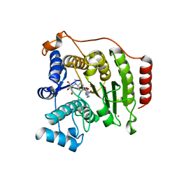
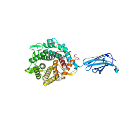
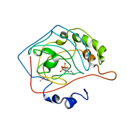
4QA6
DownloadVisualize
BU of 4qa6 by Molmil
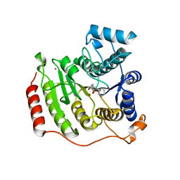
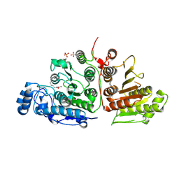
Crystal structure of I243N/Y306F HDAC8 in complex with a tetrapeptide substrate
Descriptor: 7-AMINO-4-METHYL-CHROMEN-2-ONE, GLYCEROL, Histone deacetylase 8, ...
Authors:Decroos, C, Bowman, C.B, Moser, J.-A.S, Christianson, K.E, Deardorff, M.A, Christianson, D.W.
Deposit date:2014-05-02
Release date:2014-08-06
Last modified:2023-12-06
Method:X-RAY DIFFRACTION (2.053 Å)
Cite:Compromised Structure and Function of HDAC8 Mutants Identified in Cornelia de Lange Syndrome Spectrum Disorders.
Acs Chem.Biol., 9, 2014
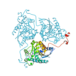
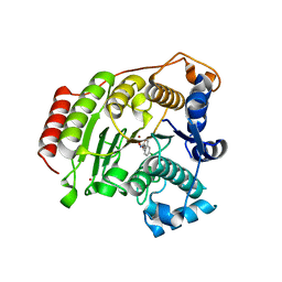
4MU4
DownloadVisualize
BU of 4mu4 by Molmil
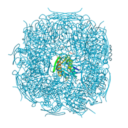
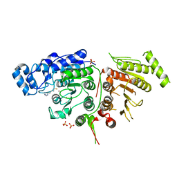
The form B structure of an E21Q catalytic mutant of A. thaliana IGPD2 in complex with Mn2+ and its substrate, 2R3S-IGP, to 1.41 A resolution
Descriptor: (2R,3S)-2,3-dihydroxy-3-(1H-imidazol-5-yl)propyl dihydrogen phosphate, 1,2-ETHANEDIOL, Imidazoleglycerol-phosphate dehydratase 2, ...
Authors:Bisson, C, Britton, K.L, Sedelnikova, S.E, Baker, P.J, Rice, D.W.
Deposit date:2013-09-20
Release date:2014-09-24
Last modified:2023-09-20
Method:X-RAY DIFFRACTION (1.41 Å)
Cite:Crystal Structures Reveal that the Reaction Mechanism of Imidazoleglycerol-Phosphate Dehydratase Is Controlled by Switching Mn(II) Coordination.
Structure, 23, 2015
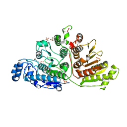
4MYL
DownloadVisualize
BU of 4myl by Molmil
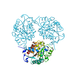
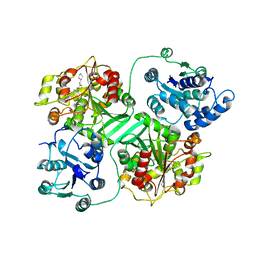
Crystal structure of Trypanosoma cruzi Formiminoglutamase (oxidized) at pH 4.6
Descriptor: Formiminoglutamase
Authors:Hai, Y, Dugery, R.J, Healy, D, Christianson, D.W.
Deposit date:2013-09-27
Release date:2013-11-27
Last modified:2024-11-06
Method:X-RAY DIFFRACTION (1.531 Å)
Cite:Formiminoglutamase from trypanosoma cruzi is an arginase-like manganese metalloenzyme.
Biochemistry, 52, 2013
4Q3Q
DownloadVisualize
BU of 4q3q by Molmil
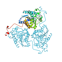
Crystal structure of Schistosoma mansoni arginase in complex with inhibitor ABH
Descriptor: 2(S)-AMINO-6-BORONOHEXANOIC ACID, Arginase, GLYCEROL, ...
Authors:Hai, Y, Edwards, J.E, Van Zandt, M.C, Hoffmann, K.F, Christianson, D.W.
Deposit date:2014-04-12
Release date:2014-07-16
Last modified:2023-09-20
Method:X-RAY DIFFRACTION (2.001 Å)
Cite:Crystal Structure of Schistosoma mansoni Arginase, a Potential Drug Target for the Treatment of Schistosomiasis.
Biochemistry, 53, 2014
4QA2
DownloadVisualize
BU of 4qa2 by Molmil
Crystal structure of I243N HDAC8 in complex with SAHA
Descriptor: GLYCEROL, Histone deacetylase 8, OCTANEDIOIC ACID HYDROXYAMIDE PHENYLAMIDE, ...
Authors:Decroos, C, Bowman, C.B, Moser, J.-A.S, Christianson, K.E, Deardorff, M.A, Christianson, D.W.
Deposit date:2014-05-02
Release date:2014-08-06
Last modified:2023-09-20
Method:X-RAY DIFFRACTION (2.377 Å)
Cite:Compromised Structure and Function of HDAC8 Mutants Identified in Cornelia de Lange Syndrome Spectrum Disorders.
Acs Chem.Biol., 9, 2014
4Q3S
DownloadVisualize
BU of 4q3s by Molmil
Crystal structure of Schistosoma mansoni arginase in complex with inhibitor ABHPE
Descriptor: Arginase, GLYCEROL, MANGANESE (II) ION, ...
Authors:Hai, Y, Edwards, J.E, Van Zandt, M.C, Hoffmann, K.F, Christianson, D.W.
Deposit date:2014-04-12
Release date:2014-07-16
Last modified:2023-09-20
Method:X-RAY DIFFRACTION (2.11 Å)
Cite:Crystal Structure of Schistosoma mansoni Arginase, a Potential Drug Target for the Treatment of Schistosomiasis.
Biochemistry, 53, 2014
4Q3T
DownloadVisualize
BU of 4q3t by Molmil
Crystal structure of Schistosoma mansoni arginase in complex with inhibitor NOHA
Descriptor: Arginase, GLYCEROL, MANGANESE (II) ION, ...
Authors:Hai, Y, Edwards, J.E, Van Zandt, M.C, Hoffmann, K.F, Christianson, D.W.
Deposit date:2014-04-12
Release date:2014-07-16
Last modified:2023-09-20
Method:X-RAY DIFFRACTION (2.142 Å)
Cite:Crystal Structure of Schistosoma mansoni Arginase, a Potential Drug Target for the Treatment of Schistosomiasis.
Biochemistry, 53, 2014
4QA1
DownloadVisualize
BU of 4qa1 by Molmil
Crystal structure of A188T HDAC8 in complex with M344
Descriptor: 4-(dimethylamino)-N-[7-(hydroxyamino)-7-oxoheptyl]benzamide, GLYCEROL, Histone deacetylase 8, ...
Authors:Decroos, C, Bowman, C.B, Moser, J.-A.S, Christianson, K.E, Deardorff, M.A, Christianson, D.W.
Deposit date:2014-05-02
Release date:2014-08-06
Last modified:2023-09-20
Method:X-RAY DIFFRACTION (1.92 Å)
Cite:Compromised Structure and Function of HDAC8 Mutants Identified in Cornelia de Lange Syndrome Spectrum Disorders.
Acs Chem.Biol., 9, 2014
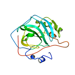
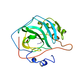
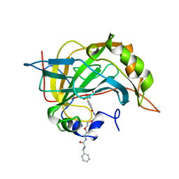
1CVF
DownloadVisualize
BU of 1cvf by Molmil
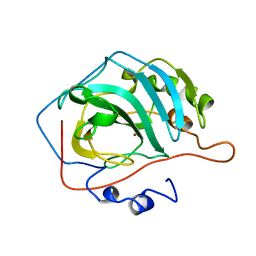
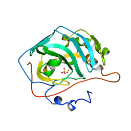
STRUCTURAL CONSEQUENCES OF REDESIGNING A PROTEIN-ZINC BINDING SITE
Descriptor: CARBONIC ANHYDRASE II
Authors:Ippolito, J.A, Christianson, D.W.
Deposit date:1994-06-21
Release date:1994-11-30
Last modified:2024-02-07
Method:X-RAY DIFFRACTION (2.25 Å)
Cite:Structural consequences of redesigning a protein-zinc binding site.
Biochemistry, 33, 1994
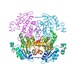
1CWU
DownloadVisualize
BU of 1cwu by Molmil
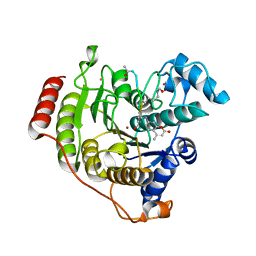
BRASSICA NAPUS ENOYL ACP REDUCTASE A138G MUTANT COMPLEXED WITH NAD+ AND THIENODIAZABORINE
Descriptor: 6-METHYL-2(PROPANE-1-SULFONYL)-2H-THIENO[3,2-D][1,2,3]DIAZABORININ-1-OL, ENOYL ACP REDUCTASE, NICOTINAMIDE-ADENINE-DINUCLEOTIDE
Authors:Roujeinikova, A, Rafferty, J.B, Rice, D.W.
Deposit date:1999-08-26
Release date:1999-09-02
Last modified:2024-02-07
Method:X-RAY DIFFRACTION (2.5 Å)
Cite:Inhibitor binding studies on enoyl reductase reveal conformational changes related to substrate recognition.
J.Biol.Chem., 274, 1999
6U4Z
DownloadVisualize
BU of 6u4z by Molmil
Crystal Structure of a family 76 glycoside hydrolase from a bovine Bacteroides thetaiotaomicron strain
Descriptor: 1,2-ETHANEDIOL, Alpha-1,6-mannanase, HEXAETHYLENE GLYCOL
Authors:Jones, D.R, Abbott, D.W.
Deposit date:2019-08-26
Release date:2020-04-29
Last modified:2023-10-11
Method:X-RAY DIFFRACTION (1.4 Å)
Cite:Analysis of Active Site Architecture and Reaction Product Linkage Chemistry Reveals a Conserved Cleavage Substrate for an Endo-alpha-mannanase within Diverse Yeast Mannans.
J.Mol.Biol., 432, 2020
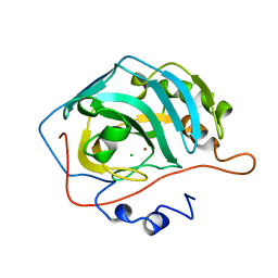
1CVA
DownloadVisualize
BU of 1cva by Molmil
STRUCTURAL AND FUNCTIONAL IMPORTANCE OF A CONSERVED HYDROGEN BOND NETWORK IN HUMAN CARBONIC ANHYDRASE II
Descriptor: AZIDE ION, CARBONIC ANHYDRASE II, ZINC ION
Authors:Ippolito, J.A, Christianson, D.W.
Deposit date:1993-02-04
Release date:1993-10-31
Last modified:2024-02-07
Method:X-RAY DIFFRACTION (2.25 Å)
Cite:Structural and functional importance of a conserved hydrogen bond network in human carbonic anhydrase II.
J.Biol.Chem., 268, 1993
1CNX
DownloadVisualize
BU of 1cnx by Molmil
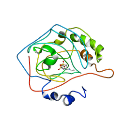
SECONDARY INTERACTIONS SIGNIFICANTLY REMOVED FROM THE SULFONAMIDE BINDING POCKET OF CARBONIC ANHYDRASE II INFLUENCE BINDING CONSTANTS
Descriptor: AMINODI(ETHYLOXY)ETHYLAMINOCARBONYLBENZENESULFONAMIDE, CARBONIC ANHYDRASE II, MERCURY (II) ION, ...
Authors:Boriack, P.A, Christianson, D.W.
Deposit date:1995-07-21
Release date:1995-11-14
Last modified:2024-02-07
Method:X-RAY DIFFRACTION (1.9 Å)
Cite:Secondary interactions significantly removed from the sulfonamide binding pocket of carbonic anhydrase II influence inhibitor binding constants.
J.Med.Chem., 38, 1995
5BWZ
DownloadVisualize
BU of 5bwz by Molmil
Crystal structure of S39E HDAC8 in complex with Droxinostat
Descriptor: Droxinostat, GLYCEROL, Histone deacetylase 8, ...
Authors:Decroos, C, Christianson, D.W.
Deposit date:2015-06-08
Release date:2016-06-08
Last modified:2023-09-27
Method:X-RAY DIFFRACTION (1.59 Å)
Cite:Phosphorylation of Histone Deacetylase 8: Structural and Mechanistic Analysis of the Phosphomimetic S39E Mutant.
Biochemistry, 58, 2019
6UFN
DownloadVisualize
BU of 6ufn by Molmil
Crystal Structure of Danio rerio Histone Deacetylase 10 in Complex with 7-[(3-aminopropyl)amino]heptan-2-one
Descriptor: 7-[(3-aminopropyl)amino]heptane-2,2-diol, PHOSPHATE ION, POTASSIUM ION, ...
Authors:Herbst-Gervasoni, C.J, Christianson, D.W.
Deposit date:2019-09-24
Release date:2019-12-04
Last modified:2023-10-11
Method:X-RAY DIFFRACTION (2.7 Å)
Cite:Binding ofN8-Acetylspermidine Analogues to Histone Deacetylase 10 Reveals Molecular Strategies for Blocking Polyamine Deacetylation.
Biochemistry, 58, 2019
6UHV
DownloadVisualize
BU of 6uhv by Molmil
Crystal Structure of Danio rerio Histone Deacetylase 10 in Complex with 6-[(3-aminopropyl)amino]-N-hydroxyhexanamide
Descriptor: 6-[(3-aminopropyl)amino]-N-hydroxyhexanamide, PHOSPHATE ION, POTASSIUM ION, ...
Authors:Herbst-Gervasoni, C.J, Christianson, D.W.
Deposit date:2019-09-28
Release date:2019-12-04
Last modified:2024-10-16
Method:X-RAY DIFFRACTION (2.53 Å)
Cite:Binding ofN8-Acetylspermidine Analogues to Histone Deacetylase 10 Reveals Molecular Strategies for Blocking Polyamine Deacetylation.
Biochemistry, 58, 2019
6UII
DownloadVisualize
BU of 6uii by Molmil
Crystal Structure of Danio rerio Histone Deacetylase 10 in Complex with 5-[(3-aminopropyl)amino]pentane-1-thiol
Descriptor: 5-[(3-aminopropyl)amino]pentane-1-thiol, PHOSPHATE ION, POTASSIUM ION, ...
Authors:Herbst-Gervasoni, C.J, Christianson, D.W.
Deposit date:2019-10-01
Release date:2019-12-04
Last modified:2023-10-11
Method:X-RAY DIFFRACTION (2.65 Å)
Cite:Binding ofN8-Acetylspermidine Analogues to Histone Deacetylase 10 Reveals Molecular Strategies for Blocking Polyamine Deacetylation.
Biochemistry, 58, 2019
1CR6
DownloadVisualize
BU of 1cr6 by Molmil
CRYSTAL STRUCTURE OF MURINE SOLUBLE EPOXIDE HYDROLASE COMPLEXED WITH CPU INHIBITOR
Descriptor: EPOXIDE HYDROLASE, N-CYCLOHEXYL-N'-(PROPYL)PHENYL UREA
Authors:Argiriadi, M.A, Morisseau, C, Hammock, B.D, Christianson, D.W.
Deposit date:1999-08-13
Release date:1999-11-19
Last modified:2024-02-07
Method:X-RAY DIFFRACTION (2.8 Å)
Cite:Detoxification of environmental mutagens and carcinogens: structure, mechanism, and evolution of liver epoxide hydrolase.
Proc.Natl.Acad.Sci.USA, 96, 1999
1CVC
DownloadVisualize
BU of 1cvc by Molmil
REDESIGNING THE ZINC BINDING SITE OF HUMAN CARBONIC ANHYDRASE II: STRUCTURE OF A HIS2ASP-ZN2+ METAL COORDINATION POLYHEDRON
Descriptor: CARBONIC ANHYDRASE II, ZINC ION
Authors:Ippolito, J.A, Christianson, D.W.
Deposit date:1993-09-22
Release date:1994-01-31
Last modified:2024-02-07
Method:X-RAY DIFFRACTION (2.3 Å)
Cite:REDESIGNING THE ZINC-BINDING SITE OF HUMAN CARBONIC ANHYDRASE-II - STRUCTURE OF A HIS2ASP-ZN2+ METAL COORDINATION POLYHEDRON.
J.Am.Chem.Soc., 115, 1993
1CIN
DownloadVisualize
BU of 1cin by Molmil
THE POSITIONS OF HIS-64 AND A BOUND WATER IN HUMAN CARBONIC ANHYDRASE II UPON BINDING THREE STRUCTURALLY RELATED INHIBITORS
Descriptor: (4S-TRANS)-4-(METHYLAMINO)-5,6-DIHYDRO-6-METHYL-4H-THIENO(2,3-B)THIOPYRAN-2-SULFONAMIDE-7,7-DIOXIDE, CARBONIC ANHYDRASE II, METHYL MERCURY ION, ...
Authors:Smith, G.M, Alexander, R.S, Christianson, D.W, Mckeever, B.M, Ponticello, G.S, Springer, J.P, Randall, W.C, Baldwin, J.J, Habecker, C.N.
Deposit date:1993-10-20
Release date:1994-01-31
Last modified:2024-02-07
Method:X-RAY DIFFRACTION (2.1 Å)
Cite:Positions of His-64 and a bound water in human carbonic anhydrase II upon binding three structurally related inhibitors.
Protein Sci., 3, 1994
1CNY
DownloadVisualize
BU of 1cny by Molmil
SECONDARY INTERACTIONS SIGNIFICANTLY REMOVED FROM THE SULFONAMIDE BINDING POCKET OF CARBONIC ANHYDRASE II INFLUENCE BINDING CONSTANTS
Descriptor: CARBONIC ANHYDRASE II, MERCURY (II) ION, PHENYLALANYLAMINODI(ETHYLOXY)ETHYL BENZENESULFONAMIDEAMINOCARBONYLBENZENESULFONAMIDE, ...
Authors:Boriack, P.A, Christianson, D.W.
Deposit date:1995-07-21
Release date:1995-11-14
Last modified:2024-02-07
Method:X-RAY DIFFRACTION (2.3 Å)
Cite:Secondary interactions significantly removed from the sulfonamide binding pocket of carbonic anhydrase II influence inhibitor binding constants.
J.Med.Chem., 38, 1995
1CNG
DownloadVisualize
BU of 1cng by Molmil
X-RAY CRYSTALLOGRAPHIC STUDIES OF ENGINEERED HYDROGEN BOND NETWORKS IN A PROTEIN-ZINC BINDING SITE
Descriptor: CARBONIC ANHYDRASE II, CHLORIDE ION, ZINC ION
Authors:Lesburg, C.A, Christianson, D.W.
Deposit date:1995-04-03
Release date:1995-07-10
Last modified:2024-02-07
Method:X-RAY DIFFRACTION (1.9 Å)
Cite:X-Ray Crystallographic Studies of Engineered Hydrogen Bond Networks in a Protein-Zinc Binding Site
J.Am.Chem.Soc., 117, 1995
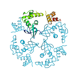
1CUK
DownloadVisualize
BU of 1cuk by Molmil
ESCHERICHIA COLI RUVA PROTEIN AT PH 4.9 AND ROOM TEMPERATURE
Descriptor: RUVA PROTEIN
Authors:Rafferty, J.B, Rice, D.W.
Deposit date:1996-08-28
Release date:1997-10-22
Last modified:2024-02-07
Method:X-RAY DIFFRACTION (1.9 Å)
Cite:Crystal structure of DNA recombination protein RuvA and a model for its binding to the Holliday junction.
Science, 274, 1996
1CNK
DownloadVisualize
BU of 1cnk by Molmil
X-RAY CRYSTALLOGRAPHIC STUDIES OF ENGINEERED HYDROGEN BOND NETWORKS IN A PROTEIN-ZINC BINDING SITE
Descriptor: CARBONIC ANHYDRASE II, SULFATE ION, ZINC ION
Authors:Lesburg, C.A, Christianson, D.W.
Deposit date:1995-04-03
Release date:1995-07-10
Last modified:2024-02-07
Method:X-RAY DIFFRACTION (2.15 Å)
Cite:X-Ray Crystallographic Studies of Engineered Hydrogen Bond Networks in a Protein-Zinc Binding Site
J.Am.Chem.Soc., 117, 1995
1CIL
DownloadVisualize
BU of 1cil by Molmil
THE POSITIONS OF HIS-64 AND A BOUND WATER IN HUMAN CARBONIC ANHYDRASE II UPON BINDING THREE STRUCTURALLY RELATED INHIBITORS
Descriptor: (4S-TRANS)-4-(ETHYLAMINO)-5,6-DIHYDRO-6-METHYL-4H-THIENO(2,3-B)THIOPYRAN-2-SULFONAMIDE-7,7-DIOXIDE, CARBONIC ANHYDRASE II, ZINC ION
Authors:Smith, G.M, Alexander, R.S, Christianson, D.W, Mckeever, B.M, Ponticello, G.S, Springer, J.P, Randall, W.C, Baldwin, J.J, Habecker, C.N.
Deposit date:1993-10-20
Release date:1994-01-31
Last modified:2024-02-07
Method:X-RAY DIFFRACTION (1.6 Å)
Cite:Positions of His-64 and a bound water in human carbonic anhydrase II upon binding three structurally related inhibitors.
Protein Sci., 3, 1994

227111

건을2024-11-06부터공개중

PDB statisticsPDBj update infoContact PDBjnumon