1R1P
 
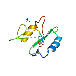 | |
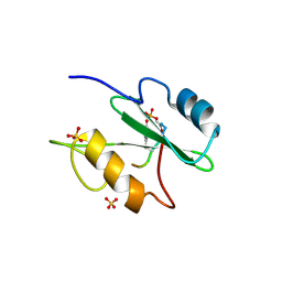
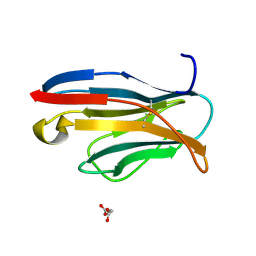
2EAX
 | | Crystal structure of human PGRP-IBETAC in complex with glycosamyl muramyl pentapeptide | | 分子名称: | 2-acetamido-2-deoxy-beta-D-glucopyranose-(1-4)-methyl 2-acetamido-3-O-[(1R)-1-carboxyethyl]-2-deoxy-beta-D-glucopyranoside, GLYCOSAMYL MURAMYL PENTAPEPTIDE, Peptidoglycan recognition protein-I-beta | | 著者 | Cho, S. | | 登録日 | 2007-02-03 | | 公開日 | 2007-10-02 | | 最終更新日 | 2023-10-25 | | 実験手法 | X-RAY DIFFRACTION (2.1 Å) | | 主引用文献 | Structural insights into the bactericidal mechanism of human peptidoglycan recognition proteins
Proc.Natl.Acad.Sci.Usa, 104, 2007
|
|
1R1S
 | |
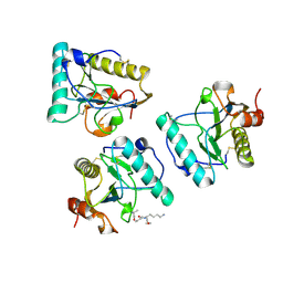
3CAD
 | | Crystal structure of Natural Killer Cell Receptor, Ly49G | | 分子名称: | Lectin-related NK cell receptor LY49G1 | | 著者 | Cho, S. | | 登録日 | 2008-02-19 | | 公開日 | 2008-04-08 | | 最終更新日 | 2011-07-13 | | 実験手法 | X-RAY DIFFRACTION (2.6 Å) | | 主引用文献 | Molecular Architecture of the Major Histocompatibility Complex Class I-binding Site of Ly49 Natural Killer Cell Receptors.
J.Biol.Chem., 283, 2008
|
|
2EAV
 | |
8J56
 | |
1R1Q
 | |
1IA5
 | | POLYGALACTURONASE FROM ASPERGILLUS ACULEATUS | | 分子名称: | POLYGALACTURONASE, alpha-D-mannopyranose, alpha-D-mannopyranose-(1-4)-2-acetamido-2-deoxy-beta-D-glucopyranose-(1-4)-2-acetamido-2-deoxy-beta-D-glucopyranose | | 著者 | Cho, S.W, Lee, S, Shin, W. | | 登録日 | 2001-03-22 | | 公開日 | 2001-09-19 | | 最終更新日 | 2020-07-29 | | 実験手法 | X-RAY DIFFRACTION (2 Å) | | 主引用文献 | The X-ray structure of Aspergillus aculeatus polygalacturonase and a modeled structure of the polygalacturonase-octagalacturonate complex.
J.Mol.Biol., 311, 2001
|
|
2APF
 | | Crystal Structure of the A52V/S54N/K66E variant of the murine T cell receptor V beta 8.2 domain | | 分子名称: | MALONIC ACID, T cell receptor beta chain V | | 著者 | Cho, S, Swaminathan, C.P, Yang, J, Kerzic, M.C, Guan, R, Kieke, M.C, Kranz, D.M, Mariuzza, R.A, Sundberg, E.J. | | 登録日 | 2005-08-16 | | 公開日 | 2006-03-21 | | 最終更新日 | 2018-04-04 | | 実験手法 | X-RAY DIFFRACTION (1.8 Å) | | 主引用文献 | Structural basis of affinity maturation and intramolecular cooperativity in a protein-protein interaction.
Structure, 13, 2005
|
|
2APT
 | | Crystal Structure of the G17E/S54N/K66E/Q72H/E80V/L81S/T87S/G96V variant of the murine T cell receptor V beta 8.2 domain | | 分子名称: | MALONIC ACID, T-cell receptor beta chain V | | 著者 | Cho, S, Swaminathan, C.P, Yang, J, Kerzic, M.C, Guan, R, Kieke, M.C, Kranz, D.M, Mariuzza, R.A, Sundberg, E.J. | | 登録日 | 2005-08-16 | | 公開日 | 2006-03-21 | | 最終更新日 | 2018-04-04 | | 実験手法 | X-RAY DIFFRACTION (2 Å) | | 主引用文献 | Structural basis of affinity maturation and intramolecular cooperativity in a protein-protein interaction.
Structure, 13, 2005
|
|
2APW
 | | Crystal Structure of the G17E/A52V/S54N/K66E/E80V/L81S/T87S/G96V variant of the murine T cell receptor V beta 8.2 domain | | 分子名称: | MALONIC ACID, T cell receptor beta chain V | | 著者 | Cho, S, Swaminathan, C.P, Yang, J, Kerzic, M.C, Guan, R, Kieke, M.C, Kranz, D.M, Mariuzza, R.A, Sundberg, E.J. | | 登録日 | 2005-08-16 | | 公開日 | 2006-03-21 | | 最終更新日 | 2018-04-04 | | 実験手法 | X-RAY DIFFRACTION (2 Å) | | 主引用文献 | Structural basis of affinity maturation and intramolecular cooperativity in a protein-protein interaction.
Structure, 13, 2005
|
|
2APV
 | | Crystal Structure of the G17E/A52V/S54N/Q72H/E80V/L81S/T87S/G96V variant of the murine T cell receptor V beta 8.2 domain | | 分子名称: | MALONIC ACID, T cell receptor beta chain V | | 著者 | Cho, S, Swaminathan, C.P, Yang, J, Kerzic, M.C, Guan, R, Kieke, M.C, Kranz, D.M, Mariuzza, R.A, Sundberg, E.J. | | 登録日 | 2005-08-16 | | 公開日 | 2006-03-21 | | 最終更新日 | 2018-04-04 | | 実験手法 | X-RAY DIFFRACTION (1.9 Å) | | 主引用文献 | Structural basis of affinity maturation and intramolecular cooperativity in a protein-protein interaction.
Structure, 13, 2005
|
|
2AQ1
 | | Crystal structure of T-cell receptor V beta domain variant complexed with superantigen SEC3 mutant | | 分子名称: | Enterotoxin type C-3, T-cell receptor beta chain V | | 著者 | Cho, S, Swaminathan, C.P, Yang, J, Kerzic, M.C, Guan, R, Kieke, M.C, Kranz, D.M, Mariuzza, R.A, Sundberg, E.J. | | 登録日 | 2005-08-17 | | 公開日 | 2006-03-21 | | 最終更新日 | 2017-10-11 | | 実験手法 | X-RAY DIFFRACTION (2.1 Å) | | 主引用文献 | Structural basis of affinity maturation and intramolecular cooperativity in a protein-protein interaction.
Structure, 13, 2005
|
|
1K81
 | |
1IBQ
 | | ASPERGILLOPEPSIN FROM ASPERGILLUS PHOENICIS | | 分子名称: | ASPERGILLOPEPSIN, ZINC ION, alpha-D-mannopyranose | | 著者 | Cho, S.W, Shin, W. | | 登録日 | 2001-03-28 | | 公開日 | 2001-07-04 | | 最終更新日 | 2020-07-29 | | 実験手法 | X-RAY DIFFRACTION (2.14 Å) | | 主引用文献 | Structure of aspergillopepsin I from Aspergillus phoenicis: variations of the S1'-S2 subsite in aspartic proteinases.
Acta Crystallogr.,Sect.D, 57, 2001
|
|
1K8B
 | |
7F2H
 | |
7F2G
 | |
7E90
 | |
7E92
 | |
7ZCW
 | | Cryo-EM structure of GMPCPP-microtubules in complex with VASH2-SVBP | | 分子名称: | GUANOSINE-5'-TRIPHOSPHATE, MAGNESIUM ION, PHOSPHOMETHYLPHOSPHONIC ACID GUANYLATE ESTER, ... | | 著者 | Choi, S.R, Blum, T, Steinmetz, M.O. | | 登録日 | 2022-03-29 | | 公開日 | 2022-12-14 | | 最終更新日 | 2023-06-28 | | 実験手法 | ELECTRON MICROSCOPY (3.6 Å) | | 主引用文献 | VASH1-SVBP and VASH2-SVBP generate different detyrosination profiles on microtubules.
J.Cell Biol., 222, 2023
|
|
5HTO
 | |
5HS4
 | | Plasmdoium Vivax Lactate dehydrogenase | | 分子名称: | L-lactate dehydrogenase | | 著者 | Choi, S.J, Ban, C. | | 登録日 | 2016-01-25 | | 公開日 | 2016-10-19 | | 最終更新日 | 2023-11-08 | | 実験手法 | X-RAY DIFFRACTION (1.339 Å) | | 主引用文献 | Crystal structure of a DNA aptamer bound to PvLDH elucidates novel single-stranded DNA structural elements for folding and recognition
Sci Rep, 6, 2016
|
|
5HRU
 | |
2GIO
 | |


