2RDV
 
 | |
7R6P
 | |
3WUX
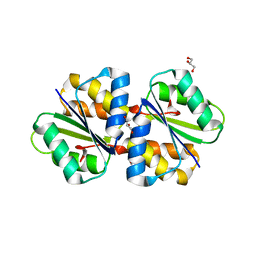
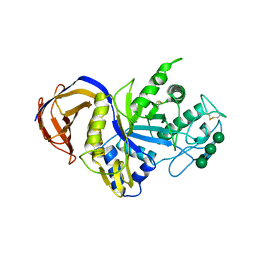
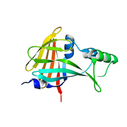
 | | Crystal structure of unsaturated glucuronyl hydrolase mutant D115N/K370S from Streptococcus agalactiae | | 分子名称: | 1,2-ETHANEDIOL, Unsaturated chondroitin disaccharide hydrolase | | 著者 | Nakamichi, Y, Oiki, S, Mikami, B, Murata, K, Hashimoto, W. | | 登録日 | 2014-05-08 | | 公開日 | 2014-05-28 | | 最終更新日 | 2023-11-08 | | 実験手法 | X-RAY DIFFRACTION (1.792 Å) | | 主引用文献 | Crystal structure of unsaturated glucuronyl hydrolase mutant D115N/K370S from Streptococcus agalactiae
to be published
|
|
7E9U
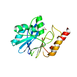
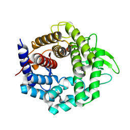
 | | Trehalase of Arabidopsis thaliana | | 分子名称: | GLYCEROL, PHOSPHATE ION, SODIUM ION, ... | | 著者 | Taguchi, Y, Saburi, W, Yu, J, Imai, R, Yao, M, Mori, H. | | 登録日 | 2021-03-05 | | 公開日 | 2022-03-09 | | 最終更新日 | 2023-11-29 | | 実験手法 | X-RAY DIFFRACTION (2.1 Å) | | 主引用文献 | pH-dependent alteration of substrate specificity of plant trehalase and its molecular mechanism
To Be Published
|
|
7E9X
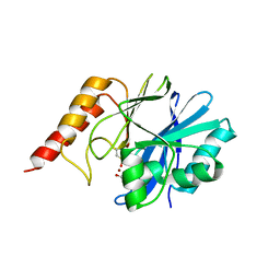
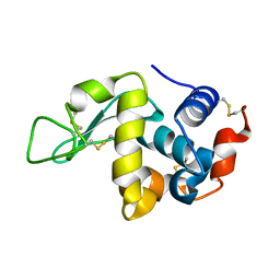
 | | Trehalase of Arabidopsis thaliana acid mutant -D380A | | 分子名称: | GLYCEROL, Trehalase | | 著者 | Taguchi, Y, Saburi, W, Yu, J, Imai, R, Yao, M, Mori, H. | | 登録日 | 2021-03-05 | | 公開日 | 2022-03-09 | | 最終更新日 | 2023-11-29 | | 実験手法 | X-RAY DIFFRACTION (1.88 Å) | | 主引用文献 | pH-dependent alteration of substrate specificity of plant trehalase and its molecular mechanism
To Be Published
|
|
7EAW
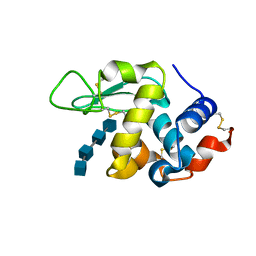
 | | Trehalase of Arabidopsis thaliana acid mutant -D380A trehalose complex | | 分子名称: | GLYCEROL, Trehalase, alpha-D-glucopyranose-(1-1)-alpha-D-glucopyranose | | 著者 | Taguchi, Y, Saburi, W, Yu, J, Imai, R, Yao, M, Mori, H. | | 登録日 | 2021-03-08 | | 公開日 | 2022-03-16 | | 最終更新日 | 2023-11-29 | | 実験手法 | X-RAY DIFFRACTION (1.8 Å) | | 主引用文献 | pH-dependent alteration of substrate specificity of plant trehalase and its molecular mechanism
To Be Published
|
|
2ZJ9
 | | X-ray crystal structure of AmpC beta-Lactamase (AmpC(D)) from an Escherichia coli with a Tripeptide Deletion (Gly286 Ser287 Asp288) on the H10 Helix | | 分子名称: | AmpC, ISOPROPYL ALCOHOL, SODIUM ION | | 著者 | Yamaguchi, Y, Sato, G, Yamagata, Y, Wachino, J, Arakawa, Y, Kurosaki, H. | | 登録日 | 2008-02-29 | | 公開日 | 2009-03-10 | | 最終更新日 | 2023-11-01 | | 実験手法 | X-RAY DIFFRACTION (1.7 Å) | | 主引用文献 | Structure of AmpC beta-lactamase (AmpCD) from an Escherichia coli clinical isolate with a tripeptide deletion (Gly286-Ser287-Asp288) in the H10 helix
Acta Crystallogr.,Sect.F, 65, 2009
|
|
1V47
 | | Crystal structure of ATP sulfurylase from Thermus thermophillus HB8 in complex with APS | | 分子名称: | ADENOSINE-5'-PHOSPHOSULFATE, ATP sulfurylase, CHLORIDE ION, ... | | 著者 | Taguchi, Y, Sugishima, M, Fukuyama, K, RIKEN Structural Genomics/Proteomics Initiative (RSGI) | | 登録日 | 2003-11-11 | | 公開日 | 2004-04-06 | | 最終更新日 | 2023-10-25 | | 実験手法 | X-RAY DIFFRACTION (2.49 Å) | | 主引用文献 | Crystal structure of a novel zinc-binding ATP sulfurylase from Thermus thermophilus HB8
Biochemistry, 43, 2004
|
|
1H2R
 | |
4TKZ
 | | Crystal structure of phosphotransferase system component EIIA from Streptococcus agalactiae | | 分子名称: | GLYCEROL, Putative uncharacterized protein gbs1890 | | 著者 | Nakamichi, Y, Maruyama, Y, Oiki, S, Mikami, B, Murata, K, Hashimoto, W. | | 登録日 | 2014-05-28 | | 公開日 | 2014-08-20 | | 最終更新日 | 2023-11-08 | | 実験手法 | X-RAY DIFFRACTION (1.8 Å) | | 主引用文献 | Crystal structure of phosphotransferase system component EIIA from Streptococcus agalactiae
To Be Published
|
|
1WUP
 | | Crystal structure of metallo-beta-lactamase IMP-1 mutant (D81E) | | 分子名称: | ACETIC ACID, Beta-lactamase IMP-1, ZINC ION | | 著者 | Yamaguchi, Y, Yamagata, Y, Goto, M. | | 登録日 | 2004-12-08 | | 公開日 | 2005-03-29 | | 最終更新日 | 2023-10-25 | | 実験手法 | X-RAY DIFFRACTION (3 Å) | | 主引用文献 | Probing the role of Asp-120(81) of metallo-beta-lactamase (IMP-1) by site-directed mutagenesis, kinetic studies, and X-ray crystallography.
J.Biol.Chem., 280, 2005
|
|
1WUO
 | | Crystal structure of metallo-beta-lactamase IMP-1 mutant (D81A) | | 分子名称: | ACETIC ACID, Beta-lactamase IMP-1, ZINC ION | | 著者 | Yamaguchi, Y, Yamagata, Y, Goto, M. | | 登録日 | 2004-12-08 | | 公開日 | 2005-03-29 | | 最終更新日 | 2023-10-25 | | 実験手法 | X-RAY DIFFRACTION (2.01 Å) | | 主引用文献 | Probing the role of Asp-120(81) of metallo-beta-lactamase (IMP-1) by site-directed mutagenesis, kinetic studies, and X-ray crystallography.
J.Biol.Chem., 280, 2005
|
|
6IUJ
 | | Crystal structure of GH30 xylanase B from Talaromyces cellulolyticus | | 分子名称: | 2-acetamido-2-deoxy-beta-D-glucopyranose, 2-acetamido-2-deoxy-beta-D-glucopyranose-(1-4)-2-acetamido-2-deoxy-beta-D-glucopyranose, GH30 Xylanase B, ... | | 著者 | Nakamichi, Y, Watanabe, M, Inoue, H. | | 登録日 | 2018-11-28 | | 公開日 | 2019-01-30 | | 最終更新日 | 2023-11-22 | | 実験手法 | X-RAY DIFFRACTION (2.25 Å) | | 主引用文献 | Structural and functional characterization of a bifunctional GH30-7 xylanase B from the filamentous fungusTalaromyces cellulolyticus.
J. Biol. Chem., 294, 2019
|
|
3WIW
 | | Crystal structure of unsaturated glucuronyl hydrolase specific for heparin | | 分子名称: | 4-(2-HYDROXYETHYL)-1-PIPERAZINE ETHANESULFONIC ACID, Glycosyl hydrolase family 88 | | 著者 | Nakamichi, Y, Mikami, B, Murata, K, Hashimoto, W. | | 登録日 | 2013-09-26 | | 公開日 | 2014-01-08 | | 最終更新日 | 2023-11-08 | | 実験手法 | X-RAY DIFFRACTION (1.35 Å) | | 主引用文献 | Crystal structure of a bacterial unsaturated glucuronyl hydrolase with specificity for heparin.
J.Biol.Chem., 289, 2014
|
|
3WVX
 | |
3WVY
 | | Structure of D48A hen egg white lysozyme in complex with (GlcNAc)4 | | 分子名称: | 2-acetamido-2-deoxy-beta-D-glucopyranose-(1-4)-2-acetamido-2-deoxy-beta-D-glucopyranose-(1-4)-2-acetamido-2-deoxy-beta-D-glucopyranose-(1-4)-2-acetamido-2-deoxy-beta-D-glucopyranose, Lysozyme C | | 著者 | Kawaguchi, Y, Yoneda, K, Araki, T. | | 登録日 | 2014-06-11 | | 公開日 | 2015-06-10 | | 最終更新日 | 2023-11-08 | | 実験手法 | X-RAY DIFFRACTION (1.56 Å) | | 主引用文献 | The role of Asp48 in the hydrogen bonding network involving Asp52 of hen egg white lysozyme
TO BE PUBLISHED
|
|
3WYH
 | |
3L6N
 | |
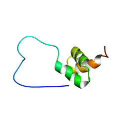
1VEK
 | | Solution Structure of RSGI RUH-011, a UBA Domain from Arabidopsis cDNA | | 分子名称: | ubiquitin-specific protease 14, putative | | 著者 | Higuchi, Y, Abe, T, Hirota, H, Saito, K, Koshiba, S, Kigawa, T, Yokoyama, S, RIKEN Structural Genomics/Proteomics Initiative (RSGI) | | 登録日 | 2004-03-31 | | 公開日 | 2004-09-30 | | 最終更新日 | 2023-12-27 | | 実験手法 | SOLUTION NMR | | 主引用文献 | Solution Structure of RSGI RUH-011, a UBA Domain from Arabidopsis cDNA
To be Published
|
|
1VEJ
 | | Solution Structure of RSGI RUH-016, a UBA Domain from mouse cDNA | | 分子名称: | RIKEN cDNA 4931431F19 | | 著者 | Higuchi, Y, Abe, T, Hirota, H, Hayashi, F, Yokoyama, S, RIKEN Structural Genomics/Proteomics Initiative (RSGI) | | 登録日 | 2004-03-31 | | 公開日 | 2005-05-31 | | 最終更新日 | 2023-12-27 | | 実験手法 | SOLUTION NMR | | 主引用文献 | Solution Structure of RSGI RUH-016, a UBA Domain from mouse cDNA
To be Published
|
|
5B0U
 | |
2E1Q
 | | Crystal Structure of Human Xanthine Oxidoreductase mutant, Glu803Val | | 分子名称: | 2-HYDROXYBENZOIC ACID, BICARBONATE ION, CALCIUM ION, ... | | 著者 | Yamaguchi, Y, Matsumura, T, Ichida, K, Okamoto, K, Nishino, T. | | 登録日 | 2006-10-27 | | 公開日 | 2007-09-18 | | 最終更新日 | 2023-10-25 | | 実験手法 | X-RAY DIFFRACTION (2.6 Å) | | 主引用文献 | Human xanthine oxidase changes its substrate specificity to aldehyde oxidase type upon mutation of amino acid residues in the active site: roles of active site residues in binding and activation of purine substrate
J.Biochem.(Tokyo), 141, 2007
|
|
1WIV
 | | solution structure of RSGI RUH-023, a UBA domain from Arabidopsis cDNA | | 分子名称: | ubiquitin-specific protease 14 | | 著者 | Higuchi, Y, Abe, T, Hirota, H, Izumi, K, Yoshida, M, Yokoyama, S, RIKEN Structural Genomics/Proteomics Initiative (RSGI) | | 登録日 | 2004-05-28 | | 公開日 | 2004-11-28 | | 最終更新日 | 2024-05-29 | | 実験手法 | SOLUTION NMR | | 主引用文献 | solution structure of RSGI RUH-023, a UBA domain from Arabidopsis cDNA
To be Published
|
|
1WMX
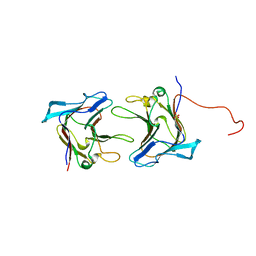 | | Crystal Structure of Family 30 Carbohydrate Binding Module | | 分子名称: | COG3291: FOG: PKD repeat, SULFATE ION | | 著者 | Horiguchi, Y, Kono, M, Suzuki, A, Yamane, T, Arai, M, Sakka, K, Omiya, K. | | 登録日 | 2004-07-21 | | 公開日 | 2004-08-03 | | 最終更新日 | 2024-03-13 | | 実験手法 | X-RAY DIFFRACTION (2 Å) | | 主引用文献 | Crystal Structure of Family 30 Carbohydrate Binding Module
To be Published
|
|
1WZX
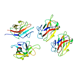 | | Crystal Structure of Family 30 Carbohydrate Binding Module. | | 分子名称: | COG3291: FOG: PKD repeat | | 著者 | Horiguchi, Y, Kono, M, Suzuki, A, Yamane, T, Arai, M, Sakka, K, Omiya, K. | | 登録日 | 2005-03-10 | | 公開日 | 2005-03-22 | | 最終更新日 | 2023-10-25 | | 実験手法 | X-RAY DIFFRACTION (3.52 Å) | | 主引用文献 | Crystal Structure of Family 30 Carbohydrate Binding Module
To be Published
|
|


