6K7P
 
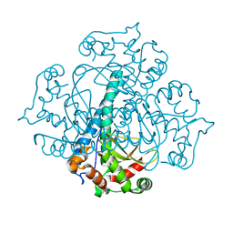
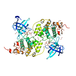
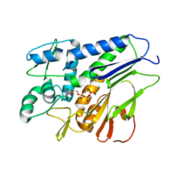
 | | Crystal structure of human AFF4-THD domain | | 分子名称: | AF4/FMR2 family member 4 | | 著者 | Tang, D, Xue, Y, Li, S, Cheng, W, Duan, J, Wang, J, Qi, S. | | 登録日 | 2019-06-08 | | 公開日 | 2020-03-11 | | 最終更新日 | 2024-03-27 | | 実験手法 | X-RAY DIFFRACTION (2.4 Å) | | 主引用文献 | Structural and functional insight into the effect of AFF4 dimerization on activation of HIV-1 proviral transcription.
Cell Discov, 6, 2020
|
|
8WSW
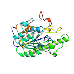
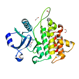
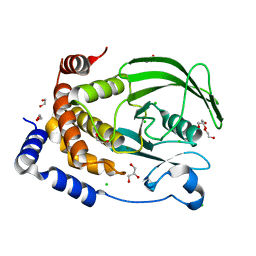
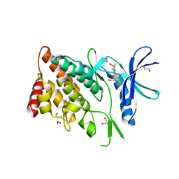 | | The Crystal Structure of LIMK2a from Biortus | | 分子名称: | 1,2-ETHANEDIOL, LIM domain kinase 2, ~{N}-[5-[2-[2,6-bis(chloranyl)phenyl]-5-[bis(fluoranyl)methyl]pyrazol-3-yl]-1,3-thiazol-2-yl]-2-methyl-propanamide | | 著者 | Wang, F, Cheng, W, Yuan, Z, Lin, D, Pan, W. | | 登録日 | 2023-10-17 | | 公開日 | 2023-11-15 | | 実験手法 | X-RAY DIFFRACTION (2.5 Å) | | 主引用文献 | The Crystal Structure of LIMK2a from Biortus.
To Be Published
|
|
8X23
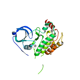
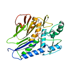
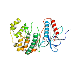 | | The Crystal Structure of MAPK13 from Biortus. | | 分子名称: | 1,2-ETHANEDIOL, GLYCEROL, Mitogen-activated protein kinase 13 | | 著者 | Wang, F, Cheng, W, Yuan, Z, Lin, D, Pan, W. | | 登録日 | 2023-11-09 | | 公開日 | 2023-12-27 | | 実験手法 | X-RAY DIFFRACTION (1.5 Å) | | 主引用文献 | The Crystal Structure of MAPK13 from Biortus.
To Be Published
|
|
8X2T
 | |
8X2A
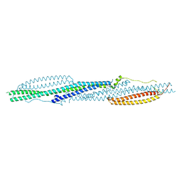
 | | The Crystal Structure of BMX from Biortus. | | 分子名称: | 1,2-ETHANEDIOL, 4-[(3S)-3-{[(2E)-but-2-enoyl]amino}piperidin-1-yl]-5-fluoro-2,3-dimethyl-1H-indole-7-carboxamide, CHLORIDE ION, ... | | 著者 | Wang, F, Cheng, W, Yuan, Z, Lin, D, Pan, W. | | 登録日 | 2023-11-09 | | 公開日 | 2023-12-27 | | 実験手法 | X-RAY DIFFRACTION (1.3 Å) | | 主引用文献 | The Crystal Structure of BMX from Biortus.
To Be Published
|
|
8X5L
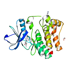
 | | The Crystal Structure of PRKACA from Biortus. | | 分子名称: | (2S)-2-(4-chlorophenyl)-2-hydroxy-2-[4-(1H-pyrazol-4-yl)phenyl]ethanaminium, SODIUM ION, cAMP-dependent protein kinase catalytic subunit alpha | | 著者 | Wang, F, Cheng, W, Lv, Z, Lin, D, Pan, W. | | 登録日 | 2023-11-17 | | 公開日 | 2023-12-27 | | 実験手法 | X-RAY DIFFRACTION (2.75 Å) | | 主引用文献 | The Crystal Structure of PRKACA from Biortus.
To Be Published
|
|
8X70
 | | The Crystal Structure of IFI16 from Biortus. | | 分子名称: | 1,2-ETHANEDIOL, BROMIDE ION, Gamma-interferon-inducible protein 16, ... | | 著者 | Wang, F, Cheng, W, Lv, Z, Meng, Q, Wang, J. | | 登録日 | 2023-11-22 | | 公開日 | 2023-12-27 | | 実験手法 | X-RAY DIFFRACTION (1.7 Å) | | 主引用文献 | The Crystal Structure of IFI16 from Biortus.
To Be Published
|
|
3RN4
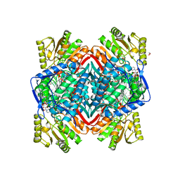
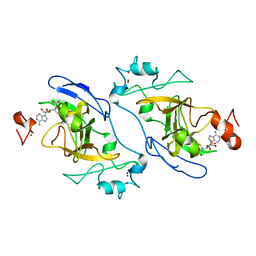
 | | Crystal structure of iron-substituted Sod2 from Saccharomyces cerevisiae | | 分子名称: | FE (III) ION, Superoxide dismutase [Mn], mitochondrial | | 著者 | Kang, Y, He, Y.-X, Cheng, W, Zhou, C.-Z, Li, W.-F. | | 登録日 | 2011-04-21 | | 公開日 | 2011-11-23 | | 最終更新日 | 2023-11-01 | | 実験手法 | X-RAY DIFFRACTION (1.79 Å) | | 主引用文献 | Structures of native and Fe-substituted SOD2 from Saccharomyces cerevisiae
Acta Crystallogr.,Sect.F, 67, 2011
|
|
5ZZU
 | |
6A83
 | |
8GYF
 | | Crystal structure of a bright green fluorescent protein (StayGold) with single mutation (K192Y) in jellyfish Cytaeis uchidae from Biortus | | 分子名称: | 1,2-ETHANEDIOL, staygold(K192Y) | | 著者 | Wu, J, Wang, F, Gui, W, Cheng, W, Yang, Y. | | 登録日 | 2022-09-22 | | 公開日 | 2023-10-04 | | 最終更新日 | 2023-11-15 | | 実験手法 | X-RAY DIFFRACTION (2 Å) | | 主引用文献 | Crystal structure of a bright green fluorescent protein (StayGold) with single mutation (K192Y) in jellyfish Cytaeis uchidae from Biortus
To Be Published
|
|
6A82
 | |
8H4R
 | | The Crystal Structure of CDK3 and CyclinE1 Complex with Dinaciclib from Biortus | | 分子名称: | 2-(N-MORPHOLINO)-ETHANESULFONIC ACID, 3-[({3-ethyl-5-[(2S)-2-(2-hydroxyethyl)piperidin-1-yl]pyrazolo[1,5-a]pyrimidin-7-yl}amino)methyl]-1-hydroxypyridinium, G1/S-specific cyclin-E1, ... | | 著者 | Gui, W, Wang, F, Cheng, W, Gao, J, Huang, Y, Ouyang, Z. | | 登録日 | 2022-10-11 | | 公開日 | 2023-10-11 | | 実験手法 | X-RAY DIFFRACTION (2.75 Å) | | 主引用文献 | The Crystal Structure of CDK3 and CyclinE1 Complex with Dinaciclib from Biortus
To Be Published
|
|
8WFQ
 | | The Crystal Structure of RALDH1 from Biortus. | | 分子名称: | 1,2-ETHANEDIOL, 1,4-DIHYDRONICOTINAMIDE ADENINE DINUCLEOTIDE, Aldehyde dehydrogenase 1A1 | | 著者 | Wang, F, Cheng, W, Lv, Z, Qi, J, Shen, Z. | | 登録日 | 2023-09-20 | | 公開日 | 2023-11-22 | | 実験手法 | X-RAY DIFFRACTION (3.5 Å) | | 主引用文献 | The Crystal Structure of RALDH1 from Biortus.
To Be Published
|
|
8XU4
 | | The Crystal Structure of MAPK2 from Biortus. | | 分子名称: | MALONIC ACID, MAP kinase-activated protein kinase 2 | | 著者 | Wang, F, Cheng, W, Yuan, Z, Qi, J, Shen, Z. | | 登録日 | 2024-01-12 | | 公開日 | 2024-01-24 | | 実験手法 | X-RAY DIFFRACTION (3.4 Å) | | 主引用文献 | The Crystal Structure of MAPK2 from Biortus.
To Be Published
|
|
8XN6
 | | The Crystal Structure of GSK3b from Biortus. | | 分子名称: | 1,2-ETHANEDIOL, DI(HYDROXYETHYL)ETHER, Glycogen synthase kinase-3 beta, ... | | 著者 | Wang, F, Cheng, W, Yuan, Z, Qi, J, Wu, B. | | 登録日 | 2023-12-29 | | 公開日 | 2024-01-24 | | 実験手法 | X-RAY DIFFRACTION (2.4 Å) | | 主引用文献 | The Crystal Structure of GSK3b from Biortus.
To Be Published
|
|
8XPT
 | | The Crystal Structure of EHMT1 from Biortus. | | 分子名称: | Histone-lysine N-methyltransferase EHMT1, S-ADENOSYL-L-HOMOCYSTEINE, SULFATE ION, ... | | 著者 | Wang, F, Cheng, W, Yuan, Z, Lin, D, Bao, C. | | 登録日 | 2024-01-04 | | 公開日 | 2024-01-24 | | 実験手法 | X-RAY DIFFRACTION (3.35 Å) | | 主引用文献 | The Crystal Structure of EHMT1 from Biortus.
To Be Published
|
|
8WD2
 | | The Crystal Structure of p53 from Biortus. | | 分子名称: | 1,2-ETHANEDIOL, Cellular tumor antigen p53, PHOSPHATE ION, ... | | 著者 | Wang, F, Cheng, W, Yuan, Z, Qi, J, Lu, Y. | | 登録日 | 2023-09-14 | | 公開日 | 2023-10-04 | | 実験手法 | X-RAY DIFFRACTION (1.85 Å) | | 主引用文献 | The Crystal Structure of p53 from Biortus.
To Be Published
|
|
8WTF
 | | The Crystal Structure of IRAK4 from Biortus | | 分子名称: | 1,2-ETHANEDIOL, 1-{[(2S,3S,4S)-3-ethyl-4-fluoro-5-oxopyrrolidin-2-yl]methoxy}-7-methoxyisoquinoline-6-carboxamide, Interleukin-1 receptor-associated kinase 4, ... | | 著者 | Wang, F, Cheng, W, Lv, Z, Meng, Q, Xu, Y. | | 登録日 | 2023-10-18 | | 公開日 | 2023-11-15 | | 実験手法 | X-RAY DIFFRACTION (2 Å) | | 主引用文献 | The Crystal Structure of IRAK4 from Biortus
To Be Published
|
|
8XP5
 | | The Crystal Structure of p53/BCL-xL fusion complex from Biortus. | | 分子名称: | Bcl-2-like protein 1,Cellular tumor antigen p53, ZINC ION | | 著者 | Wang, F, Cheng, W, Yuan, Z, Lin, D, Bao, C. | | 登録日 | 2024-01-03 | | 公開日 | 2024-03-06 | | 実験手法 | X-RAY DIFFRACTION (2.55 Å) | | 主引用文献 | The Crystal Structure of p53/BCL-xL fusion complex from Biortus.
To Be Published
|
|
7ESA
 | |
7ESB
 | | FmnB complexed with ATP | | 分子名称: | ADENOSINE-5'-TRIPHOSPHATE, FAD:protein FMN transferase, MAGNESIUM ION | | 著者 | Zheng, Y.H, Cheng, W. | | 登録日 | 2021-05-09 | | 公開日 | 2021-11-03 | | 最終更新日 | 2023-11-29 | | 実験手法 | X-RAY DIFFRACTION (1.7 Å) | | 主引用文献 | Structural insights into the catalytic and inhibitory mechanisms of the flavin transferase FmnB in Listeria monocytogenes.
MedComm (2020), 3, 2022
|
|
8XOY
 | | The Crystal Structure of PTP1B from Biortus. | | 分子名称: | 1,2-ETHANEDIOL, CHLORIDE ION, GLYCEROL, ... | | 著者 | Wang, F, Cheng, W, Lv, Z, Meng, Q, Li, J. | | 登録日 | 2024-01-02 | | 公開日 | 2024-01-24 | | 実験手法 | X-RAY DIFFRACTION (1.55 Å) | | 主引用文献 | The Crystal Structure of PTP1B from Biortus.
To Be Published
|
|
8WF7
 | | The Crystal Structure of integrase from Biortus | | 分子名称: | ACETATE ION, Integrase, SULFATE ION | | 著者 | Wang, F, Cheng, W, Yuan, Z, Qi, J, Li, J. | | 登録日 | 2023-09-19 | | 公開日 | 2023-10-04 | | 実験手法 | X-RAY DIFFRACTION (1.55 Å) | | 主引用文献 | The Crystal Structure of integrase from Biortus
To Be Published
|
|
8WR8
 | |


