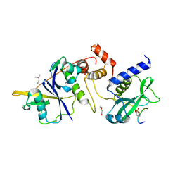
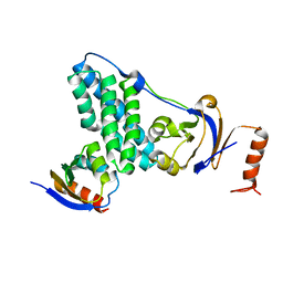
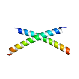
3CRN
 
 | | Crystal structure of response regulator receiver domain protein (CheY-like) from Methanospirillum hungatei JF-1 | | 分子名称: | GLYCEROL, Response regulator receiver domain protein, CheY-like, ... | | 著者 | Hiner, R.L, Toro, R, Patskovsky, Y, Freeman, J, Chang, S, Smith, D, Groshong, C, Wasserman, S.R, Sauder, J.M, Burley, S.K, Almo, S.C, New York SGX Research Center for Structural Genomics (NYSGXRC) | | 登録日 | 2008-04-07 | | 公開日 | 2008-04-22 | | 最終更新日 | 2021-02-03 | | 実験手法 | X-RAY DIFFRACTION (1.58 Å) | | 主引用文献 | Crystal structure of response regulator receiver domain protein (CheY-like) from Methanospirillum hungatei JF-1.
To be Published
|
|
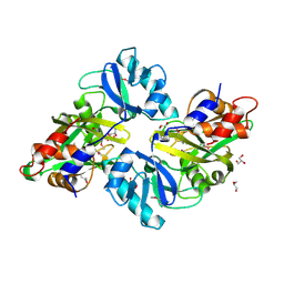
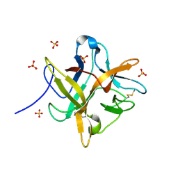
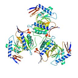
2RJZ
 | | Crystal structure of the type 4 fimbrial biogenesis protein PilO from Pseudomonas aeruginosa | | 分子名称: | PilO protein, SULFATE ION | | 著者 | Bonanno, J.B, Freeman, J, Bain, K.T, Chang, S, Ozyurt, S, Smith, D, Wasserman, S, Sauder, J.M, Burley, S.K, Almo, S.C, New York SGX Research Center for Structural Genomics (NYSGXRC) | | 登録日 | 2007-10-16 | | 公開日 | 2007-11-06 | | 最終更新日 | 2024-02-21 | | 実験手法 | X-RAY DIFFRACTION (2.2 Å) | | 主引用文献 | Periplasmic domains of Pseudomonas aeruginosa PilN and PilO form a stable heterodimeric complex.
J.Mol.Biol., 394, 2009
|
|
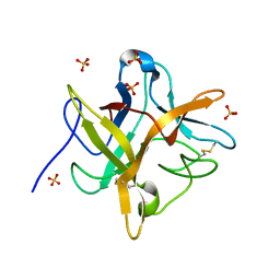
6I5N
 | | Crystal structure of SOCS2:Elongin C:Elongin B in complex with growth hormone receptor peptide | | 分子名称: | COBALT (II) ION, Elongin-B, Elongin-C, ... | | 著者 | Kung, W.W, Ramachandran, S, Makukhin, N, Bruno, E, Ciulli, A. | | 登録日 | 2018-11-14 | | 公開日 | 2019-05-29 | | 最終更新日 | 2019-06-19 | | 実験手法 | X-RAY DIFFRACTION (1.98 Å) | | 主引用文献 | Structural insights into substrate recognition by the SOCS2 E3 ubiquitin ligase.
Nat Commun, 10, 2019
|
|
6I4X
 | | Crystal structure of SOCS2:Elongin C:Elongin B in complex with erythropoietin receptor peptide | | 分子名称: | DI(HYDROXYETHYL)ETHER, Elongin-B, Elongin-C, ... | | 著者 | Kung, W.W, Ramachandran, S, Makukhin, N, Bruno, E, Ciulli, A. | | 登録日 | 2018-11-12 | | 公開日 | 2019-05-29 | | 最終更新日 | 2019-06-19 | | 実験手法 | X-RAY DIFFRACTION (2.69 Å) | | 主引用文献 | Structural insights into substrate recognition by the SOCS2 E3 ubiquitin ligase.
Nat Commun, 10, 2019
|
|
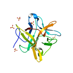
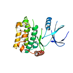
3CG0
 | | Crystal structure of signal receiver domain of modulated diguanylate cyclase from Desulfovibrio desulfuricans G20, an example of alternate folding | | 分子名称: | Response regulator receiver modulated diguanylate cyclase with PAS/PAC sensor | | 著者 | Patskovsky, Y, Bonanno, J.B, Romero, R, Gilmore, M, Chang, S, Groshong, C, Koss, J, Wasserman, S.R, Sauder, J.M, Burley, S.K, Almo, S.C, New York SGX Research Center for Structural Genomics (NYSGXRC) | | 登録日 | 2008-03-04 | | 公開日 | 2008-03-18 | | 最終更新日 | 2024-02-21 | | 実験手法 | X-RAY DIFFRACTION (2.15 Å) | | 主引用文献 | Crystal Structure of Signal Receiver Domain of Modulated Diguanylate Cyclase from Desulfovibrio desulfuricans.
To be Published
|
|
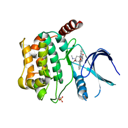
3DUP
 | | Crystal structure of mutt/nudix family hydrolase from rhodospirillum rubrum atcc 11170 | | 分子名称: | GLYCEROL, MutT/nudix family protein, PHOSPHATE ION | | 著者 | Patskovsky, Y, Ramagopal, U.A, Toro, R, Freeman, J, Chang, S, Groshong, C, Sauder, J.M, Burley, S.K, Almo, S.C, New York SGX Research Center for Structural Genomics (NYSGXRC) | | 登録日 | 2008-07-17 | | 公開日 | 2008-09-02 | | 最終更新日 | 2021-02-10 | | 実験手法 | X-RAY DIFFRACTION (1.8 Å) | | 主引用文献 | Crystal Structure of Mutt/Nudix Family Hydrolase from Rhodospirillum Rubrum
To be Published
|
|
1EYL
 | | STRUCTURE OF A RECOMBINANT WINGED BEAN CHYMOTRYPSIN INHIBITOR | | 分子名称: | CHYMOTRYPSIN INHIBITOR, SULFATE ION | | 著者 | Dattagupta, J.K, Chakrabarti, C, Ravichandran, S, Ghosh, S. | | 登録日 | 2000-05-07 | | 公開日 | 2000-05-24 | | 最終更新日 | 2022-12-21 | | 実験手法 | X-RAY DIFFRACTION (1.9 Å) | | 主引用文献 | The role of Asn14 in the stability and conformation of the reactive-site loop of winged bean chymotrypsin inhibitor: crystal structures of two point mutants Asn14-->Lys and Asn14-->Asp.
Protein Eng., 14, 2001
|
|
5E38
 | | Structural basis of mapping the spontaneous mutations with 5-flourouracil in uracil phosphoribosyltransferase from Mycobacterium tuberculosis | | 分子名称: | Uracil phosphoribosyltransferase | | 著者 | Ghode, P, Jobichen, C, Ramachandran, S, Bifani, P, Sivaraman, J. | | 登録日 | 2015-10-02 | | 公開日 | 2015-10-21 | | 最終更新日 | 2023-11-08 | | 実験手法 | X-RAY DIFFRACTION (3 Å) | | 主引用文献 | Structural basis of mapping the spontaneous mutations with 5-flurouracil in uracil phosphoribosyltransferase from Mycobacterium tuberculosis
Biochem.Biophys.Res.Commun., 467, 2015
|
|
7E4J
 | | X-ray crystal structure of VapB12 antitoxin from mycobacterium tuberculosis in space group P41. | | 分子名称: | Antitoxin, ZINC ION | | 著者 | Pratap, S, Megta, A.K, Talwar, S, Chandresh, S, Pandey, A.K, Krishnan, V. | | 登録日 | 2021-02-13 | | 公開日 | 2022-02-16 | | 最終更新日 | 2024-05-29 | | 実験手法 | X-RAY DIFFRACTION (1.63 Å) | | 主引用文献 | X-ray crystal structure of VapB12 antitoxin from mycobacterium tuberculosis in space group P41.
To be published
|
|
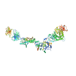
5T1O
 | | Solution-state NMR and SAXS structural ensemble of NPr (1-85) in complex with EIN-Ntr (170-424) | | 分子名称: | Phosphocarrier protein NPr, Phosphoenolpyruvate-protein phosphotransferase PtsP | | 著者 | Strickland, M, Stanley, A.M, Wang, G, Schwieters, C.D, Buchanan, S, Peterkofsky, A, Tjandra, N. | | 登録日 | 2016-08-19 | | 公開日 | 2016-11-16 | | 最終更新日 | 2024-05-15 | | 実験手法 | SOLUTION NMR, SOLUTION SCATTERING | | 主引用文献 | Structure of the NPr:EIN(Ntr) Complex: Mechanism for Specificity in Paralogous Phosphotransferase Systems.
Structure, 24, 2016
|
|
1FMZ
 | | CRYSTAL STRUCTURE OF A MUTANT WINGED BEAN CHYMOTRYPSIN INHIBITOR PROTEIN, N14K. | | 分子名称: | CHYMOTRYPSIN INHIBITOR 3, SULFATE ION | | 著者 | Dattagupta, J.K, Chakrabarti, C, Ravichandran, S, Dasgupta, J, Ghosh, S. | | 登録日 | 2000-08-19 | | 公開日 | 2001-02-19 | | 最終更新日 | 2021-11-03 | | 実験手法 | X-RAY DIFFRACTION (2.05 Å) | | 主引用文献 | The role of Asn14 in the stability and conformation of the reactive-site loop of winged bean chymotrypsin inhibitor: crystal structures of two point mutants Asn14-->Lys and Asn14-->Asp.
PROTEIN ENG., 14, 2001
|
|
1FN0
 | | STRUCTURE OF A MUTANT WINGED BEAN CHYMOTRYPSIN INHIBITOR PROTEIN, N14D. | | 分子名称: | CHYMOTRYPSIN INHIBITOR 3, SULFATE ION | | 著者 | Dattagupta, J.K, Chakrabarti, C, Ravichandran, S, Dasgupta, J, Ghosh, S. | | 登録日 | 2000-08-19 | | 公開日 | 2001-02-19 | | 最終更新日 | 2021-11-03 | | 実験手法 | X-RAY DIFFRACTION (2 Å) | | 主引用文献 | The role of Asn14 in the stability and conformation of the reactive-site loop of winged bean chymotrypsin inhibitor: crystal structures of two point mutants Asn14-->Lys and Asn14-->Asp.
PROTEIN ENG., 14, 2001
|
|
5VOD
 | |
5VOB
 | |
5VOC
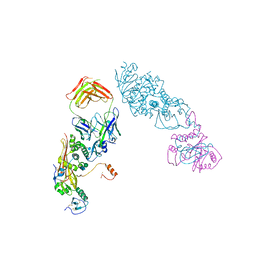 | |
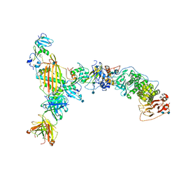
7JSN
 | | Structure of the Visual Signaling Complex between Transducin and Phosphodiesterase 6 | | 分子名称: | 2-{2-ETHOXY-5-[(4-ETHYLPIPERAZIN-1-YL)SULFONYL]PHENYL}-5-METHYL-7-PROPYLIMIDAZO[5,1-F][1,2,4]TRIAZIN-4(1H)-ONE, GUANOSINE-3',5'-MONOPHOSPHATE, GUANOSINE-5'-TRIPHOSPHATE, ... | | 著者 | Gao, Y, Eskici, G, Ramachandran, S, Skiniotis, G, Cerione, R.A. | | 登録日 | 2020-08-15 | | 公開日 | 2020-10-21 | | 最終更新日 | 2024-05-29 | | 実験手法 | ELECTRON MICROSCOPY (3.2 Å) | | 主引用文献 | Structure of the Visual Signaling Complex between Transducin and Phosphodiesterase 6.
Mol.Cell, 80, 2020
|
|
2OG2
 | | Crystal structure of chloroplast FtsY from Arabidopsis thaliana | | 分子名称: | MAGNESIUM ION, MALONATE ION, Putative signal recognition particle receptor | | 著者 | Chartron, J, Chandrasekar, S, Ampornpan, P.J, Shan, S. | | 登録日 | 2007-01-04 | | 公開日 | 2007-12-11 | | 最終更新日 | 2023-08-30 | | 実験手法 | X-RAY DIFFRACTION (2 Å) | | 主引用文献 | Structure of the Chloroplast Signal Recognition Particle (SRP) Receptor: Domain Arrangement Modulates SRP-Receptor Interaction.
J.Mol.Biol., 375, 2007
|
|
3PKO
 | | Crystal structure of geranylgeranyl pyrophosphate synthase from lactobacillus brevis atcc 367 complexed with citrate | | 分子名称: | CITRIC ACID, GLYCEROL, Geranylgeranyl pyrophosphate synthase | | 著者 | Patskovsky, Y, Toro, R, Rutter, M, Chang, S, Sauder, J.M, Poulter, C.D, Burley, S.K, Gerlt, J.A, Almo, S.C, New York SGX Research Center for Structural Genomics (NYSGXRC) | | 登録日 | 2010-11-11 | | 公開日 | 2010-11-24 | | 最終更新日 | 2024-02-21 | | 実験手法 | X-RAY DIFFRACTION (1.98 Å) | | 主引用文献 | Prediction of function for the polyprenyl transferase subgroup in the isoprenoid synthase superfamily.
Proc.Natl.Acad.Sci.USA, 110, 2013
|
|
3MZV
 | | Crystal structure of a decaprenyl diphosphate synthase from Rhodobacter capsulatus | | 分子名称: | Decaprenyl diphosphate synthase | | 著者 | Quartararo, C.E, Patskovsky, Y, Bonanno, J.B, Rutter, M, Bain, K.T, Chang, S, Toro, R, Sauder, J.M, Burley, S.K, Almo, S.C, New York SGX Research Center for Structural Genomics (NYSGXRC) | | 登録日 | 2010-05-13 | | 公開日 | 2010-06-09 | | 最終更新日 | 2024-02-21 | | 実験手法 | X-RAY DIFFRACTION (1.9 Å) | | 主引用文献 | Prediction of function for the polyprenyl transferase subgroup in the isoprenoid synthase superfamily.
Proc.Natl.Acad.Sci.USA, 110, 2013
|
|
1ET1
 | | CRYSTAL STRUCTURE OF HUMAN PARATHYROID HORMONE 1-34 AT 0.9 A RESOLUTION | | 分子名称: | PARATHYROID HORMONE, SODIUM ION | | 著者 | Jin, L, Briggs, S.L, Chandrasekhar, S, Chirgadze, N.Y, Clawson, D.K, Schevitz, R.W, Smiley, D.L, Tashjian, A.H, Zhang, F. | | 登録日 | 2000-04-12 | | 公開日 | 2000-09-06 | | 最終更新日 | 2024-02-07 | | 実験手法 | X-RAY DIFFRACTION (0.9 Å) | | 主引用文献 | Crystal structure of human parathyroid hormone 1-34 at 0.9-A resolution.
J.Biol.Chem., 275, 2000
|
|
5IAO
 | |
3CKW
 | | Crystal structure of sterile 20-like kinase 3 (MST3, STK24) | | 分子名称: | MERCURY (II) ION, Serine/threonine-protein kinase 24 | | 著者 | Antonysamy, S.S, Burley, S.K, Buchanan, S, Chau, F, Feil, I, Wu, L, Sauder, J.M, New York SGX Research Center for Structural Genomics (NYSGXRC) | | 登録日 | 2008-03-17 | | 公開日 | 2008-04-29 | | 最終更新日 | 2024-02-21 | | 実験手法 | X-RAY DIFFRACTION (1.96 Å) | | 主引用文献 | Crystal structure of sterile 20-like kinase 3 (MST3, STK24).
To be Published
|
|
3CKX
 | | Crystal structure of sterile 20-like kinase 3 (MST3, STK24) in complex with staurosporine | | 分子名称: | STAUROSPORINE, Serine/threonine-protein kinase 24 | | 著者 | Antonysamy, S.S, Burley, S.K, Buchanan, S, Chau, F, Feil, I, Wu, L, Sauder, J.M, New York SGX Research Center for Structural Genomics (NYSGXRC) | | 登録日 | 2008-03-17 | | 公開日 | 2008-04-29 | | 最終更新日 | 2023-08-30 | | 実験手法 | X-RAY DIFFRACTION (2.7 Å) | | 主引用文献 | Crystal structure of sterile 20-like kinase 3 (MST3, STK24) in complex with staurosporine.
To be Published
|
|
2PLT
 | |
4LR6
 | | Structure of BRD4 bromodomain 1 with a 3-methyl-4-phenylisoxazol-5-amine fragment | | 分子名称: | 3-methyl-4-phenyl-1,2-oxazol-5-amine, Bromodomain-containing protein 4, FORMIC ACID | | 著者 | Jayaram, H, Poy, F, Gehling, V, Hewitt, M, Vaswani, R, Leblanc, Y, Cote, A, Nasveschuk, C, Taylor, A, Harmange, J.-C, Audia, J, Pardo, E, Joshi, S, Sandy, P, Mertz, J, Sims, R, Bergeron, L, Bryant, B, Ravichandran, S, Yellapuntala, S, Nandana, B.S, Birudukota, S, Albrecht, B, Bellon, S. | | 登録日 | 2013-07-19 | | 公開日 | 2013-08-07 | | 最終更新日 | 2023-09-20 | | 実験手法 | X-RAY DIFFRACTION (1.29 Å) | | 主引用文献 | Discovery, Design, and Optimization of Isoxazole Azepine BET Inhibitors.
ACS Med Chem Lett, 4, 2013
|
|


