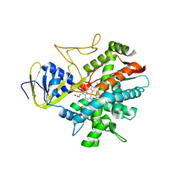
1RPS
 
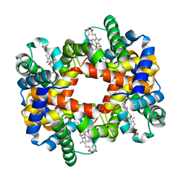 | | Crystallographic Analysis of the Interaction of Nitric Oxide with Quaternary-T Human Hemoglobin. Hemoglobin exposed to NO under anerobic conditions | | 分子名称: | Hemoglobin alpha chain, Hemoglobin beta chain, NITRIC OXIDE, ... | | 著者 | Chan, N.-L, Kavanaugh, J.S, Rogers, P.H, Arnone, A. | | 登録日 | 2003-12-03 | | 公開日 | 2003-12-23 | | 最終更新日 | 2023-08-23 | | 実験手法 | X-RAY DIFFRACTION (2.11 Å) | | 主引用文献 | Crystallographic analysis of the interaction of nitric oxide with quaternary-T human hemoglobin.
Biochemistry, 43, 2004
|
|
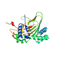
1BUW
 | |
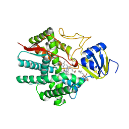
1RQA
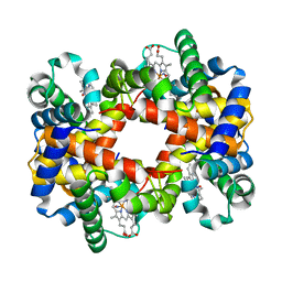 | | Crystallographic Analysis of the Interaction of Nitric Oxide with Quaternary-T Human Hemoglobin. Beta W73E hemoglobin exposed to NO under anaerobic conditions | | 分子名称: | Hemoglobin alpha chain, Hemoglobin beta chain, NITRIC OXIDE, ... | | 著者 | Chan, N.-L, Kavanaugh, J.S, Rogers, P.H, Arnone, A. | | 登録日 | 2003-12-04 | | 公開日 | 2005-01-04 | | 最終更新日 | 2024-04-03 | | 実験手法 | X-RAY DIFFRACTION (2.11 Å) | | 主引用文献 | Crystallographic analysis of the interaction of nitric oxide with quaternary-T human hemoglobin.
Biochemistry, 43, 2004
|
|
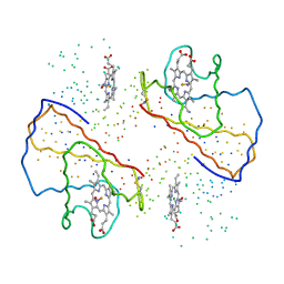
7E38
 | | Crystal structure of deoxypodophyllotoxin synthase from Sinopodophyllum hexandrum in complex with yatein and succinate | | 分子名称: | (3~{R},4~{R})-4-(1,3-benzodioxol-5-ylmethyl)-3-[(3,4,5-trimethoxyphenyl)methyl]oxolan-2-one, (3~{S},4~{S})-4-(1,3-benzodioxol-5-ylmethyl)-3-[(3,4,5-trimethoxyphenyl)methyl]oxolan-2-one, Deoxypodophyllotoxin synthase, ... | | 著者 | Wu, M.-H, Chang, W.-c, Chien, T.-C, Chan, N.-L. | | 登録日 | 2021-02-08 | | 公開日 | 2021-12-15 | | 最終更新日 | 2023-11-29 | | 実験手法 | X-RAY DIFFRACTION (2.05 Å) | | 主引用文献 | Mechanistic analysis of carbon-carbon bond formation by deoxypodophyllotoxin synthase.
Proc.Natl.Acad.Sci.USA, 119, 2022
|
|
7E37
 | | Crystal structure of deoxypodophyllotoxin synthase from Sinopodophyllum hexandrum in complex with 2-oxoglutarate | | 分子名称: | 2-OXOGLUTARIC ACID, Deoxypodophyllotoxin synthase, FE (III) ION | | 著者 | Wu, M.-H, Lin, H.-Y, Chang, W.-c, Chien, T.-C, Chan, N.-L. | | 登録日 | 2021-02-08 | | 公開日 | 2021-12-15 | | 最終更新日 | 2023-11-29 | | 実験手法 | X-RAY DIFFRACTION (2.09 Å) | | 主引用文献 | Mechanistic analysis of carbon-carbon bond formation by deoxypodophyllotoxin synthase.
Proc.Natl.Acad.Sci.USA, 119, 2022
|
|
2D27
 | | Structure of the N-terminal domain of XpsE (crystal form I4122) | | 分子名称: | type II secretion ATPase XpsE | | 著者 | Chen, Y, Shiue, S.-J, Huang, C.-W, Chang, J.-L, Chien, Y.-L, Hu, N.-T, Chan, N.-L. | | 登録日 | 2005-09-03 | | 公開日 | 2005-09-20 | | 最終更新日 | 2011-07-13 | | 実験手法 | X-RAY DIFFRACTION (2.21 Å) | | 主引用文献 | Structure and Function of the XpsE N-Terminal Domain, an Essential Component of the Xanthomonas campestris Type II Secretion System
J.Biol.Chem., 280, 2005
|
|
2D28
 | | Structure of the N-terminal domain of XpsE (crystal form P43212) | | 分子名称: | CACODYLATE ION, type II secretion ATPase XpsE | | 著者 | Chen, Y, Shiue, S.-J, Huang, C.-W, Chang, J.-L, Chien, Y.-L, Hu, N.-T, Chan, N.-L. | | 登録日 | 2005-09-03 | | 公開日 | 2005-09-20 | | 最終更新日 | 2021-11-10 | | 実験手法 | X-RAY DIFFRACTION (2 Å) | | 主引用文献 | Structure and Function of the XpsE N-Terminal Domain, an Essential Component of the Xanthomonas campestris Type II Secretion System
J.Biol.Chem., 280, 2005
|
|
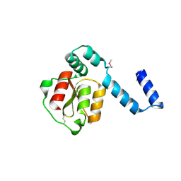
1WP5
 | |
3B98
 | | Crystal structure of zebrafish prostacyclin synthase (cytochrome P450 8A1) | | 分子名称: | PROTOPORPHYRIN IX CONTAINING FE, Prostaglandin I2 synthase | | 著者 | Li, Y.-C, Chiang, C.-W, Yeh, H.-C, Hsu, P.-Y, Whitby, F.G, Wang, L.-H, Chan, N.-L. | | 登録日 | 2007-11-03 | | 公開日 | 2007-11-20 | | 最終更新日 | 2023-11-01 | | 実験手法 | X-RAY DIFFRACTION (2.08 Å) | | 主引用文献 | Structures of Prostacyclin Synthase and Its Complexes with Substrate Analog and Inhibitor Reveal a Ligand-specific Heme Conformation Change
J.Biol.Chem., 283, 2008
|
|
3B6H
 | | Crystal structure of human prostacyclin synthase in complex with inhibitor minoxidil | | 分子名称: | 6-PIPERIDIN-1-YLPYRIMIDINE-2,4-DIAMINE 3-OXIDE, PROTOPORPHYRIN IX CONTAINING FE, Prostacyclin synthase, ... | | 著者 | Li, Y.-C, Chiang, C.-W, Yeh, H.-C, Hsu, P.-Y, Whitby, F.G, Wang, L.-H, Chan, N.-L. | | 登録日 | 2007-10-29 | | 公開日 | 2007-11-20 | | 最終更新日 | 2023-11-01 | | 実験手法 | X-RAY DIFFRACTION (1.62 Å) | | 主引用文献 | Structures of Prostacyclin Synthase and Its Complexes with Substrate Analog and Inhibitor Reveal a Ligand-specific Heme Conformation Change
J.Biol.Chem., 283, 2008
|
|
3B99
 | | Crystal structure of zebrafish prostacyclin synthase (cytochrome P450 8A1) in complex with substrate analog U51605 | | 分子名称: | (5Z)-7-{(1R,4S,5R,6R)-6-[(1E)-oct-1-en-1-yl]-2,3-diazabicyclo[2.2.1]hept-2-en-5-yl}hept-5-enoic acid, PROTOPORPHYRIN IX CONTAINING FE, Prostaglandin I2 synthase | | 著者 | Li, Y.-C, Chiang, C.-W, Yeh, H.-C, Hsu, P.-Y, Whitby, F.G, Wang, L.-H, Chan, N.-L. | | 登録日 | 2007-11-03 | | 公開日 | 2007-11-20 | | 最終更新日 | 2023-11-01 | | 実験手法 | X-RAY DIFFRACTION (2.5 Å) | | 主引用文献 | Structures of Prostacyclin Synthase and Its Complexes with Substrate Analog and Inhibitor Reveal a Ligand-specific Heme Conformation Change
J.Biol.Chem., 283, 2008
|
|
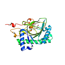
2E11
 | | The Crystal Structure of XC1258 from Xanthomonas campestris: A CN-hydrolase Superfamily Protein with an Arsenic Adduct in the Active Site | | 分子名称: | CACODYLATE ION, Hydrolase | | 著者 | Chin, K.-H, Tsai, Y.-D, Chan, N.-L, Huang, K.-F, Wang, A.H.-J, Chou, S.-H. | | 登録日 | 2006-10-17 | | 公開日 | 2007-08-07 | | 最終更新日 | 2024-03-13 | | 実験手法 | X-RAY DIFFRACTION (1.73 Å) | | 主引用文献 | The crystal structure of XC1258 from Xanthomonas campestris: A putative procaryotic Nit protein with an arsenic adduct in the active site
Proteins, 69, 2007
|
|
2IAG
 | | Crystal structure of human prostacyclin synthase | | 分子名称: | PROTOPORPHYRIN IX CONTAINING FE, Prostacyclin synthase, SODIUM ION | | 著者 | Chiang, C.-W, Yeh, H.-C, Wang, L.-H, Chan, N.-L. | | 登録日 | 2006-09-08 | | 公開日 | 2006-10-10 | | 最終更新日 | 2024-03-13 | | 実験手法 | X-RAY DIFFRACTION (2.15 Å) | | 主引用文献 | Crystal Structure of the Human Prostacyclin Synthase
J.Mol.Biol., 364, 2006
|
|
3WJA
 | | The crystal structure of human cytosolic NADP(+)-dependent malic enzyme in apo form | | 分子名称: | NADP-dependent malic enzyme | | 著者 | Li, S.-Y, Chen, M.-C, Yang, P.-C, Chan, N.-L, Liu, J.-H, Hung, H.-C. | | 登録日 | 2013-10-08 | | 公開日 | 2014-08-13 | | 最終更新日 | 2023-11-08 | | 実験手法 | X-RAY DIFFRACTION (2.548 Å) | | 主引用文献 | Structural characteristics of the nonallosteric human cytosolic malic enzyme.
Biochim.Biophys.Acta, 1844, 2014
|
|


