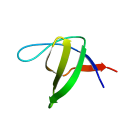
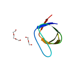
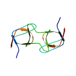
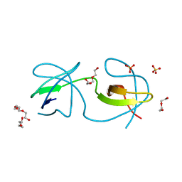
1JGW
 
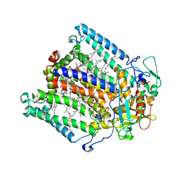 | | Photosynthetic Reaction Center Mutant With Thr M 21 Replaced With Leu | | Descriptor: | BACTERIOCHLOROPHYLL A, BACTERIOPHEOPHYTIN A, CARDIOLIPIN, ... | | Authors: | Camara-Artigas, A, Magee, C.L, Williams, J.C, Allen, J.P. | | Deposit date: | 2001-06-27 | | Release date: | 2001-09-05 | | Last modified: | 2023-08-16 | | Method: | X-RAY DIFFRACTION (2.8 Å) | | Cite: | Individual interactions influence the crystalline order for membrane proteins.
Acta Crystallogr.,Sect.D, 57, 2001
|
|
4OMM
 | |
4R61
 | |
4QT7
 | |
7PVR
 | |
7PVQ
 | |
7PVX
 | |
7PW2
 | |
7PVS
 | |
7PVV
 | |
7PW0
 | |
7PVT
 | |
7PVW
 | |
7PVY
 | |
7PVZ
 | |
4REX
 | |
4OMP
 | |
4RTY
 | |
4RTV
 | |
4RTX
 | |
4RTU
 | |
4RTZ
 | |
4RTW
 | |
4HVW
 | |
4HVV
 | |


