6SYD
 
 | |
6SYE
 | |
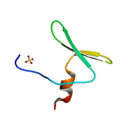
4OMO
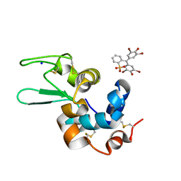
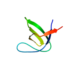
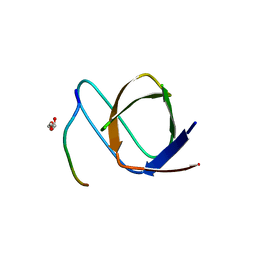
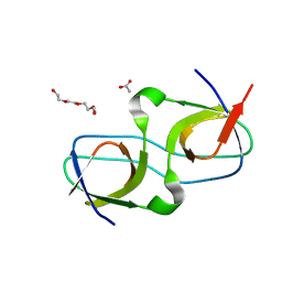
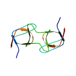
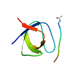
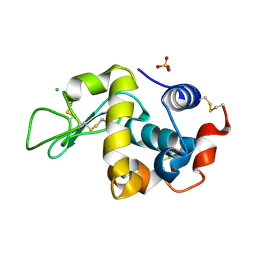
 | | Crystal structure of the c-Src tyrosine kinase SH3 domain mutant Q128E | | Descriptor: | 4-(2-HYDROXYETHYL)-1-PIPERAZINE ETHANESULFONIC ACID, NICKEL (II) ION, Proto-oncogene tyrosine-protein kinase Src | | Authors: | Camara-Artigas, A, Bacarizo, J. | | Deposit date: | 2014-01-27 | | Release date: | 2014-12-10 | | Last modified: | 2023-09-20 | | Method: | X-RAY DIFFRACTION (1.04 Å) | | Cite: | Electrostatic Effects in the Folding of the SH3 Domain of the c-Src Tyrosine Kinase: pH-Dependence in 3D-Domain Swapping and Amyloid Formation.
Plos One, 9, 2014
|
|
4OML
 | |
4OMN
 | |
4OMQ
 | |
4OMM
 | |
7PVR
 | |
7PVQ
 | |
7PVX
 | |
7PW2
 | |
7PVS
 | |
7PVV
 | |
7PVT
 | |
7PVW
 | |
7PW0
 | |
7PVY
 | |
7PVZ
 | |
4QT7
 | |
4OMP
 | |
4R61
 | |
4REX
 | |
4RTY
 | |
4RTV
 | |
6F9Y
 | |


