7Y5R
 
 | |
7Y73
 | |
7Y6E
 | |
7Y8S
 | |
7Y8I
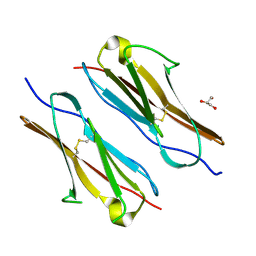
 | | Crystal structure of sDscam FNIII3 domain, isoform alpha7 | | 分子名称: | CHLORIDE ION, DI(HYDROXYETHYL)ETHER, Dscam, ... | | 著者 | Chen, Q, Yu, Y, Cheng, J. | | 登録日 | 2022-06-24 | | 公開日 | 2023-05-24 | | 最終更新日 | 2023-11-29 | | 実験手法 | X-RAY DIFFRACTION (1.9 Å) | | 主引用文献 | Structural basis for the self-recognition of sDSCAM in Chelicerata.
Nat Commun, 14, 2023
|
|
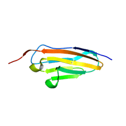
7Y95
 | | Crystal structure of sDscam Ig1 domain, isoform beta6v2 | | 分子名称: | Dscam, GLYCEROL, SODIUM ION | | 著者 | Chen, Q, Yu, Y, Cheng, J. | | 登録日 | 2022-06-24 | | 公開日 | 2023-05-24 | | 最終更新日 | 2023-11-29 | | 実験手法 | X-RAY DIFFRACTION (1.55 Å) | | 主引用文献 | Structural basis for the self-recognition of sDSCAM in Chelicerata.
Nat Commun, 14, 2023
|
|
7Y54
 | |
7Y6O
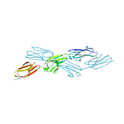 | |
7Y9A
 | | Crystal structure of sDscam Ig1-2 domains, isoform beta2v6 | | 分子名称: | Down Syndrome Cell Adhesion Molecules, beta-D-mannopyranose-(1-4)-2-acetamido-2-deoxy-beta-D-glucopyranose-(1-4)-[beta-L-fucopyranose-(1-3)][beta-L-fucopyranose-(1-6)]2-acetamido-2-deoxy-beta-D-glucopyranose | | 著者 | Chen, Q, Yu, Y, Cheng, J. | | 登録日 | 2022-06-24 | | 公開日 | 2023-05-24 | | 最終更新日 | 2023-09-06 | | 実験手法 | X-RAY DIFFRACTION (2.51 Å) | | 主引用文献 | Structural basis for the self-recognition of sDSCAM in Chelicerata.
Nat Commun, 14, 2023
|
|
7Y8H
 | |
7EPW
 | |
7EPV
 | |
2GEB
 | |
5TV1
 | | active arrestin-3 with inositol hexakisphosphate | | 分子名称: | Beta-arrestin-2, GLYCEROL, INOSITOL HEXAKISPHOSPHATE | | 著者 | Chen, Q, Gilbert, N.C, Perry, N.A, Vishniveteskiy, S, Gurevich, V.V, Iverson, T.M. | | 登録日 | 2016-11-07 | | 公開日 | 2017-11-22 | | 最終更新日 | 2023-10-04 | | 実験手法 | X-RAY DIFFRACTION (2.4 Å) | | 主引用文献 | Structural basis of arrestin-3 activation and signaling.
Nat Commun, 8, 2017
|
|
5SXU
 | | X-ray structure of 2-bromoethanol bound to a pentameric ligand gated ion channel (ELIC) in a desensitized state | | 分子名称: | 2-(N-MORPHOLINO)-ETHANESULFONIC ACID, 2-BROMOETHANOL, 3-AMINOPROPANE, ... | | 著者 | Chen, Q, Kinde, M, Cohen, A, Xu, Y, Tang, P. | | 登録日 | 2016-08-10 | | 公開日 | 2017-06-21 | | 最終更新日 | 2023-10-04 | | 実験手法 | X-RAY DIFFRACTION (3.1 Å) | | 主引用文献 | Structural Basis of Alcohol Inhibition of the Pentameric Ligand-Gated Ion Channel ELIC.
Structure, 25, 2017
|
|
5SXV
 | | X-ray structure of 2-bromoethanol bound to a pentameric ligand gated ion channel (ELIC) in a resting state | | 分子名称: | 2-(N-MORPHOLINO)-ETHANESULFONIC ACID, 2-BROMOETHANOL, Cys-loop ligand-gated ion channel | | 著者 | Chen, Q, Kinde, M, Cohen, A, Xu, Y, Tang, P. | | 登録日 | 2016-08-10 | | 公開日 | 2017-06-21 | | 最終更新日 | 2023-10-04 | | 実験手法 | X-RAY DIFFRACTION (3.4 Å) | | 主引用文献 | Structural Basis of Alcohol Inhibition of the Pentameric Ligand-Gated Ion Channel ELIC.
Structure, 25, 2017
|
|
7Y4X
 | |
3Q5Y
 | | V beta/V beta homodimerization-based pre-TCR model suggested by TCR beta crystal structures | | 分子名称: | 4-(2-HYDROXYETHYL)-1-PIPERAZINE ETHANESULFONIC ACID, DI(HYDROXYETHYL)ETHER, GLYCEROL, ... | | 著者 | Chen, Q, Zhang, H, Wang, J.-H. | | 登録日 | 2010-12-30 | | 公開日 | 2011-03-09 | | 最終更新日 | 2014-10-15 | | 実験手法 | X-RAY DIFFRACTION (1.9 Å) | | 主引用文献 | A conserved hydrophobic patch on Vbeta domains revealed by TCRbeta chain crystal structures: implications for pre-TCR dimerization
Front Immunol, 2, 2011
|
|
3Q5T
 | |
8H2F
 | |
8HI1
 | |
8H18
 | |
8J4U
 | |
6CTE
 | | 77Se-NMR probes the protein environment of selenomethionine | | 分子名称: | (4R)-2-METHYLPENTANE-2,4-DIOL, (4S)-2-METHYL-2,4-PENTANEDIOL, ACETATE ION, ... | | 著者 | Chen, Q, Rozovsky, S. | | 登録日 | 2018-03-22 | | 公開日 | 2019-07-10 | | 最終更新日 | 2023-11-15 | | 実験手法 | X-RAY DIFFRACTION (1.2 Å) | | 主引用文献 | 77Se NMR Probes the Protein Environment of Selenomethionine.
J.Phys.Chem.B, 124, 2020
|
|
6C9O
 | |


