Loading
PDBj
メニューPDBj@FacebookPDBj@TwitterPDBj@YouTubewwPDB FoundationwwPDB
RCSB PDBPDBeBMRBAdv. SearchSearch help
PDB: 39 件

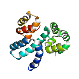
6XQJ
DownloadVisualize
BU of 6xqj by Molmil
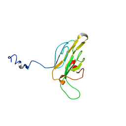
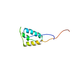
Structure of HIV-1 Vpr in complex with the human nucleotide excision repair protein hHR23A
分子名称: Protein Vpr,UV excision repair protein RAD23 homolog A, ZINC ION
著者Byeon, I.-J.L, Calero, G, Wu, Y, Byeon, C.H, Gronenborn, A.M.
登録日2020-07-09
公開日2021-11-17
最終更新日2024-05-15
実験手法SOLUTION NMR
主引用文献Structure of HIV-1 Vpr in complex with the human nucleotide excision repair protein hHR23A.
Nat Commun, 12, 2021
1QU5
DownloadVisualize
BU of 1qu5 by Molmil
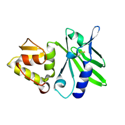
NMR STRUCTURE OF A NEW PHOSPHOTYROSINE BINDING DOMAIN CONTAINING THE FHA2 DOMAIN OF RAD 53
分子名称: PROTEIN KINASE SPK1
著者Byeon, I.-J.L, Liao, H, Tsai, M.-D.
登録日1999-07-06
公開日1999-12-17
最終更新日2024-05-22
実験手法SOLUTION NMR
主引用文献Structure and function of a new phosphopeptide-binding domain containing the FHA2 of Rad53.
J.Mol.Biol., 294, 1999
2M65
DownloadVisualize
BU of 2m65 by Molmil
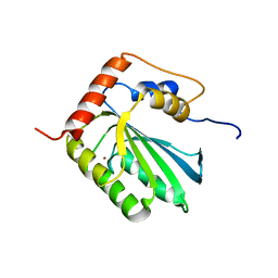
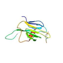
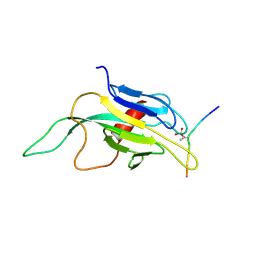
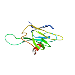
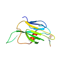
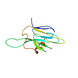
NMR structure of human restriction factor APOBEC3A
分子名称: Probable DNA dC->dU-editing enzyme APOBEC-3A, ZINC ION
著者Byeon, I.L, Byeon, C, Ahn, J, Gronenborn, A.M.
登録日2013-03-21
公開日2013-05-22
最終更新日2024-05-15
実験手法SOLUTION NMR
主引用文献NMR structure of human restriction factor APOBEC3A reveals substrate binding and enzyme specificity.
Nat Commun, 4, 2013
2NBQ
DownloadVisualize
BU of 2nbq by Molmil
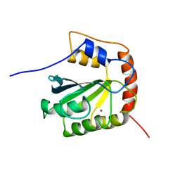
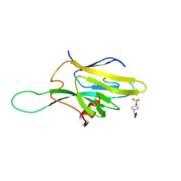
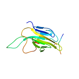
NMR Structure of the C-Terminal Domain of human APOBEC3B
分子名称: DNA dC->dU-editing enzyme APOBEC-3B, ZINC ION
著者Byeon, I.L, Byeon, C, Gronenborn, A.M.
登録日2016-03-09
公開日2016-06-01
最終更新日2024-05-15
実験手法SOLUTION NMR
主引用文献Nuclear Magnetic Resonance Structure of the APOBEC3B Catalytic Domain: Structural Basis for Substrate Binding and DNA Deaminase Activity.
Biochemistry, 55, 2016
2A5E
DownloadVisualize
BU of 2a5e by Molmil
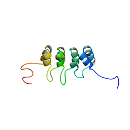
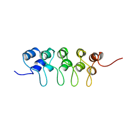
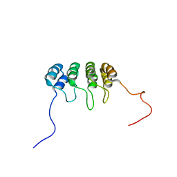
SOLUTION NMR STRUCTURE OF TUMOR SUPPRESSOR P16INK4A, RESTRAINED MINIMIZED MEAN STRUCTURE
分子名称: TUMOR SUPPRESSOR P16INK4A
著者Byeon, I.-J.L, Li, J, Ericson, K, Selby, T.L, Tevelev, A, Kim, H.-J, O'Maille, P, Tsai, M.-D.
登録日1998-02-13
公開日1999-08-13
最終更新日2024-05-22
実験手法SOLUTION NMR
主引用文献Tumor suppressor p16INK4A: determination of solution structure and analyses of its interaction with cyclin-dependent kinase 4.
Mol.Cell, 1, 1998
2AFF
DownloadVisualize
BU of 2aff by Molmil
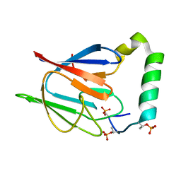
The solution structure of the Ki67FHA/hNIFK(226-269)3P complex
分子名称: Antigen KI-67, MKI67 FHA domain interacting nucleolar phosphoprotein
著者Byeon, I.-J.L, Li, H, Song, H, Gronenborn, A.M, Tsai, M.D.
登録日2005-07-25
公開日2005-10-25
最終更新日2024-10-30
実験手法SOLUTION NMR
主引用文献Sequential phosphorylation and multisite interactions characterize specific target recognition by the FHA domain of Ki67.
Nat.Struct.Mol.Biol., 12, 2005
1R21
DownloadVisualize
BU of 1r21 by Molmil
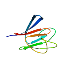
Solution Structure of human Ki67 FHA Domain
分子名称: Antigen Ki-67
著者Byeon, I.J, Li, H, Tsai, M.D.
登録日2003-09-25
公開日2003-12-30
最終更新日2024-05-22
実験手法SOLUTION NMR
主引用文献Structure of Human Ki67 FHA Domain and its Binding to a Phosphoprotein Fragment from hNIFK Reveal Unique Recognition Sites and New Views to the Structural Basis of FHA Domain Functions
J.Mol.Biol., 335, 2004
1BU9
DownloadVisualize
BU of 1bu9 by Molmil
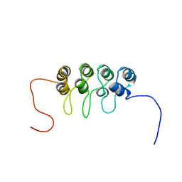
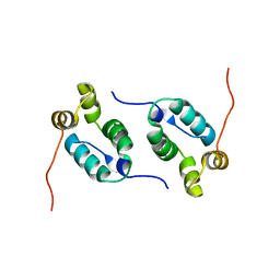
SOLUTION STRUCTURE OF P18-INK4C, 21 STRUCTURES
分子名称: PROTEIN (CYCLIN-DEPENDENT KINASE 6 INHIBITOR)
著者Byeon, I.-J.L, Li, J, Tsai, M.-D.
登録日1998-09-15
公開日1999-09-13
最終更新日2023-12-27
実験手法SOLUTION NMR
主引用文献Tumor suppressor INK4: determination of the solution structure of p18INK4C and demonstration of the functional significance of loops in p18INK4C and p16INK4A.
Biochemistry, 38, 1999
1A5E
DownloadVisualize
BU of 1a5e by Molmil
SOLUTION NMR STRUCTURE OF TUMOR SUPPRESSOR P16INK4A, 18 STRUCTURES
分子名称: TUMOR SUPPRESSOR P16INK4A
著者Byeon, I.-J.L, Li, J, Ericson, K, Selby, T.L, Tevelev, A, Kim, H.-J, O'Maille, P, Tsai, M.-D.
登録日1998-02-13
公開日1999-08-13
最終更新日2024-05-22
実験手法SOLUTION NMR
主引用文献Tumor suppressor p16INK4A: determination of solution structure and analyses of its interaction with cyclin-dependent kinase 4.
Mol.Cell, 1, 1998
1DC2
DownloadVisualize
BU of 1dc2 by Molmil
SOLUTION NMR STRUCTURE OF TUMOR SUPPRESSOR P16INK4A, 20 STRUCTURES
分子名称: CYCLIN-DEPENDENT KINASE 4 INHIBITOR A (P16INK4A)
著者Byeon, I.-J.L, Li, J, Yuan, C, Tsai, M.-D.
登録日1999-11-04
公開日1999-12-23
最終更新日2024-05-22
実験手法SOLUTION NMR
主引用文献Tumor suppressor INK4: refinement of p16INK4A structure and determination of p15INK4B structure by comparative modeling and NMR data.
Protein Sci., 9, 2000
1J4K
DownloadVisualize
BU of 1j4k by Molmil
SOLUTION STRUCTURE OF THE FHA2 DOMAIN OF RAD53 COMPLEXED WITH A PHOSPHOTYROSYL PEPTIDE DERIVED FROM RAD9
分子名称: DNA REPAIR PROTEIN RAD9, PROTEIN KINASE SPK1
著者Byeon, I.-J.L, Yongkiettrakul, S, Tsai, M.-D.
登録日2001-10-03
公開日2001-12-05
最終更新日2024-10-16
実験手法SOLUTION NMR
主引用文献Solution structure of the yeast Rad53 FHA2 complexed with a phosphothreonine peptide pTXXL: comparison with the structures of FHA2-pYXL and FHA1-pTXXD complexes.
J.Mol.Biol., 314, 2001
1J4L
DownloadVisualize
BU of 1j4l by Molmil
SOLUTION STRUCTURE OF THE FHA2 DOMAIN OF RAD53 COMPLEXED WITH A PHOSPHOTHREONYL PEPTIDE DERIVED FROM RAD9
分子名称: DNA REPAIR PROTEIN RAD9, PROTEIN KINASE SPK1
著者Byeon, I.-J.L, Yongkiettrakul, S, Tsai, M.-D.
登録日2001-10-03
公開日2001-12-05
最終更新日2023-12-27
実験手法SOLUTION NMR
主引用文献Solution structure of the yeast Rad53 FHA2 complexed with a phosphothreonine peptide pTXXL: comparison with the structures of FHA2-pYXL and FHA1-pTXXD complexes.
J.Mol.Biol., 314, 2001
1FHR
DownloadVisualize
BU of 1fhr by Molmil
SOLUTION STRUCTURE OF THE FHA2 DOMAIN OF RAD53 COMPLEXED WITH A PHOSPHOTYROSYL PEPTIDE
分子名称: DNA REPAIR PROTEIN RAD9, PROTEIN KINASE SPK1
著者Byeon, I.-J.L, Liao, H, Yongkiettrakul, S, Tsai, M.-D.
登録日2000-08-02
公開日2000-10-18
最終更新日2024-10-30
実験手法SOLUTION NMR
主引用文献II. Structure and specificity of the interaction between the FHA2 domain of Rad53 and phosphotyrosyl peptides.
J.Mol.Biol., 302, 2000
1FHQ
DownloadVisualize
BU of 1fhq by Molmil
REFINED SOLUTION STRUCTURE OF THE FHA2 DOMAIN OF RAD53
分子名称: PROTEIN KINASE SPK1
著者Byeon, I.-J.L, Liao, H, Tsai, M.-D.
登録日2000-08-02
公開日2000-10-18
最終更新日2024-05-22
実験手法SOLUTION NMR
主引用文献II. Structure and specificity of the interaction between the FHA2 domain of Rad53 and phosphotyrosyl peptides.
J.Mol.Biol., 302, 2000
1Q10
DownloadVisualize
BU of 1q10 by Molmil
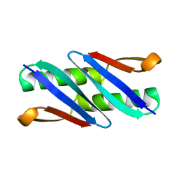
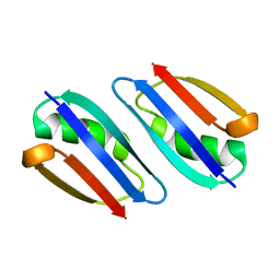
Ensemble of 40 Structures of the Dimeric Mutant of the B1 Domain of Streptococcal Protein G
分子名称: Immunoglobulin G binding protein G
著者Byeon, I.J, Louis, J.M, Gronenborn, A.M.
登録日2003-07-18
公開日2003-10-14
最終更新日2024-05-22
実験手法SOLUTION NMR
主引用文献A Protein Contortionist: Core Mutations of GB1 that Induce Dimerization and Domain Swapping
J.Mol.Biol., 333, 2003
2KOD
DownloadVisualize
BU of 2kod by Molmil
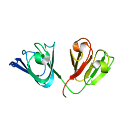
A high-resolution NMR structure of the dimeric C-terminal domain of HIV-1 CA
分子名称: HIV-1 CA C-terminal domain
著者Byeon, I.-J.L, Jung, J, Ahn, J, concel, J, Gronenborn, A.M.
登録日2009-09-18
公開日2009-11-24
最終更新日2024-05-01
実験手法SOLUTION NMR
主引用文献Structural convergence between Cryo-EM and NMR reveals intersubunit interactions critical for HIV-1 capsid function.
Cell(Cambridge,Mass.), 139, 2009
1K2N
DownloadVisualize
BU of 1k2n by Molmil
Solution Structure of the FHA2 domain of Rad53 Complexed with a Phosphothreonyl Peptide Derived from Rad9
分子名称: DNA repair protein Rad9, Protein Kinase SPK1
著者Byeon, I.-J.L, Yongkiettrakul, S, Tsai, M.-D.
登録日2001-09-28
公開日2001-12-05
最終更新日2024-10-16
実験手法SOLUTION NMR
主引用文献Solution structure of the yeast Rad53 FHA2 complexed with a phosphothreonine peptide pTXXL: comparison with the structures of FHA2-pYXL and FHA1-pTXXD complexes.
J.Mol.Biol., 314, 2001
1K2M
DownloadVisualize
BU of 1k2m by Molmil
Solution Structure of the FHA2 Domain of Rad53 Complexed with a Phosphotyrosyl Peptide Derived from Rad9
分子名称: DNA repair protein Rad9, Protein Kinase SPK1
著者Byeon, I.-J.L, Yongkiettrakul, S, Tsai, M.-D.
登録日2001-09-28
公開日2001-12-05
最終更新日2023-11-15
実験手法SOLUTION NMR
主引用文献Solution structure of the yeast Rad53 FHA2 complexed with a phosphothreonine peptide pTXXL: comparison with the structures of FHA2-pYXL and FHA1-pTXXD complexes.
J.Mol.Biol., 314, 2001
1JQR
DownloadVisualize
BU of 1jqr by Molmil
NMR structure of the African swine fever virus DNA polymerase X
分子名称: DNA POLYMERASE BETA-LIKE
著者Byeon, I.-J.L, Su, M.-I, Showalter, A.K, Tsai, M.-D.
登録日2001-08-08
公開日2001-10-26
最終更新日2024-10-30
実験手法SOLUTION NMR
主引用文献Solution structure of a viral DNA polymerase X and evidence for a mutagenic function.
Nat.Struct.Biol., 8, 2001
1DMZ
DownloadVisualize
BU of 1dmz by Molmil
A REFINED NMR STRUCTURE OF A NEW PHOPHOPEPTIDE-BINDING DOMAIN CONTAINING THE FHA2 OF RAD53
分子名称: PROTEIN (PROTEIN KINASE SPK1)
著者Byeon, I.-J.L, Liao, H, Tsai, M.-D.
登録日1999-12-15
公開日2000-01-06
最終更新日2024-05-22
実験手法SOLUTION NMR
主引用文献Structure and Function of a New Phosphopeptide-Binding Domain Containing the Fha2 of Rad53
J.Mol.Biol., 294, 1999
2KLJ
DownloadVisualize
BU of 2klj by Molmil
Solution Structure of gammaD-Crystallin with RDC and SAXS
分子名称: Gamma-crystallin D
著者Wang, J, Zuo, X, Yu, P, Byeon, I, Jung, J, Gronenborn, A.M, Wang, Y.
登録日2009-07-06
公開日2009-10-06
最終更新日2024-05-22
実験手法SOLUTION NMR, SOLUTION SCATTERING
主引用文献Determination of multicomponent protein structures in solution using global orientation and shape restraints.
J.Am.Chem.Soc., 131, 2009
5T82
DownloadVisualize
BU of 5t82 by Molmil
HIV-1 reverse transcriptase thumb subdomain
分子名称: Reverse transcriptase
著者Gronenborn, A.M, Sharaf, N.G, Byeon, I.-J.L.
登録日2016-09-06
公開日2017-01-18
最終更新日2024-05-15
実験手法SOLUTION NMR
主引用文献NMR structure of the HIV-1 reverse transcriptase thumb subdomain.
J. Biomol. NMR, 66, 2016
7TA8
DownloadVisualize
BU of 7ta8 by Molmil
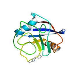
NMR structure of crosslinked cyclophilin A
分子名称: Peptidyl-prolyl cis-trans isomerase A
著者Lu, M, Toptygin, D, Xiang, Y, Shi, Y, Schwieters, C.D, Lipinski, E.C, Ahn, J, Byeon, I.-J.L, Gronenborn, A.M.
登録日2021-12-20
公開日2022-06-01
最終更新日2024-10-30
実験手法SOLUTION NMR
主引用文献The Magic of Linking Rings: Discovery of a Unique Photoinduced Fluorescent Protein Crosslink.
J.Am.Chem.Soc., 144, 2022
2RMM
DownloadVisualize
BU of 2rmm by Molmil
Solution structure of GB1 A34F mutant
分子名称: Immunoglobulin G-binding protein G
著者Jee, J, Byeon, I, Louis, J.M, Gronenborn, A.M.
登録日2007-10-30
公開日2007-12-04
最終更新日2024-05-29
実験手法SOLUTION NMR
主引用文献Solution structure of GB1 A34F mutant
To be Published
1PK2
DownloadVisualize
BU of 1pk2 by Molmil
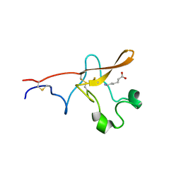
SOLUTION STRUCTURE OF THE TISSUE-TYPE PLASMINOGEN ACTIVATOR KRINGLE 2 DOMAIN COMPLEXED TO 6-AMINOHEXANOIC ACID AN ANTIFIBRINOLYTIC DRUG
分子名称: 6-AMINOHEXANOIC ACID, TISSUE-TYPE PLASMINOGEN ACTIVATOR
著者Llinas, M, Byeon, I.-J.L.
登録日1991-09-16
公開日1994-01-31
最終更新日2024-10-23
実験手法SOLUTION NMR
主引用文献Solution structure of the tissue-type plasminogen activator kringle 2 domain complexed to 6-aminohexanoic acid an antifibrinolytic drug.
J.Mol.Biol., 222, 1991


12>

226707

件を2024-10-30に公開中

PDB statisticsPDBj update infoContact PDBjnumon