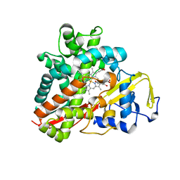
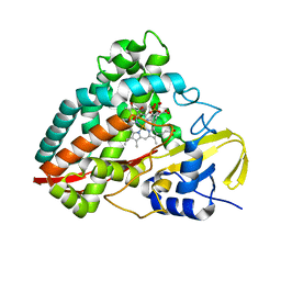
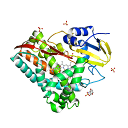
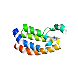
3LXH
 
 | | Crystal Structure of Cytochrome P450 CYP101D1 | | 分子名称: | 1,4-DIETHYLENE DIOXIDE, Cytochrome P450, PHOSPHATE ION, ... | | 著者 | Yang, W, Bell, S.G, Wang, H, Bartlam, M, Wong, L.L, Rao, Z. | | 登録日 | 2010-02-25 | | 公開日 | 2010-06-23 | | 最終更新日 | 2023-11-01 | | 実験手法 | X-RAY DIFFRACTION (2.2 Å) | | 主引用文献 | Molecular characterization of a class I P450 electron transfer system from Novosphingobium aromaticivorans DSM12444
J.Biol.Chem., 285, 2010
|
|
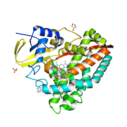
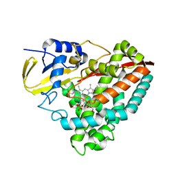
3LXD
 | | Crystal Structure of Ferredoxin Reductase ArR from Novosphingobium aromaticivorans | | 分子名称: | FAD-dependent pyridine nucleotide-disulphide oxidoreductase, FLAVIN-ADENINE DINUCLEOTIDE | | 著者 | Yang, W, Bell, S.G, Wang, H, Bartlam, M, Wong, L.L, Rao, Z. | | 登録日 | 2010-02-25 | | 公開日 | 2010-06-23 | | 最終更新日 | 2023-11-01 | | 実験手法 | X-RAY DIFFRACTION (2.5 Å) | | 主引用文献 | Molecular characterization of a class I P450 electron transfer system from Novosphingobium aromaticivorans DSM12444
J.Biol.Chem., 285, 2010
|
|
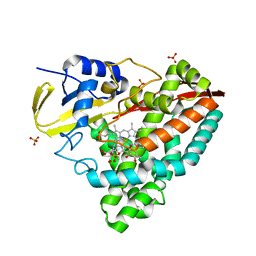
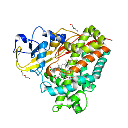
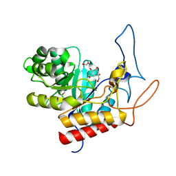
3OFT
 | | Crystal Structure of Cytochrome P450 CYP101C1 | | 分子名称: | (2R,5R)-hexane-2,5-diol, Cytochrome P450, PROTOPORPHYRIN IX CONTAINING FE | | 著者 | Zhou, W, Ma, M, Bell, S.G, Yang, W, Hao, Y, Rees, N.H, Bartlam, M, Wong, L.-L, Rao, Z. | | 登録日 | 2010-08-16 | | 公開日 | 2011-07-20 | | 最終更新日 | 2023-11-01 | | 実験手法 | X-RAY DIFFRACTION (1.9 Å) | | 主引用文献 | Structural Analysis of CYP101C1 from Novosphingobium aromaticivorans DSM12444.
Chembiochem, 12, 2011
|
|
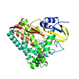
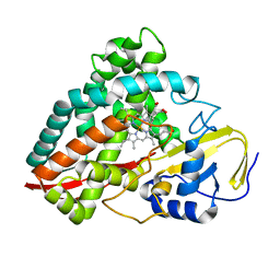
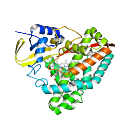
3LXI
 | | Crystal Structure of Camphor-Bound CYP101D1 | | 分子名称: | CAMPHOR, Cytochrome P450, PHOSPHATE ION, ... | | 著者 | Yang, W, Bell, S.G, Wang, H, Bartlam, M, Wong, L.L, Rao, Z. | | 登録日 | 2010-02-25 | | 公開日 | 2010-06-23 | | 最終更新日 | 2023-11-01 | | 実験手法 | X-RAY DIFFRACTION (2.2 Å) | | 主引用文献 | Molecular characterization of a class I P450 electron transfer system from Novosphingobium aromaticivorans DSM12444
J.Biol.Chem., 285, 2010
|
|
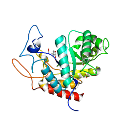
3M4V
 | | Crystal structure of the A330P mutant of cytochrome P450 BM3 | | 分子名称: | Bifunctional P-450/NADPH-P450 reductase, PROTOPORPHYRIN IX CONTAINING FE | | 著者 | Yang, W, Whitehouse, C.J.C, Bell, S.G, Bartlam, M, Wong, L.L, Rao, Z. | | 登録日 | 2010-03-12 | | 公開日 | 2011-03-23 | | 最終更新日 | 2023-11-01 | | 実験手法 | X-RAY DIFFRACTION (1.9 Å) | | 主引用文献 | Structural basis for the properties of two single-site proline mutants of CYP102A1 (P450BM3)
Chembiochem, 11, 2010
|
|
3NV5
 | | Crystal Structure of Cytochrome P450 CYP101D2 | | 分子名称: | Cytochrome P450, DI(HYDROXYETHYL)ETHER, PROTOPORPHYRIN IX CONTAINING FE | | 著者 | Yang, W, Bell, S.G, Wang, H, Zhou, W.H, Bartlam, M, Wong, L.L, Rao, Z. | | 登録日 | 2010-07-08 | | 公開日 | 2010-11-03 | | 最終更新日 | 2023-11-01 | | 実験手法 | X-RAY DIFFRACTION (2.41 Å) | | 主引用文献 | The structure of CYP101D2 unveils a potential path for substrate entry into the active site
Biochem.J., 433, 2011
|
|
4EGO
 | | The X-ray crystal structure of CYP199A4 in complex with indole-6-carboxylic acid | | 分子名称: | 1H-indole-6-carboxylic acid, CHLORIDE ION, Cytochrome P450, ... | | 著者 | Zhou, W, Bell, S.G, Yang, W, Zhou, R.M, Tan, A.B.H, Wong, L.-L. | | 登録日 | 2012-03-31 | | 公開日 | 2013-02-20 | | 最終更新日 | 2023-11-08 | | 実験手法 | X-RAY DIFFRACTION (1.76 Å) | | 主引用文献 | Investigation of the substrate range of CYP199A4: modification of the partition between hydroxylation and desaturation activities by substrate and protein engineering
Chemistry, 18, 2012
|
|
4EGM
 | | The X-ray crystal structure of CYP199A4 in complex with 4-ethylbenzoic acid | | 分子名称: | 4-ethylbenzoic acid, CHLORIDE ION, Cytochrome P450, ... | | 著者 | Zhou, W, Bell, S.G, Yang, W, Zhou, R.M, Tan, A.B.H, Wong, L.-L. | | 登録日 | 2012-03-31 | | 公開日 | 2013-02-20 | | 最終更新日 | 2023-11-08 | | 実験手法 | X-RAY DIFFRACTION (2.91 Å) | | 主引用文献 | Investigation of the substrate range of CYP199A4: modification of the partition between hydroxylation and desaturation activities by substrate and protein engineering
Chemistry, 18, 2012
|
|
3NV6
 | | Crystal Structure of Camphor-Bound CYP101D2 | | 分子名称: | CAMPHOR, Cytochrome P450, DI(HYDROXYETHYL)ETHER, ... | | 著者 | Yang, W, Bell, S.G, Wang, H, Zhou, W.H, Bartlam, M, Wong, L.L, Rao, Z. | | 登録日 | 2010-07-08 | | 公開日 | 2010-11-03 | | 最終更新日 | 2023-11-01 | | 実験手法 | X-RAY DIFFRACTION (2.2 Å) | | 主引用文献 | The structure of CYP101D2 unveils a potential path for substrate entry into the active site
Biochem.J., 433, 2011
|
|
4EGN
 | | The X-ray crystal structure of CYP199A4 in complex with veratric acid | | 分子名称: | 3,4-dimethoxybenzoic acid, CHLORIDE ION, Cytochrome P450, ... | | 著者 | Zhou, W, Bell, S.G, Yang, W, Zhou, R.M, Tan, A.B.H, Wong, L.-L. | | 登録日 | 2012-03-31 | | 公開日 | 2013-02-20 | | 最終更新日 | 2023-11-08 | | 実験手法 | X-RAY DIFFRACTION (2 Å) | | 主引用文献 | Investigation of the substrate range of CYP199A4: modification of the partition between hydroxylation and desaturation activities by substrate and protein engineering
Chemistry, 18, 2012
|
|
4EGP
 | | The X-ray crystal structure of CYP199A4 in complex with 2-naphthoic acid | | 分子名称: | CHLORIDE ION, Cytochrome P450, GLYCEROL, ... | | 著者 | Zhou, W, Bell, S.G, Yang, W, Zhou, R.M, Tan, A.B.H, Wong, L.-L. | | 登録日 | 2012-03-31 | | 公開日 | 2013-02-20 | | 最終更新日 | 2023-11-08 | | 実験手法 | X-RAY DIFFRACTION (3 Å) | | 主引用文献 | Investigation of the substrate range of CYP199A4: modification of the partition between hydroxylation and desaturation activities by substrate and protein engineering
Chemistry, 18, 2012
|
|
4DO1
 | | The crystal structures of 4-methoxybenzoate bound CYP199A4 | | 分子名称: | 4-METHOXYBENZOIC ACID, CHLORIDE ION, Cytochrome P450, ... | | 著者 | Zhou, W, Bell, S.G, Yang, W, Tan, A.B.H, Zhou, R, Johnson, E.O.D, Zhang, A, Rao, Z, Wong, L.-L. | | 登録日 | 2012-02-09 | | 公開日 | 2012-08-15 | | 最終更新日 | 2023-11-08 | | 実験手法 | X-RAY DIFFRACTION (2 Å) | | 主引用文献 | The crystal structures of 4-methoxybenzoate bound CYP199A2 and CYP199A4: structural changes on substrate binding and the identification of an anion binding site
Dalton Trans, 41, 2012
|
|
4DNZ
 | | The crystal structures of CYP199A4 | | 分子名称: | CHLORIDE ION, Cytochrome P450, GLYCEROL, ... | | 著者 | Zhou, W, Bell, S.G, Yang, W, Tan, A.B.H, Zhou, R, Johnson, E.O.D, Zhang, A, Rao, Z, Wong, L.-L. | | 登録日 | 2012-02-09 | | 公開日 | 2012-08-15 | | 最終更新日 | 2023-11-08 | | 実験手法 | X-RAY DIFFRACTION (2.6 Å) | | 主引用文献 | The crystal structures of 4-methoxybenzoate bound CYP199A2 and CYP199A4: structural changes on substrate binding and the identification of an anion binding site
Dalton Trans, 41, 2012
|
|
4DXY
 | | Crystal structures of CYP101D2 Y96A mutant | | 分子名称: | Cytochrome P450, DI(HYDROXYETHYL)ETHER, PROTOPORPHYRIN IX CONTAINING FE | | 著者 | Zhou, W, Bell, S.G, Yang, W, Dale, A, Wong, L.-L. | | 登録日 | 2012-02-28 | | 公開日 | 2012-08-15 | | 最終更新日 | 2023-11-08 | | 実験手法 | X-RAY DIFFRACTION (2 Å) | | 主引用文献 | Improving the affinity and activity of CYP101D2 for hydrophobic substrates
Appl.Microbiol.Biotechnol., 2012
|
|
4DNJ
 | | The crystal structures of 4-methoxybenzoate bound CYP199A2 | | 分子名称: | 4-METHOXYBENZOIC ACID, CHLORIDE ION, PROTOPORPHYRIN IX CONTAINING FE, ... | | 著者 | Zhou, W, Bell, S.G, Yang, W, Tan, A.B.H, Zhou, R, Johnson, E.O.D, Zhang, A, Rao, Z, Wong, L.-L. | | 登録日 | 2012-02-08 | | 公開日 | 2012-08-15 | | 最終更新日 | 2023-11-08 | | 実験手法 | X-RAY DIFFRACTION (1.8 Å) | | 主引用文献 | The crystal structures of 4-methoxybenzoate bound CYP199A2 and CYP199A4: structural changes on substrate binding and the identification of an anion binding site
Dalton Trans, 41, 2012
|
|
5YQH
 | | The crystal structure of CYP199A4 binding with 4-n-Propyl benzoic acid | | 分子名称: | 4-methoxybenzamide, CHLORIDE ION, Cytochrome P450, ... | | 著者 | Zhou, W, Zhang, T, Qiao, R, Bell, S, Coleman, T. | | 登録日 | 2017-11-06 | | 公開日 | 2019-02-27 | | 最終更新日 | 2023-11-22 | | 実験手法 | X-RAY DIFFRACTION (2.3 Å) | | 主引用文献 | The crystal structure of CYP199A4 binding with 4-n-Propyl benzoic acid
To Be Published
|
|
5YQA
 | | The crystal structure of CYP199A4 binding with 4-n-Propyl benzoic acid | | 分子名称: | 4-propylbenzoic acid, CHLORIDE ION, Cytochrome P450, ... | | 著者 | Zhou, W, Zhang, T, Qiao, R, Bell, S, Coleman, T. | | 登録日 | 2017-11-06 | | 公開日 | 2019-02-27 | | 最終更新日 | 2023-11-22 | | 実験手法 | X-RAY DIFFRACTION (1.38 Å) | | 主引用文献 | The crystal structure of CYP199A4 binding with 4-n-Propyl benzoic acid
To Be Published
|
|
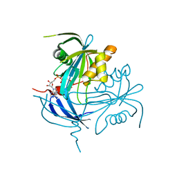
7XLD
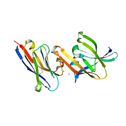 | | Crystal structure of IsdH linker-NEAT3 bound to a nanobody (VHH) | | 分子名称: | Iron-regulated surface determinant protein H, MAGNESIUM ION, Nanobody VHH6, ... | | 著者 | Caaveiro, J.M.M, Valenciano-Bellido, S, Tsumoto, K. | | 登録日 | 2022-04-21 | | 公開日 | 2023-05-31 | | 最終更新日 | 2023-11-29 | | 実験手法 | X-RAY DIFFRACTION (1.65 Å) | | 主引用文献 | Targeting hemoglobin receptors IsdH and IsdB of Staphylococcus aureus with a single VHH antibody inhibits bacterial growth.
J.Biol.Chem., 299, 2023
|
|
8STL
 | | Crystal Structure of Nanobody PIK3_Nb16 against wild-type PI3Kalpha | | 分子名称: | Nanobody PIK3_Nb16, SULFATE ION | | 著者 | Nwafor, J.N, Srinivasan, L, Chen, Z, Gabelli, S.B, Iheanacho, A, Alzogaray, V, Klinke, S. | | 登録日 | 2023-05-10 | | 公開日 | 2024-05-22 | | 実験手法 | X-RAY DIFFRACTION (1.85 Å) | | 主引用文献 | Development of isoform specific nanobodies for Class I PI3Ks
To be published
|
|
5DBM
 | | Crystal structure of the CBP bromodomain in complex with CPI703 | | 分子名称: | (4R)-6-(1-tert-butyl-1H-pyrazol-4-yl)-4-methyl-1,3,4,5-tetrahydro-2H-1,5-benzodiazepin-2-one, CREB-binding protein | | 著者 | Setser, J.W, Poy, F, Bellon, S.F. | | 登録日 | 2015-08-21 | | 公開日 | 2016-04-20 | | 最終更新日 | 2023-09-27 | | 実験手法 | X-RAY DIFFRACTION (1.86 Å) | | 主引用文献 | Regulatory T Cell Modulation by CBP/EP300 Bromodomain Inhibition.
J.Biol.Chem., 291, 2016
|
|
4V27
 | | Structure of the GH99 endo-alpha-mannanase from Bacteroides xylanisolvens in complex with mannose-alpha-1,3-isofagomine | | 分子名称: | 1,2-ETHANEDIOL, 5-HYDROXYMETHYL-3,4-DIHYDROXYPIPERIDINE, GLYCOSYL HYDROLASE FAMILY 71, ... | | 著者 | Hakki, Z, Bellmaine, S, Thompson, A.J, Speciale, G, Davies, G.J, Williams, S.J. | | 登録日 | 2014-10-07 | | 公開日 | 2014-12-24 | | 最終更新日 | 2024-01-10 | | 実験手法 | X-RAY DIFFRACTION (1.9 Å) | | 主引用文献 | Structural and Kinetic Dissection of the Endo-Alpha-1,2-Mannanase Activity of Bacterial Gh99 Glycoside Hydrolases from Bacteroides Spp.
Chemistry, 21, 2015
|
|
4V28
 | | Structure of an E333Q variant of the GH99 endo-alpha-mannanase from Bacteroides xylanisolvens in complex with Man-Man-Methylumbelliferone | | 分子名称: | 1,2-ETHANEDIOL, 7-hydroxy-4-methyl-2H-chromen-2-one, ACETATE ION, ... | | 著者 | Hakki, Z, Bellmaine, S, Thompson, A.J, Speciale, G, Davies, G.J, Williams, S.J. | | 登録日 | 2014-10-07 | | 公開日 | 2014-12-24 | | 最終更新日 | 2024-01-10 | | 実験手法 | X-RAY DIFFRACTION (1.2 Å) | | 主引用文献 | Structural and Kinetic Dissection of the Endo-Alpha-1,2-Mannanase Activity of Bacterial Gh99 Glycoside Hydrolases from Bacteroides Spp.
Chemistry, 21, 2015
|
|
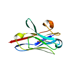
1MK1
 | | Structure of the MT-ADPRase in complex with ADPR, a Nudix enzyme | | 分子名称: | ADENOSINE-5-DIPHOSPHORIBOSE, ADPR pyrophosphatase | | 著者 | Kang, L.-W, Gabelli, S.B, Bianchet, M.A, Cunningham, J.E, O'Handley, S.F, Amzel, L.M. | | 登録日 | 2002-08-28 | | 公開日 | 2003-08-05 | | 最終更新日 | 2024-02-14 | | 実験手法 | X-RAY DIFFRACTION (2 Å) | | 主引用文献 | Structure and mechanism of MT-ADPRase, a Nudix hydrolase from Mycobacterium tuberculosis
Structure, 11, 2003
|
|
1MP2
 | | Structure of MT-ADPRase (Apoenzyme), a Nudix hydrolase from Mycobacterium tuberculosis | | 分子名称: | ADPR pyrophosphatase | | 著者 | Kang, L.-W, Gabelli, S.B, Bianchet, M.A, Cunningham, J.E, O'Handley, S.F, Amzel, L.M. | | 登録日 | 2002-09-11 | | 公開日 | 2003-08-05 | | 最終更新日 | 2024-02-14 | | 実験手法 | X-RAY DIFFRACTION (2.3 Å) | | 主引用文献 | Structure and mechanism of MT-ADPRase, a Nudix hydrolase from Mycobacterium tuberculosis
Structure, 11, 2003
|
|
1MQE
 | | Structure of the MT-ADPRase in complex with gadolidium and ADP-ribose, a Nudix enzyme | | 分子名称: | ADENOSINE-5-DIPHOSPHORIBOSE, ADPR pyrophosphatase, GADOLINIUM ION | | 著者 | Kang, L.-W, Gabelli, S.B, Bianchet, M.A, Cunningham, J.E, O'Handley, S.F, Amzel, L.M. | | 登録日 | 2002-09-16 | | 公開日 | 2003-08-05 | | 最終更新日 | 2024-02-14 | | 実験手法 | X-RAY DIFFRACTION (2 Å) | | 主引用文献 | Structure and mechanism of MT-ADPRase, a Nudix hydrolase from Mycobacterium tuberculosis
Structure, 11, 2003
|
|


