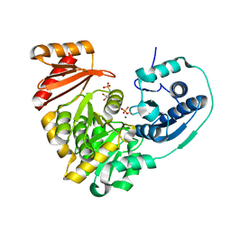
8FSD
 
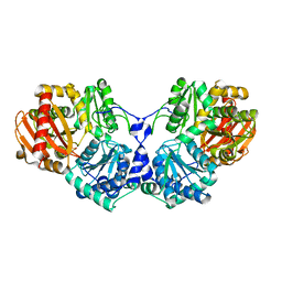
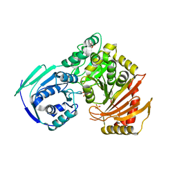
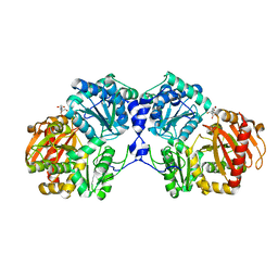
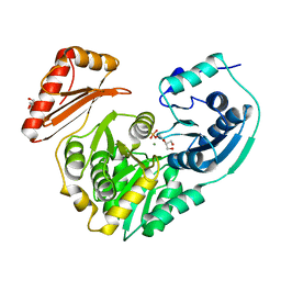
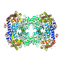 | | P130R mutant of soybean SHMT8 in complex with PLP-glycine and formylTHF | | 分子名称: | 1,2-ETHANEDIOL, N-GLYCINE-[3-HYDROXY-2-METHYL-5-PHOSPHONOOXYMETHYL-PYRIDIN-4-YL-METHANE], N-[4-({[(6S)-2-amino-5-formyl-4-oxo-3,4,5,6,7,8-hexahydropteridin-6-yl]methyl}amino)benzoyl]-L-glutamic acid, ... | | 著者 | Beamer, L.J, Korasick, D.A. | | 登録日 | 2023-01-09 | | 公開日 | 2023-10-18 | | 最終更新日 | 2024-01-31 | | 実験手法 | X-RAY DIFFRACTION (1.49 Å) | | 主引用文献 | Structural and functional analysis of two SHMT8 variants associated with soybean cyst nematode resistance.
Febs J., 291, 2024
|
|
9CE6
 | |
9CG8
 | |
5JN5
 | |
5EPC
 | |
5F9C
 | |
1LMB
 | |
3OLP
 | |
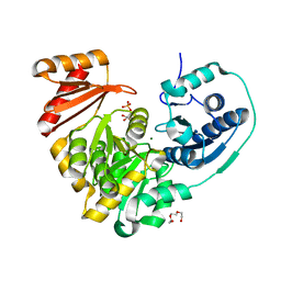
1BP1
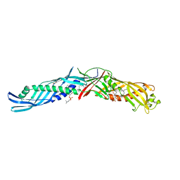 | | CRYSTAL STRUCTURE OF BPI, THE HUMAN BACTERICIDAL PERMEABILITY-INCREASING PROTEIN | | 分子名称: | 1,2-DIACYL-SN-GLYCERO-3-PHOSPHOCHOLINE, BACTERICIDAL/PERMEABILITY-INCREASING PROTEIN | | 著者 | Beamer, L.J, Carroll, S.F, Eisenberg, D. | | 登録日 | 1997-04-08 | | 公開日 | 1997-09-04 | | 最終更新日 | 2024-11-06 | | 実験手法 | X-RAY DIFFRACTION (2.4 Å) | | 主引用文献 | Crystal structure of human BPI and two bound phospholipids at 2.4 angstrom resolution.
Science, 276, 1997
|
|
6UO6
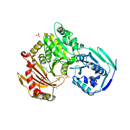 | |
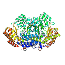
2H4L
 | | Complex of PMM/PGM with ribose 1-phosphate | | 分子名称: | 1-O-phosphono-alpha-D-ribofuranose, Phosphomannomutase/phosphoglucomutase, ZINC ION | | 著者 | Beamer, L.J. | | 登録日 | 2006-05-24 | | 公開日 | 2006-08-08 | | 最終更新日 | 2024-10-09 | | 実験手法 | X-RAY DIFFRACTION (2.4 Å) | | 主引用文献 | Complexes of the enzyme phosphomannomutase/phosphoglucomutase with a slow substrate and an inhibitor.
Acta Crystallogr.,Sect.F, 62, 2006
|
|
5TR2
 | |
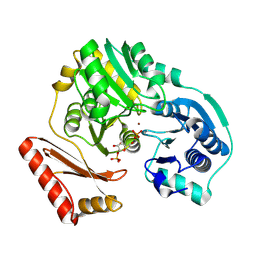
6MNV
 | | Crystal structure of X. citri phosphoglucomutase in complex with CH2FG1P | | 分子名称: | 1-deoxy-1-fluoro-2-O-phosphono-alpha-D-gluco-hept-2-ulopyranose, MAGNESIUM ION, Phosphomannomutase/phosphoglucomutase, ... | | 著者 | Beamer, L, Stiers, K. | | 登録日 | 2018-10-03 | | 公開日 | 2019-07-31 | | 最終更新日 | 2023-10-11 | | 実験手法 | X-RAY DIFFRACTION (1.65 Å) | | 主引用文献 | Inhibitory Evaluation of alpha PMM/PGM fromPseudomonas aeruginosa: Chemical Synthesis, Enzyme Kinetics, and Protein Crystallographic Study.
J.Org.Chem., 84, 2019
|
|
6MLF
 | |
6MLW
 | | Crystal structure of X. citri phosphoglucomutase in complex with 2-fluoro mannosyl-1-methyl-phosphonic acid | | 分子名称: | 2,6-anhydro-5,7-dideoxy-5-fluoro-7-phosphono-D-glycero-D-manno-heptitol, MAGNESIUM ION, Phosphoglucomutase | | 著者 | Beamer, L, Stiers, K. | | 登録日 | 2018-09-28 | | 公開日 | 2019-07-31 | | 最終更新日 | 2024-10-23 | | 実験手法 | X-RAY DIFFRACTION (1.9 Å) | | 主引用文献 | Inhibitory Evaluation of alpha PMM/PGM fromPseudomonas aeruginosa: Chemical Synthesis, Enzyme Kinetics, and Protein Crystallographic Study.
J.Org.Chem., 84, 2019
|
|
6MLH
 | | Crystal structure of X. citri phosphoglucomutase in complex with GLUCOPYRANOSYL-1-METHYL-PHOSPHONIC ACID | | 分子名称: | (1S)-1,5-anhydro-1-(phosphonomethyl)-D-glucitol, MAGNESIUM ION, Phosphoglucomutase | | 著者 | Beamer, L, Stiers, K. | | 登録日 | 2018-09-27 | | 公開日 | 2019-07-31 | | 最終更新日 | 2024-11-06 | | 実験手法 | X-RAY DIFFRACTION (1.65 Å) | | 主引用文献 | Inhibitory Evaluation of alpha PMM/PGM fromPseudomonas aeruginosa: Chemical Synthesis, Enzyme Kinetics, and Protein Crystallographic Study.
J.Org.Chem., 84, 2019
|
|
6N1E
 | | Crystal structure of X. citri phosphoglucomutase in complex with 1-methyl-glucose 6-phosphate | | 分子名称: | 1-deoxy-7-O-phosphono-alpha-D-gluco-hept-2-ulopyranose, MAGNESIUM ION, Phosphomannomutase/phosphoglucomutase | | 著者 | Beamer, L.J, Stiers, K.M. | | 登録日 | 2018-11-08 | | 公開日 | 2019-05-01 | | 最終更新日 | 2023-10-11 | | 実験手法 | X-RAY DIFFRACTION (1.7 Å) | | 主引用文献 | Synthesis, Derivatization, and Structural Analysis of Phosphorylated Mono-, Di-, and Trifluorinated d-Gluco-heptuloses by Glucokinase: Tunable Phosphoglucomutase Inhibition.
Acs Omega, 4, 2019
|
|
1U6D
 | |
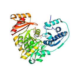
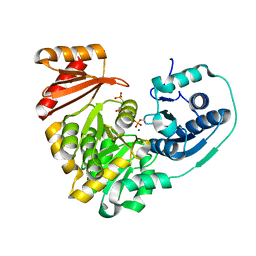
1EWF
 | | THE 1.7 ANGSTROM CRYSTAL STRUCTURE OF BPI | | 分子名称: | 1,2-DIACYL-SN-GLYCERO-3-PHOSPHOCHOLINE, BACTERICIDAL/PERMEABILITY-INCREASING PROTEIN | | 著者 | Kleiger, G, Beamer, L.J, Grothe, R, Mallick, P, Eisenberg, D. | | 登録日 | 2000-04-25 | | 公開日 | 2000-06-21 | | 最終更新日 | 2024-10-30 | | 実験手法 | X-RAY DIFFRACTION (1.7 Å) | | 主引用文献 | The 1.7 A crystal structure of BPI: a study of how two dissimilar amino acid sequences can adopt the same fold.
J.Mol.Biol., 299, 2000
|
|
8DOM
 | |
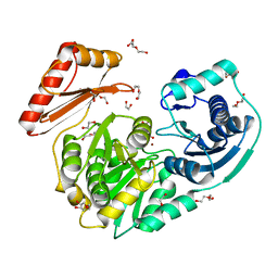
8TQF
 | | Crystal structure of Soybean SHMT8 in complex with PLP-glycine and diglutamylated 5-formyltetrahydrofolate | | 分子名称: | 1,2-ETHANEDIOL, N-GLYCINE-[3-HYDROXY-2-METHYL-5-PHOSPHONOOXYMETHYL-PYRIDIN-4-YL-METHANE], N-[4-({[(6R)-2-amino-5-formyl-4-hydroxy-5,6,7,8-tetrahydropteridin-6-yl]methyl}amino)benzoyl]-L-gamma-glutamyl-L-glutamic acid, ... | | 著者 | Owuocha, L.F, Beamer, L.J. | | 登録日 | 2023-08-07 | | 公開日 | 2024-08-21 | | 実験手法 | X-RAY DIFFRACTION (1.69 Å) | | 主引用文献 | Structural insights into high affinity binding of polyglutamylated tetrahydrofolate to soybean serine hydroxymethyltransferase
To Be Published
|
|
3NA5
 | | Crystal structure of a bacterial phosphoglucomutase, an enzyme important in the virulence of several human pathogens. | | 分子名称: | 2-[BIS-(2-HYDROXY-ETHYL)-AMINO]-2-HYDROXYMETHYL-PROPANE-1,3-DIOL, MAGNESIUM ION, Phosphoglucomutase | | 著者 | Mehra-Chaudhary, R, Beamer, L.J. | | 登録日 | 2010-06-01 | | 公開日 | 2011-02-16 | | 最終更新日 | 2023-11-22 | | 実験手法 | X-RAY DIFFRACTION (1.7 Å) | | 主引用文献 | Crystal structure of a bacterial phosphoglucomutase, an enzyme involved in the virulence of multiple human pathogens.
Proteins, 79, 2011
|
|
4MRQ
 | | Crystal Structure of wild-type unphosphorylated PMM/PGM | | 分子名称: | 1,2-ETHANEDIOL, DI(HYDROXYETHYL)ETHER, L(+)-TARTARIC ACID, ... | | 著者 | Lee, Y, Beamer, L. | | 登録日 | 2013-09-17 | | 公開日 | 2014-01-08 | | 最終更新日 | 2023-09-20 | | 実験手法 | X-RAY DIFFRACTION (1.9 Å) | | 主引用文献 | Promotion of enzyme flexibility by dephosphorylation and coupling to the catalytic mechanism of a phosphohexomutase.
J.Biol.Chem., 289, 2014
|
|
1ZGK
 | | 1.35 angstrom structure of the Kelch domain of Keap1 | | 分子名称: | Kelch-like ECH-associated protein 1 | | 著者 | Li, X, Bottoms, C.A, Hannink, M, Beamer, L.J. | | 登録日 | 2005-04-21 | | 公開日 | 2005-10-04 | | 最終更新日 | 2011-07-13 | | 実験手法 | X-RAY DIFFRACTION (1.35 Å) | | 主引用文献 | Conserved solvent and side-chain interactions in the 1.35 Angstrom structure of the Kelch domain of Keap1.
Acta Crystallogr.,Sect.D, 61, 2005
|
|
4IL8
 | | Crystal structure of an H329A mutant of p. aeruginosa PMM/PGM | | 分子名称: | GLYCEROL, MAGNESIUM ION, Phosphomannomutase/phosphoglucomutase | | 著者 | Lee, Y, Mehra-Chaudhary, R, Furdui, C, Beamer, L. | | 登録日 | 2012-12-29 | | 公開日 | 2013-08-14 | | 最終更新日 | 2023-11-29 | | 実験手法 | X-RAY DIFFRACTION (1.8 Å) | | 主引用文献 | Identification of an essential active-site residue in the alpha-D-phosphohexomutase enzyme superfamily.
Febs J., 280, 2013
|
|


