5IZ2
 
 | |
5UM6
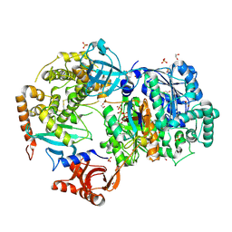
 | | Crystal Structure of S. pombe Uba1 in a closed conformation | | 分子名称: | 2-(decylamino)ethane-1-thiol, N-(2-{[(4-chlorophenyl)methyl]disulfanyl}ethyl)decan-1-amine, SULFATE ION, ... | | 著者 | Lv, Z, Yuan, L, Aldana-Masangkay, G, Atkison, J.H, Chen, Y, Olsen, S.K. | | 登録日 | 2017-01-26 | | 公開日 | 2017-06-14 | | 最終更新日 | 2023-10-04 | | 実験手法 | X-RAY DIFFRACTION (2.794 Å) | | 主引用文献 | Domain alternation and active site remodeling are conserved structural features of ubiquitin E1.
J. Biol. Chem., 292, 2017
|
|
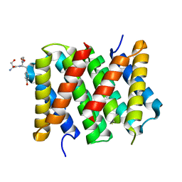
6CWY
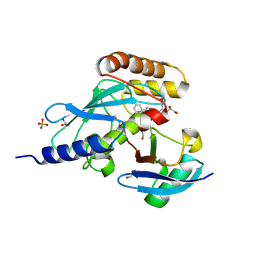
 | | Crystal structure of SUMO E1 in complex with an allosteric inhibitor | | 分子名称: | GLYCEROL, MAGNESIUM ION, SULFATE ION, ... | | 著者 | Lv, Z, Yuan, L, Atkison, J.H, Williams, K.M, Olsen, S.K. | | 登録日 | 2018-04-01 | | 公開日 | 2019-01-16 | | 最終更新日 | 2023-10-04 | | 実験手法 | X-RAY DIFFRACTION (2.462 Å) | | 主引用文献 | Molecular mechanism of a covalent allosteric inhibitor of SUMO E1 activating enzyme.
Nat Commun, 9, 2018
|
|
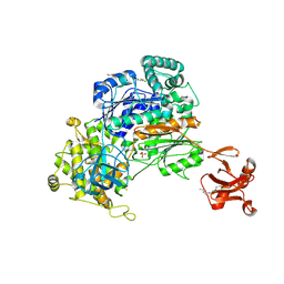
6CWZ
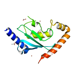
 | | Crystal structure of apo SUMO E1 | | 分子名称: | SUMO-activating enzyme subunit 1, SUMO-activating enzyme subunit 2, ZINC ION | | 著者 | Lv, Z, Yuan, L, Atkison, J.H, Williams, K.M, Olsen, S.K. | | 登録日 | 2018-04-01 | | 公開日 | 2019-01-16 | | 最終更新日 | 2023-10-04 | | 実験手法 | X-RAY DIFFRACTION (3.1 Å) | | 主引用文献 | Molecular mechanism of a covalent allosteric inhibitor of SUMO E1 activating enzyme.
Nat Commun, 9, 2018
|
|
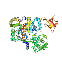
6DC6
 | | Crystal structure of human ubiquitin activating enzyme E1 (Uba1) in complex with ubiquitin | | 分子名称: | MAGNESIUM ION, PYROPHOSPHATE 2-, Ubiquitin, ... | | 著者 | Lv, Z, Yuan, L, Williams, K.M, Atkison, J.H, Olsen, S.K. | | 登録日 | 2018-05-04 | | 公開日 | 2018-10-10 | | 最終更新日 | 2023-10-11 | | 実験手法 | X-RAY DIFFRACTION (3.14 Å) | | 主引用文献 | Crystal structure of a human ubiquitin E1-ubiquitin complex reveals conserved functional elements essential for activity.
J. Biol. Chem., 293, 2018
|
|
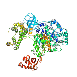
6NYA
 | | Crystal Structure of ubiquitin E1 (Uba1) in complex with Ubc3 (Cdc34) and ubiquitin | | 分子名称: | 1,2-ETHANEDIOL, ADENOSINE-5'-TRIPHOSPHATE, MAGNESIUM ION, ... | | 著者 | Olsen, S.K, Williams, K.M, Atkison, J.H. | | 登録日 | 2019-02-11 | | 公開日 | 2019-08-07 | | 最終更新日 | 2023-10-11 | | 実験手法 | X-RAY DIFFRACTION (2.065 Å) | | 主引用文献 | Structural insights into E1 recognition and the ubiquitin-conjugating activity of the E2 enzyme Cdc34.
Nat Commun, 10, 2019
|
|
6NYO
 | | Crystal structure of a human Cdc34-ubiquitin thioester mimetic | | 分子名称: | 1,2-ETHANEDIOL, 4,5-dideoxy-5-(3',5'-dichlorobiphenyl-4-yl)-4-[(methoxyacetyl)amino]-L-arabinonic acid, PHOSPHATE ION, ... | | 著者 | Olsen, S.K, Williams, K.M, Atkison, J.H. | | 登録日 | 2019-02-11 | | 公開日 | 2019-08-07 | | 最終更新日 | 2023-10-11 | | 実験手法 | X-RAY DIFFRACTION (1.502 Å) | | 主引用文献 | Structural insights into E1 recognition and the ubiquitin-conjugating activity of the E2 enzyme Cdc34.
Nat Commun, 10, 2019
|
|
6NYD
 | |


