Loading
PDBj
MenuPDBj@FacebookPDBj@TwitterPDBj@YouTubewwPDB FoundationwwPDB
RCSB PDBPDBeBMRBAdv. SearchSearch help
PDB: 566 results

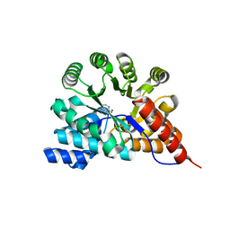
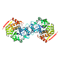
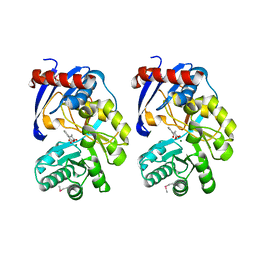
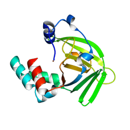
1YV9
DownloadVisualize
BU of 1yv9 by Molmil
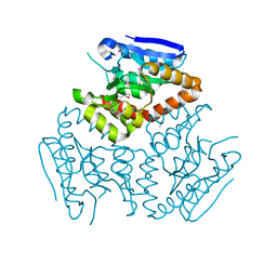
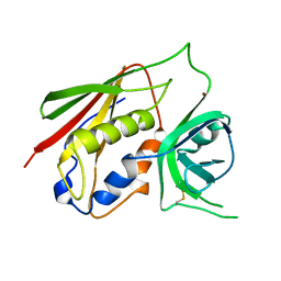
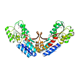
Crystal structure of a HAD-like phosphatase from Enterococcus faecalis V583
Descriptor: PHOSPHATE ION, hydrolase, haloacid dehalogenase family
Authors:Seetharaman, J, Swaminathan, S, Burley, S.K, New York SGX Research Center for Structural Genomics (NYSGXRC)
Deposit date:2005-02-15
Release date:2005-03-15
Last modified:2023-08-23
Method:X-RAY DIFFRACTION (2.8 Å)
Cite:Crystal Structure of a Hypothetical protein, hydrolase haloacid dehalogenase-like family
To be Published
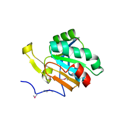
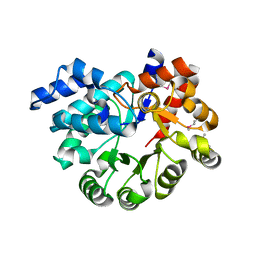
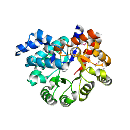
1TXL
DownloadVisualize
BU of 1txl by Molmil
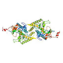
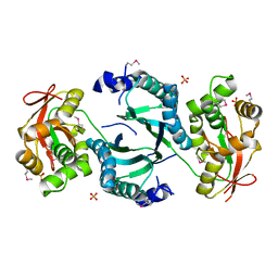
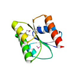
Crystal structure of metal-binding protein yodA from E. coli, Pfam DUF149
Descriptor: Metal-binding protein yodA, ZINC ION
Authors:Eswaramoorthy, S, Swaminathan, S, Burley, S.K, New York SGX Research Center for Structural Genomics (NYSGXRC)
Deposit date:2004-07-05
Release date:2004-07-20
Last modified:2021-02-03
Method:X-RAY DIFFRACTION (1.7 Å)
Cite:Crystal structure of a hypothetical protein yodA
To be Published
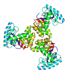
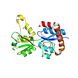
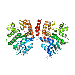
3RYS
DownloadVisualize
BU of 3rys by Molmil
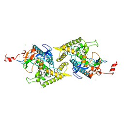
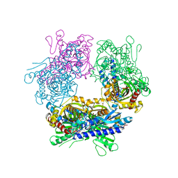
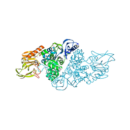
The crystal structure of adenine deaminase (AAur1117) from Arthrobacter aurescens
Descriptor: ADENINE, Adenosine deaminase 1, ZINC ION
Authors:Zhang, Z, Goble, A.M, Raushel, F.M, Swaminathan, S.
Deposit date:2011-05-11
Release date:2011-05-25
Last modified:2023-09-13
Method:X-RAY DIFFRACTION (2.601 Å)
Cite:The crystal structure of adenine deaminase (AAur1117) from Arthrobacter aurescens
To be Published
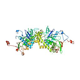
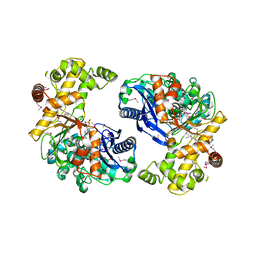
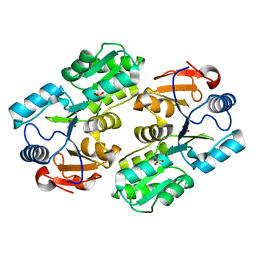
3UMA
DownloadVisualize
BU of 3uma by Molmil
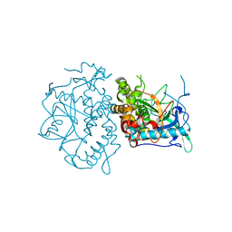
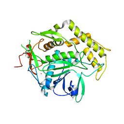
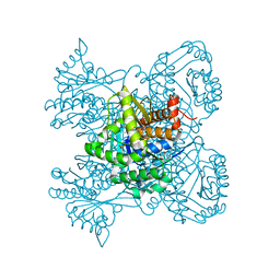
Crystal structure of a hypothetical peroxiredoxin protein frm Sinorhizobium meliloti
Descriptor: Hypothetical peroxiredoxin protein, SULFATE ION
Authors:Eswaramoorthy, S, Chamala, S, Evans, B, Foti, R, Gizzi, A, Hillerich, B, Kar, A, LaFleur, J, Seidel, R, Villigas, G, Zencheck, W, Almo, S.C, Swaminathan, S, New York Structural Genomics Research Consortium (NYSGRC)
Deposit date:2011-11-12
Release date:2011-11-23
Last modified:2024-10-09
Method:X-RAY DIFFRACTION (2.2 Å)
Cite:Crystal structure of a hypothetical peroxiredoxin protein from Sinorhizobium meliloti
To be Published
1SGJ
DownloadVisualize
BU of 1sgj by Molmil
Crystal structure of citrate lyase beta subunit
Descriptor: MAGNESIUM ION, OXALOACETATE ION, citrate lyase, ...
Authors:Eswaramoorthy, S, Swaminathan, S, Burley, S.K, New York SGX Research Center for Structural Genomics (NYSGXRC)
Deposit date:2004-02-23
Release date:2004-08-17
Last modified:2024-02-14
Method:X-RAY DIFFRACTION (1.84 Å)
Cite:Crystal structure of citrate lyase beta subunit
To be Published
1ZL5
DownloadVisualize
BU of 1zl5 by Molmil
Crystal structure of Glu335Gln mutant of Clostridium botulinum neurotoxin E catalytic domain
Descriptor: CHLORIDE ION, botulinum neurotoxin type E
Authors:Agarwal, R, Binz, T, Swaminathan, S.
Deposit date:2005-05-05
Release date:2005-07-05
Last modified:2023-08-23
Method:X-RAY DIFFRACTION (2.6 Å)
Cite:Analysis of Active Site Residues of Botulinum Neurotoxin E by Mutational, Functional, and Structural Studies: Glu335Gln Is an Apoenzyme.
Biochemistry, 44, 2005
4KD6
DownloadVisualize
BU of 4kd6 by Molmil
Crystal structure of a Enoyl-CoA hydratase/isomerase from Burkholderia graminis C4D1M
Descriptor: Enoyl-CoA hydratase/isomerase
Authors:Kumaran, D, Chamala, S, Evans, B, Foti, R, Gizzi, A, Hillerich, B, Kar, A, Lafleur, J, Seidel, R, Villigas, G, Zencheck, W, Al Obaidi, N, Almo, S.C, Swaminathan, S, New York Structural Genomics Research Consortium (NYSGRC)
Deposit date:2013-04-24
Release date:2013-07-24
Last modified:2024-10-30
Method:X-RAY DIFFRACTION (2.25 Å)
Cite:Crystal structure of a Enoyl-CoA hydratase/isomerase from Burkholderia graminis C4D1M
To be Published
1T3C
DownloadVisualize
BU of 1t3c by Molmil
Clostridium botulinum type E catalytic domain E212Q mutant
Descriptor: CHLORIDE ION, ZINC ION, neurotoxin type E
Authors:Agarwal, R, Eswaramoorthy, S, Kumaran, D, Binz, T, Swaminathan, S.
Deposit date:2004-04-26
Release date:2004-06-29
Last modified:2023-08-23
Method:X-RAY DIFFRACTION (1.9 Å)
Cite:Structural analysis of botulinum neurotoxin type E catalytic domain and its mutant Glu212-->Gln reveals the pivotal role of the Glu212 carboxylate in the catalytic pathway
Biochemistry, 43, 2004
1ZKW
DownloadVisualize
BU of 1zkw by Molmil
Crystal structure of Arg347Ala mutant of botulinum neurotoxin E catalytic domain
Descriptor: CHLORIDE ION, ZINC ION, botulinum neurotoxin type E
Authors:Agarwal, R, Binz, T, Swaminathan, S.
Deposit date:2005-05-04
Release date:2005-06-28
Last modified:2023-08-23
Method:X-RAY DIFFRACTION (2.17 Å)
Cite:Analysis of Active Site Residues of Botulinum Neurotoxin E by Mutational, Functional, and Structural Studies: Glu335Gln Is an Apoenzyme.
Biochemistry, 44, 2005
1YVG
DownloadVisualize
BU of 1yvg by Molmil
Structural analysis of the catalytic domain of tetanus neurotoxin
Descriptor: Tetanus toxin, light chain, ZINC ION
Authors:Rao, K.N, Kumaran, D, Binz, T, Swaminathan, S.
Deposit date:2005-02-15
Release date:2005-03-22
Last modified:2023-08-23
Method:X-RAY DIFFRACTION (2.6 Å)
Cite:Structural analysis of the catalytic domain of tetanus neurotoxin.
Toxicon, 45, 2005
1CQV
DownloadVisualize
BU of 1cqv by Molmil
CRYSTAL STRUCTURE OF STAPHYLOCOCCAL ENTEROTOXIN C2 AT 100K CRYSTALLIZED AT PH 5.0
Descriptor: PROTEIN (STAPHYLOCOCCAL ENTEROTOXIN C2), ZINC ION
Authors:Kumaran, D, Swaminathan, S.
Deposit date:1999-08-11
Release date:1999-08-22
Last modified:2023-08-09
Method:X-RAY DIFFRACTION (2.06 Å)
Cite:Structure of staphylococcal enterotoxin C2 at various pH levels.
Acta Crystallogr.,Sect.D, 57, 2001
1VQV
DownloadVisualize
BU of 1vqv by Molmil
Crystal Structure of Thiamine Monophosphate Kinase (thil) from Aquifex Aeolicus
Descriptor: PHOSPHATE ION, thiamine monophosphate kinase
Authors:Eswaramoorthy, S, Swaminathan, S, Burley, S.K, New York SGX Research Center for Structural Genomics (NYSGXRC)
Deposit date:2005-01-05
Release date:2005-01-11
Last modified:2024-10-16
Method:X-RAY DIFFRACTION (2.65 Å)
Cite:Crystal Structure of Thiamine Monophosphate Kinase (Thil) from Aquifex Aeolicus
To be Published
2GUW
DownloadVisualize
BU of 2guw by Molmil
Crystal structure of AMP Nucleosidase from Salmonella typhimurium LT2
Descriptor: AMP nucleosidase
Authors:Rao, K.N, Swaminathan, S, Burley, S.K, New York SGX Research Center for Structural Genomics (NYSGXRC)
Deposit date:2006-05-01
Release date:2006-06-06
Last modified:2024-02-14
Method:X-RAY DIFFRACTION (2.64 Å)
Cite:Crystal structure of AMP Nucleosidase from Salmonella typhimurium LT2
To be Published
2A8A
DownloadVisualize
BU of 2a8a by Molmil
Crystal structure of Clostridium botulinum neurotoxin serotype F light chain
Descriptor: Botulinum neurotoxin type F, CADMIUM ION, ZINC ION
Authors:Agarwal, R, Binz, T, Swaminathan, S.
Deposit date:2005-07-07
Release date:2005-09-20
Last modified:2023-08-23
Method:X-RAY DIFFRACTION (2 Å)
Cite:Structural analysis of botulinum neurotoxin serotype f light chain: implications on substrate binding and inhibitor design
Biochemistry, 44, 2005
2QVC
DownloadVisualize
BU of 2qvc by Molmil
Crystal structure of a periplasmic sugar ABC transporter from Thermotoga maritima
Descriptor: Sugar ABC transporter, periplasmic sugar-binding protein, beta-D-glucopyranose
Authors:Palani, K, Kumaran, D, Burley, S.K, Swaminathan, S, New York SGX Research Center for Structural Genomics (NYSGXRC)
Deposit date:2007-08-08
Release date:2007-08-28
Last modified:2024-10-30
Method:X-RAY DIFFRACTION (2.4 Å)
Cite:Structure of a periplasmic glucose-binding protein from Thermotoga maritima.
Acta Crystallogr.,Sect.F, 68, 2012
3KTO
DownloadVisualize
BU of 3kto by Molmil
Crystal structure of response regulator receiver protein from Pseudoalteromonas atlantica
Descriptor: Response regulator receiver protein
Authors:Damodharan, L, Burley, S.K, Swaminathan, S, New York SGX Research Center for Structural Genomics (NYSGXRC)
Deposit date:2009-11-25
Release date:2009-12-15
Last modified:2024-10-16
Method:X-RAY DIFFRACTION (1.98 Å)
Cite:Crystal structure of response regulator receiver protein from Pseudoalteromonas atlantica
To be Published
3NQB
DownloadVisualize
BU of 3nqb by Molmil
Crystal Structure of Adenine Deaminase from Agrobacterium tumefaciens (str. C 58)
Descriptor: Adenine deaminase 2, MANGANESE (II) ION
Authors:Bagaria, A, Kumaran, D, Burley, S.K, Swaminathan, S, New York SGX Research Center for Structural Genomics (NYSGXRC)
Deposit date:2010-06-29
Release date:2010-07-28
Last modified:2023-12-27
Method:X-RAY DIFFRACTION (2.21 Å)
Cite:Crystal Structure of Adenine Deaminase from Agrobacterium tumefaciens (str. C 58)
To be Published
3KTN
DownloadVisualize
BU of 3ktn by Molmil
Crystal Structure of a putative 2-Keto-3-deoxygluconate Kinase from Enterococcus faecalis
Descriptor: Carbohydrate kinase, pfkB family, MAGNESIUM ION, ...
Authors:Satyanarayana, L, Burley, S.K, Swaminathan, S, New York SGX Research Center for Structural Genomics (NYSGXRC)
Deposit date:2009-11-25
Release date:2009-12-15
Last modified:2024-10-30
Method:X-RAY DIFFRACTION (2.26 Å)
Cite:Crystal Structure of a putative 2-Keto-3-deoxygluconate Kinase from Enterococcus faecalis
To be Published
3KZH
DownloadVisualize
BU of 3kzh by Molmil
Crystal structure of a putative sugar kinase from Clostridium perfringens
Descriptor: Probable sugar kinase, beta-D-glucopyranose
Authors:Syed Ibrahim, B, Kumaran, D, Burley, S.K, Swaminathan, S, New York SGX Research Center for Structural Genomics (NYSGXRC)
Deposit date:2009-12-08
Release date:2009-12-22
Last modified:2024-10-30
Method:X-RAY DIFFRACTION (2.45 Å)
Cite:Crystal structure of a putative sugar kinase from Clostridium perfringens
To be Published
3OU8
DownloadVisualize
BU of 3ou8 by Molmil
The crystal structure of adenosine deaminase from Pseudomonas aeruginosa
Descriptor: Adenosine deaminase, ZINC ION
Authors:Zhang, Z, Burley, S.K, Swaminathan, S, New York SGX Research Center for Structural Genomics (NYSGXRC)
Deposit date:2010-09-14
Release date:2010-10-13
Last modified:2021-02-10
Method:X-RAY DIFFRACTION (2.51 Å)
Cite:The crystal structure of adenosine deaminase from Pseudomonas aeruginosa
To be Published
3KZG
DownloadVisualize
BU of 3kzg by Molmil
Crystal structure of an arginine 3rd transport system periplasmic binding protein from Legionella pneumophila
Descriptor: Arginine 3rd transport system periplasmic binding protein
Authors:Palani, K, Burley, S.K, Swaminathan, S, New York SGX Research Center for Structural Genomics (NYSGXRC)
Deposit date:2009-12-08
Release date:2009-12-22
Last modified:2021-02-10
Method:X-RAY DIFFRACTION (2.06 Å)
Cite:Crystal structure of an arginine 3rd transport system periplasmic binding protein from Legionella pneumophila
To be Published
3L0Q
DownloadVisualize
BU of 3l0q by Molmil
The crystal structure of xlylulose kinase from Yersinia pseudotuberculosis
Descriptor: 4-(2-HYDROXYETHYL)-1-PIPERAZINE ETHANESULFONIC ACID, D-XYLULOSE, GLYCEROL, ...
Authors:Zhang, Z, Burley, S.K, Swaminathan, S, New York SGX Research Center for Structural Genomics (NYSGXRC)
Deposit date:2009-12-10
Release date:2010-01-05
Last modified:2023-11-22
Method:X-RAY DIFFRACTION (1.61 Å)
Cite:The crystal structure of xylulose kinase from Yersinia pseudotuberculosis
To be Published
2QVH
DownloadVisualize
BU of 2qvh by Molmil
Crystal structure of O-succinylbenzoate synthase complexed with O-succinyl benzoate (OSB)
Descriptor: 2-SUCCINYLBENZOATE, MAGNESIUM ION, O-succinylbenzoate-CoA synthase
Authors:Sugadev, R, Burley, S.K, Swaminathan, S, New York SGX Research Center for Structural Genomics (NYSGXRC)
Deposit date:2007-08-08
Release date:2007-08-28
Last modified:2023-11-15
Method:X-RAY DIFFRACTION (1.76 Å)
Cite:Crystal structure of O-succinylbenzoate synthase complexed with O-succinyl benzoate.
To be Published
3PAO
DownloadVisualize
BU of 3pao by Molmil
The crystal structure of adenosine deaminase with adenine bound from Pseudomonas aeruginosa
Descriptor: ADENINE, Adenosine deaminase, ZINC ION
Authors:Zhang, Z, Burley, S.K, Swaminathan, S, New York SGX Research Center for Structural Genomics (NYSGXRC)
Deposit date:2010-10-19
Release date:2010-12-22
Last modified:2024-10-30
Method:X-RAY DIFFRACTION (2.493 Å)
Cite:The crystal structure of adenosine deaminase with adenine bound from Pseudomonas aeruginosa
To be Published
3LJS
DownloadVisualize
BU of 3ljs by Molmil
Crystal structure of Fructokinase from Xylella fastidiosa
Descriptor: Fructokinase, PHOSPHATE ION
Authors:Satyanarayana, L, Burley, S.K, Swaminathan, S, New York SGX Research Center for Structural Genomics (NYSGXRC)
Deposit date:2010-01-26
Release date:2010-03-09
Last modified:2024-10-16
Method:X-RAY DIFFRACTION (1.97 Å)
Cite:Crystal structure of Fructokinase from Xylella fastidiosa
To be Published

226707

數據於2024-10-30公開中

PDB statisticsPDBj update infoContact PDBjnumon