1RSB
 
 | |
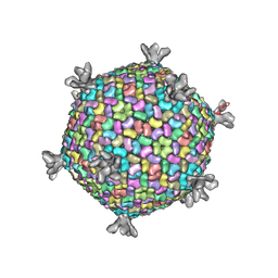
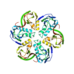
6H82
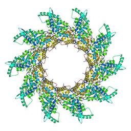
 | | Cryo-EM structure of the archaeal extremophilic internal membrane containing Haloarcula hispanica icosahedral virus 2 (HHIV-2) at 3.78 Angstroms resolution. | | 分子名称: | GPS III, Uncharacterized protein, VP16 (vertex complex), ... | | 著者 | Abrescia, N.G, Santos-Perez, I, Charro, D. | | 登録日 | 2018-08-01 | | 公開日 | 2019-04-03 | | 最終更新日 | 2024-05-15 | | 実験手法 | ELECTRON MICROSCOPY (3.78 Å) | | 主引用文献 | Structural basis for assembly of vertical single beta-barrel viruses.
Nat Commun, 10, 2019
|
|
6H9C
 | | Cryo-EM structure of archaeal extremophilic internal membrane-containing Haloarcula californiae icosahedral virus 1 (HCIV-1) at 3.74 Angstroms resolution. | | 分子名称: | (Half) GPS-II protein located underneath the two-tower capsomer sitting ON the icosahedral 2-fold axis., GPS-II protein located underneath the two-tower capsomer NOT sitting on the icosahedral 2-fold axis., GPS-III molecule located underneath the capsomer close to the icosahedral three-fold axis., ... | | 著者 | Abrescia, N.G, Santos-Perez, I, Charro, D. | | 登録日 | 2018-08-03 | | 公開日 | 2019-03-27 | | 最終更新日 | 2024-07-10 | | 実験手法 | ELECTRON MICROSCOPY (3.74 Å) | | 主引用文献 | Structural basis for assembly of vertical single beta-barrel viruses.
Nat Commun, 10, 2019
|
|
1W8X
 | | Structural analysis of PRD1 | | 分子名称: | MAJOR CAPSID PROTEIN (PROTEIN P3), PROTEIN P16, PROTEIN P30, ... | | 著者 | Abrescia, N.G.A, Cockburn, J.J.B, Grimes, J.M, Sutton, G.C, Diprose, J.M, Butcher, S.J, Fuller, S.D, San Martin, C, Burnett, R.M, Stuart, D.I, Bamford, D.H, Bamford, J.K.H. | | 登録日 | 2004-10-01 | | 公開日 | 2004-11-11 | | 最終更新日 | 2024-05-08 | | 実験手法 | X-RAY DIFFRACTION (4.2 Å) | | 主引用文献 | Insights Into Assembly from Structural Analysis of Bacteriophage Prd1.
Nature, 432, 2004
|
|
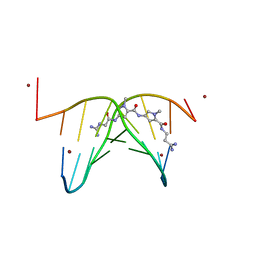
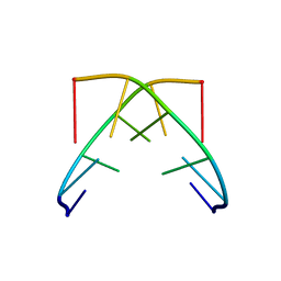
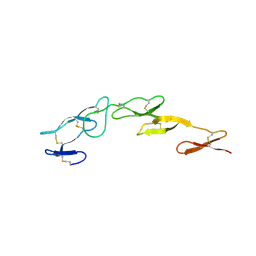
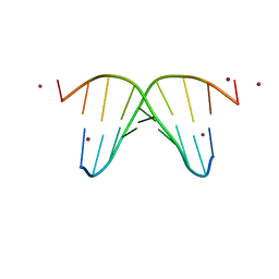
446D
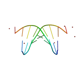 | | STRUCTURE OF THE OLIGONUCLEOTIDE D(CGTATATACG) AS A SITE SPECIFIC COMPLEX WITH NICKEL IONS | | 分子名称: | DNA (5'-D(*CP*GP*TP*AP*TP*AP*TP*AP*CP*G)-3'), NICKEL (II) ION | | 著者 | Abrescia, N.G.A, Malinina, L, Gonzaga, L.F, Huynh-Dinh, T, Neidle, S, Subirana, J.A. | | 登録日 | 1999-01-15 | | 公開日 | 1999-03-18 | | 最終更新日 | 2024-02-28 | | 実験手法 | X-RAY DIFFRACTION (3 Å) | | 主引用文献 | Structure of the oligonucleotide d(CGTATATACG) as a site-specific complex with nickel ions.
Nucleic Acids Res., 27, 1999
|
|
473D
 | |
2W0C
 | | X-ray structure of the entire lipid-containing bacteriophage PM2 | | 分子名称: | CALCIUM ION, MAJOR CAPSID PROTEIN P2, PROTEIN 2, ... | | 著者 | Abrescia, N.G.A, Grimes, J.M, Kivela, H.M, Assenberg, R, Sutton, G.C, Butcher, S.J, Bamford, J.K.H, Bamford, D.H, Stuart, D.I. | | 登録日 | 2008-08-13 | | 公開日 | 2008-09-23 | | 最終更新日 | 2024-05-08 | | 実験手法 | X-RAY DIFFRACTION (7 Å) | | 主引用文献 | Insights Into Virus Evolution and Membrane Biogenesis from the Structure of the Marine Lipid-Containing Bacteriophage Pm2
Mol.Cell, 31, 2008
|
|
1GQU
 | |
2VVD
 | | Crystal structure of the receptor binding domain of the spike protein P1 from bacteriophage PM2 | | 分子名称: | CALCIUM ION, SPIKE PROTEIN P1 | | 著者 | Abrescia, N.G.A, Grimes, J.M, Kivela, H.K, Assenberg, R, Sutton, G.C, Butcher, S.J, Bamford, J.K.H, Bamford, D.H, Stuart, D.I. | | 登録日 | 2008-06-06 | | 公開日 | 2008-09-16 | | 最終更新日 | 2011-07-13 | | 実験手法 | X-RAY DIFFRACTION (2.26 Å) | | 主引用文献 | Insights Into Virus Evolution and Membrane Biogenesis from the Structure of the Marine Lipid-Containing Bacteriophage Pm2.
Mol.Cell, 31, 2008
|
|
2VVE
 | | Crystal structure of the stem and receptor binding domain of the spike protein P1 from bacteriophage PM2 | | 分子名称: | CALCIUM ION, CHLORIDE ION, SPIKE PROTEIN P1 | | 著者 | Abrescia, N.G.A, Grimes, J.M, Kivela, H.K, Assenberg, R, Sutton, G.C, Butcher, S.J, Bamford, J.K.H, Bamford, D.H, Stuart, D.I. | | 登録日 | 2008-06-06 | | 公開日 | 2008-09-16 | | 最終更新日 | 2023-12-13 | | 実験手法 | X-RAY DIFFRACTION (1.77 Å) | | 主引用文献 | Insights Into Virus Evolution and Membrane Biogenesis from the Structure of the Marine Lipid-Containing Bacteriophage Pm2.
Mol.Cell, 31, 2008
|
|
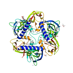
2VVF
 | | Crystal structure of the major capsid protein P2 from Bacteriophage PM2 | | 分子名称: | CALCIUM ION, MAJOR CAPSID PROTEIN P2 | | 著者 | Abrescia, N.G.A, Grimes, J.M, Kivela, H.K, Assenberg, R, Sutton, G.C, Butcher, S.J, Bamford, J.K.H, Bamford, D.H, Stuart, D.I. | | 登録日 | 2008-06-06 | | 公開日 | 2008-09-16 | | 最終更新日 | 2024-05-08 | | 実験手法 | X-RAY DIFFRACTION (2.5 Å) | | 主引用文献 | Insights Into Virus Evolution and Membrane Biogenesis from the Structure of the Marine Lipid-Containing Bacteriophage Pm2.
Mol.Cell, 31, 2008
|
|
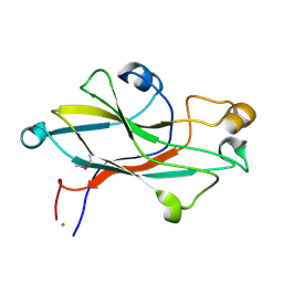
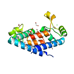
5M3D
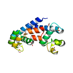 | | Structural tuning of CD81LEL (space group P31) | | 分子名称: | 1,2-ETHANEDIOL, CD81 antigen, PHOSPHATE ION | | 著者 | Cunha, E.S, Sfriso, P, Rojas, A.L, Roversi, P, Hospital, A, Orozco, M, Abrescia, N.G. | | 登録日 | 2016-10-14 | | 公開日 | 2016-12-14 | | 最終更新日 | 2024-01-17 | | 実験手法 | X-RAY DIFFRACTION (2.38 Å) | | 主引用文献 | Mechanism of Structural Tuning of the Hepatitis C Virus Human Cellular Receptor CD81 Large Extracellular Loop.
Structure, 25, 2017
|
|
5M2C
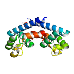 | | Structural tuning of CD81LEL (space group P32 1 2) | | 分子名称: | CD81 antigen, PHOSPHATE ION | | 著者 | Cunha, E.S, Sfriso, P, Rojas, A.L, Roversi, P, Hospital, A, Orozco, M, Abrescia, N.G. | | 登録日 | 2016-10-12 | | 公開日 | 2016-12-14 | | 最終更新日 | 2024-01-17 | | 実験手法 | X-RAY DIFFRACTION (1.961 Å) | | 主引用文献 | Mechanism of Structural Tuning of the Hepatitis C Virus Human Cellular Receptor CD81 Large Extracellular Loop.
Structure, 25, 2017
|
|
5M33
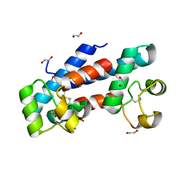 | | Structural tuning of CD81LEL (space group P21) | | 分子名称: | 1,2-ETHANEDIOL, CD81 antigen | | 著者 | Cunha, E.S, Sfriso, P, Rojas, A.L, Roversi, P, Hospital, A, Orozco, M, Abrescia, N.G. | | 登録日 | 2016-10-14 | | 公開日 | 2016-12-14 | | 最終更新日 | 2024-01-17 | | 実験手法 | X-RAY DIFFRACTION (1.28 Å) | | 主引用文献 | Mechanism of Structural Tuning of the Hepatitis C Virus Human Cellular Receptor CD81 Large Extracellular Loop.
Structure, 25, 2017
|
|
5M3T
 | | Structural tuning of CD81LEL (space group P64) | | 分子名称: | 1,2-ETHANEDIOL, CD81 antigen, CHLORIDE ION | | 著者 | Cunha, E.S, Sfriso, P, Rojas, A.L, Roversi, P, Hospital, A, Orozco, M, Abrescia, N.G. | | 登録日 | 2016-10-17 | | 公開日 | 2016-12-14 | | 最終更新日 | 2024-01-17 | | 実験手法 | X-RAY DIFFRACTION (2.021 Å) | | 主引用文献 | Mechanism of Structural Tuning of the Hepatitis C Virus Human Cellular Receptor CD81 Large Extracellular Loop.
Structure, 25, 2017
|
|
5M4R
 | | Structural tuning of CD81LEL (space group C2) | | 分子名称: | 1,2-ETHANEDIOL, CD81 antigen, SULFATE ION | | 著者 | Cunha, E.S, Sfriso, P, Rojas, A.L, Roversi, P, Hospital, A, Orozco, M, Abrescia, N.G. | | 登録日 | 2016-10-19 | | 公開日 | 2016-12-14 | | 最終更新日 | 2024-01-17 | | 実験手法 | X-RAY DIFFRACTION (3.1 Å) | | 主引用文献 | Mechanism of Structural Tuning of the Hepatitis C Virus Human Cellular Receptor CD81 Large Extracellular Loop.
Structure, 25, 2017
|
|
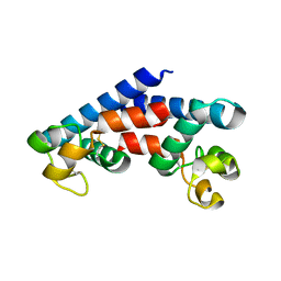
2UWI
 | | Structure of CrmE, a poxvirus TNF receptor | | 分子名称: | CRME PROTEIN | | 著者 | Graham, S.C, Bahar, M.W, Abrescia, N.G, Smith, G.L, Stuart, D.I, Grimes, J.M. | | 登録日 | 2007-03-22 | | 公開日 | 2007-07-10 | | 最終更新日 | 2019-05-15 | | 実験手法 | X-RAY DIFFRACTION (2 Å) | | 主引用文献 | Structure of Crme, a Virus-Encoded Tumour Necrosis Factor Receptor.
J.Mol.Biol., 372, 2007
|
|
7OOK
 | | Bacteriophage PRD1 Major Capsid Protein P3 in complex with CPZ | | 分子名称: | (4S)-2-METHYL-2,4-PENTANEDIOL, 3-(2-chloro-10H-phenothiazin-10-yl)-N,N-dimethylpropan-1-amine, CHLORIDE ION, ... | | 著者 | Duyvesteyn, H.M.E, Peccati, F, Martinez-Castillo, A, Jimenez-Oses, G, Oksanen, H.M, Stuart, D.I, Abrescia, N.G.A. | | 登録日 | 2021-05-27 | | 公開日 | 2022-06-08 | | 最終更新日 | 2024-01-31 | | 実験手法 | X-RAY DIFFRACTION (2.23 Å) | | 主引用文献 | Bacteriophage PRD1 as a nanoscaffold for drug loading
Nanoscale, 13, 2021
|
|
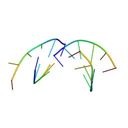
4V8S
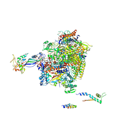
 | | Archaeal RNAP-DNA binary complex at 4.32Ang | | 分子名称: | 5'-D(*AP*TP*AP*GP*AP*GP*TP*AP*TP*AP*AP*GP*AP*TP *AP*G)-3', 5'-D(*TP*CP*TP*TP*AP*TP*AP*CP*TP*CP*TP*AP*TP*CP)-3', DNA-DIRECTED RNA POLYMERASE, ... | | 著者 | Wojtas, M.N, Mogni, M, Millet, O, Bell, S.D, Abrescia, N.G.A. | | 登録日 | 2012-07-12 | | 公開日 | 2014-07-09 | | 最終更新日 | 2024-01-10 | | 実験手法 | X-RAY DIFFRACTION (4.323 Å) | | 主引用文献 | Structural and Functional Analyses of the Interaction of Archaeal RNA Polymerase with DNA.
Nucleic Acids Res., 40, 2012
|
|
4V5V
 | | Structure of respiratory syncytial virus nucleocapsid protein, P1 crystal form | | 分子名称: | RESPIRATORY SYNCYTIAL VIRUS NUCLEOCAPSID PROTEIN, RNA | | 著者 | El Omari, K, Dhaliwal, B, Ren, J, Abrescia, N.G.A, Lockyer, M, Powell, K.L, Hawkins, A.R, Stammers, D.K. | | 登録日 | 2011-05-04 | | 公開日 | 2014-07-09 | | 最終更新日 | 2024-01-10 | | 実験手法 | X-RAY DIFFRACTION (3.6 Å) | | 主引用文献 | Structures of Respiratory Syncytial Virus Nucleocapsid Protein from Two Crystal Forms: Details of Potential Packing Interactions in the Native Helical Form.
Acta Crystallogr.,Sect.F, 67, 2011
|
|
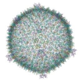
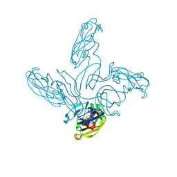
8A9U
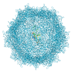 | | Full AAV3B-VP1KO virion | | 分子名称: | Capsid protein VP1 | | 著者 | Arriaga, I, Abrescia, N.G.A. | | 登録日 | 2022-06-29 | | 公開日 | 2022-09-21 | | 最終更新日 | 2022-11-23 | | 実験手法 | ELECTRON MICROSCOPY (3.1 Å) | | 主引用文献 | Cellular and Structural Characterization of VP1 and VP2 Knockout Mutants of AAV3B Serotype and Implications for AAV Manufacturing.
Hum Gene Ther, 33, 2022
|
|
3DUZ
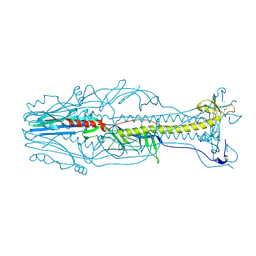 | | Crystal structure of the postfusion form of baculovirus fusion protein GP64 | | 分子名称: | MERCURY (II) ION, Major envelope glycoprotein | | 著者 | Kadlec, J, Loureiro, S, Abrescia, N.G.A, Jones, I.M, Stuart, D.I. | | 登録日 | 2008-07-18 | | 公開日 | 2008-09-16 | | 最終更新日 | 2011-07-13 | | 実験手法 | X-RAY DIFFRACTION (2.95 Å) | | 主引用文献 | The postfusion structure of baculovirus gp64 supports a unified view of viral fusion machines.
Nat.Struct.Mol.Biol., 15, 2008
|
|
4AYB
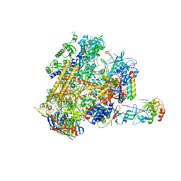
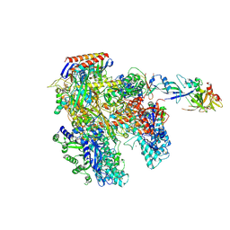 | | RNAP at 3.2Ang | | 分子名称: | DNA-DIRECTED RNA POLYMERASE, IRON/SULFUR CLUSTER, MAGNESIUM ION, ... | | 著者 | Wojtas, M.N, Mogni, M, Millet, O, Bell, S.D, Abrescia, N.G.A. | | 登録日 | 2012-06-19 | | 公開日 | 2012-08-08 | | 最終更新日 | 2023-12-20 | | 実験手法 | X-RAY DIFFRACTION (3.202 Å) | | 主引用文献 | Structural and Functional Analyses of the Interaction of Archaeal RNA Polymerase with DNA.
Nucleic Acids Res., 40, 2012
|
|
1G3V
 | |
2Y0S
 | | Crystal structure of Sulfolobus shibatae RNA polymerase in P21 space group | | 分子名称: | DNA-DIRECTED RNA POLYMERASE, DNA-DIRECTED RNA POLYMERASE SUBUNIT A'', DNA-DIRECTED RNA POLYMERASE SUBUNIT D, ... | | 著者 | Wojtas, M, Peralta, B, Ondiviela, M, Mogni, M, Bell, S.D, Abrescia, N.G.A. | | 登録日 | 2010-12-07 | | 公開日 | 2011-02-16 | | 最終更新日 | 2023-12-20 | | 実験手法 | X-RAY DIFFRACTION (3.8 Å) | | 主引用文献 | Archaeal RNA polymerase: the influence of the protruding stalk in crystal packing and preliminary biophysical analysis of the Rpo13 subunit.
Biochem. Soc. Trans., 39, 2011
|
|


