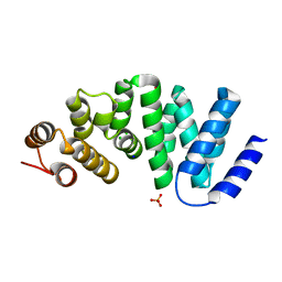
7YCN
 
 | |
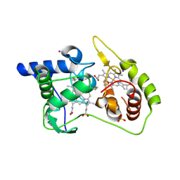
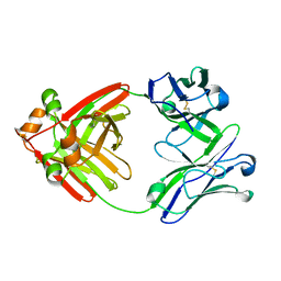
5KXJ
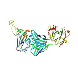
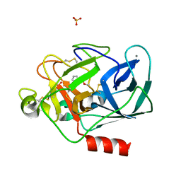
 | | Crystal Structure of L-Aspartate Oxidase from Salmonella typhimurium in the Complex with Substrate L-Aspartate | | 分子名称: | 1,2-ETHANEDIOL, ASPARTIC ACID, GLYCEROL, ... | | 著者 | Kim, Y, Osipiuk, J, Mulligan, R, Makowska-Grzyska, M, Maltseva, N, Shatsman, S, Gu, M, Anderson, W.F, Joachimiak, A, Center for Structural Genomics of Infectious Diseases (CSGID) | | 登録日 | 2016-07-20 | | 公開日 | 2016-08-03 | | 最終更新日 | 2024-03-06 | | 実験手法 | X-RAY DIFFRACTION (1.87 Å) | | 主引用文献 | Crystal Structure of L-Aspartate Oxidase from Salmonella typhimurium in the Complex with Substrate L-Aspartate
To Be Published
|
|
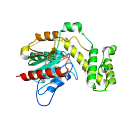
5IQT
 | | WelO5 bound to Fe(II), Cl, 2-oxoglutarate, and 12-epifischerindole U | | 分子名称: | (6aS,9R,10R,10aS)-9-ethyl-10-isocyano-6,6,9-trimethyl-5,6,6a,7,8,9,10,10a-octahydroindeno[2,1-b]indole, 2-OXOGLUTARIC ACID, CHLORIDE ION, ... | | 著者 | Mitchell, A.J, Boal, A.K. | | 登録日 | 2016-03-11 | | 公開日 | 2016-06-29 | | 最終更新日 | 2023-09-27 | | 実験手法 | X-RAY DIFFRACTION (2.4 Å) | | 主引用文献 | Structural basis for halogenation by iron- and 2-oxo-glutarate-dependent enzyme WelO5.
Nat.Chem.Biol., 12, 2016
|
|
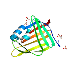
7RMY
 | | De Novo designed tunable protein pockets, D_3-337 | | 分子名称: | De Novo designed tunable homodimer, D_3-337 | | 著者 | Bera, A.K, Hicks, D.R, Kang, A, Sankaran, B, Baker, D. | | 登録日 | 2021-07-28 | | 公開日 | 2022-08-03 | | 最終更新日 | 2024-04-03 | | 実験手法 | X-RAY DIFFRACTION (3.17 Å) | | 主引用文献 | De novo design of protein homodimers containing tunable symmetric protein pockets.
Proc.Natl.Acad.Sci.USA, 119, 2022
|
|
6UGE
 | |
6MFQ
 | | Crystal structure of a PMS2 variant | | 分子名称: | Mismatch repair endonuclease PMS2 | | 著者 | D'Arcy, B.M, Prakash, A. | | 登録日 | 2018-09-11 | | 公開日 | 2019-02-06 | | 最終更新日 | 2023-10-11 | | 実験手法 | X-RAY DIFFRACTION (2.6 Å) | | 主引用文献 | Biochemical and structural characterization of two variants of uncertain significance in the PMS2 gene.
Hum. Mutat., 40, 2019
|
|
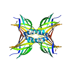
7RMX
 | | Structure of De Novo designed tunable symmetric protein pockets | | 分子名称: | Tunable symmetric protein, D_3_212 | | 著者 | Bera, A.K, Hicks, D.R, Kang, A, Sankaran, B, Baker, D. | | 登録日 | 2021-07-28 | | 公開日 | 2022-08-03 | | 最終更新日 | 2024-04-03 | | 実験手法 | X-RAY DIFFRACTION (1.65 Å) | | 主引用文献 | De novo design of protein homodimers containing tunable symmetric protein pockets.
Proc.Natl.Acad.Sci.USA, 119, 2022
|
|
6QQY
 | | Crystal structure of TrmD, a tRNA-(N1G37) methyltransferase, from Mycobacterium abscessus in complex with inhibitor | | 分子名称: | 5-azanyl-3-[1-(pyridin-2-ylmethyl)indol-6-yl]-1~{H}-pyrazole-4-carbonitrile, tRNA (guanine-N(1)-)-methyltransferase | | 著者 | Thomas, S.E, Whitehouse, A.J, Coyne, A.G, Abell, C, Mendes, V, Blundell, T.L. | | 登録日 | 2019-02-19 | | 公開日 | 2020-03-18 | | 最終更新日 | 2024-01-24 | | 実験手法 | X-RAY DIFFRACTION (1.49 Å) | | 主引用文献 | Fragment-based discovery of a new class of inhibitors targeting mycobacterial tRNA modification.
Nucleic Acids Res., 48, 2020
|
|
6OT6
 | | Rat ERK2 D319N | | 分子名称: | Mitogen-activated protein kinase 1, SULFATE ION | | 著者 | Taylor, C.A, Goldsmith, E.J, Cobb, M.H. | | 登録日 | 2019-05-02 | | 公開日 | 2019-07-10 | | 最終更新日 | 2023-10-11 | | 実験手法 | X-RAY DIFFRACTION (1.65 Å) | | 主引用文献 | Functional divergence caused by mutations in an energetic hotspot in ERK2.
Proc.Natl.Acad.Sci.USA, 116, 2019
|
|
7RK7
 | | The complex between TIL 1383i TCR and human Class I MHC HLA-A2 with the bound Tyrosinase(369-377)(N371D) nonameric peptide | | 分子名称: | Beta-2-microglobulin, HLA class I histocompatibility antigen, A alpha chain, ... | | 著者 | Singh, N.K, Davancaze, L.M, Arbuiso, A, Weiss, L.I, Keller, G.L.J, Baker, B.M. | | 登録日 | 2021-07-22 | | 公開日 | 2022-11-02 | | 最終更新日 | 2023-10-18 | | 実験手法 | X-RAY DIFFRACTION (2.54 Å) | | 主引用文献 | The complex between TIL 1383i TCR and human Class I MHC HLA-A2 with the bound Tyrosinase(369-377)(N371D) nonameric peptide
To Be Published
|
|
6QQQ
 | | Crystal structure of TrmD, a tRNA-(N1G37) methyltransferase, from Mycobacterium abscessus in complex with inhibitor | | 分子名称: | (3-methoxy-5-pyridin-3-yl-phenyl)methyl 1~{H}-pyrazole-4-carboxylate, SULFATE ION, tRNA (guanine-N(1)-)-methyltransferase | | 著者 | Thomas, S.E, Whitehouse, A.J, Coyne, A.G, Abell, C, Mendes, V, Blundell, T.L. | | 登録日 | 2019-02-19 | | 公開日 | 2019-09-18 | | 最終更新日 | 2024-01-24 | | 実験手法 | X-RAY DIFFRACTION (1.85 Å) | | 主引用文献 | Development of Inhibitors againstMycobacterium abscessustRNA (m1G37) Methyltransferase (TrmD) Using Fragment-Based Approaches.
J.Med.Chem., 62, 2019
|
|
6QQU
 | | Crystal structure of TrmD, a tRNA-(N1G37) methyltransferase, from Mycobacterium abscessus in complex with inhibitor | | 分子名称: | 5-azanyl-3-(1~{H}-indol-6-yl)-1~{H}-pyrazole-4-carbonitrile, tRNA (guanine-N(1)-)-methyltransferase | | 著者 | Thomas, S.E, Whitehouse, A.J, Coyne, A.G, Abell, C, Mendes, V, Blundell, T.L. | | 登録日 | 2019-02-19 | | 公開日 | 2019-09-18 | | 最終更新日 | 2024-01-24 | | 実験手法 | X-RAY DIFFRACTION (1.59 Å) | | 主引用文献 | Development of Inhibitors againstMycobacterium abscessustRNA (m1G37) Methyltransferase (TrmD) Using Fragment-Based Approaches.
J.Med.Chem., 62, 2019
|
|
4WQE
 | | Thiosulfate dehydrogenase (TsdA) from Allochromatium vinosum - K208G mutant | | 分子名称: | HEME C, IODIDE ION, SULFATE ION, ... | | 著者 | Brito, J.A, Denkmann, K, Pereira, I.A.C, Dahl, C, Archer, M. | | 登録日 | 2014-10-21 | | 公開日 | 2015-02-18 | | 最終更新日 | 2015-04-15 | | 実験手法 | X-RAY DIFFRACTION (1.4 Å) | | 主引用文献 | Thiosulfate Dehydrogenase (TsdA) from Allochromatium vinosum: STRUCTURAL AND FUNCTIONAL INSIGHTS INTO THIOSULFATE OXIDATION.
J.Biol.Chem., 290, 2015
|
|
4WQ7
 | | Thiosulfate dehydrogenase (TsdA) from Allochromatium vinosum - "as isolated" form | | 分子名称: | HEME C, IODIDE ION, SULFATE ION, ... | | 著者 | Brito, J.A, Denkmann, K, Pereira, I.A.C, Dahl, C, Archer, M. | | 登録日 | 2014-10-21 | | 公開日 | 2015-02-18 | | 最終更新日 | 2015-04-15 | | 実験手法 | X-RAY DIFFRACTION (1.98 Å) | | 主引用文献 | Thiosulfate Dehydrogenase (TsdA) from Allochromatium vinosum: STRUCTURAL AND FUNCTIONAL INSIGHTS INTO THIOSULFATE OXIDATION.
J.Biol.Chem., 290, 2015
|
|
6E5W
 | | Crystal structure of human cellular retinol binding protein 3 in complex with abnormal-cannabidiol (abn-CBD) | | 分子名称: | (1'R,2'R)-5'-methyl-6-pentyl-2'-(prop-1-en-2-yl)-1',2',3',4'-tetrahydro[1,1'-biphenyl]-2,4-diol, GLYCEROL, Retinol-binding protein 5 | | 著者 | Silvaroli, J.A, Banerjee, S, Kiser, P.D, Golczak, M. | | 登録日 | 2018-07-23 | | 公開日 | 2019-02-13 | | 最終更新日 | 2023-10-11 | | 実験手法 | X-RAY DIFFRACTION (2.5 Å) | | 主引用文献 | Abnormal Cannabidiol Modulates Vitamin A Metabolism by Acting as a Competitive Inhibitor of CRBP1.
Acs Chem.Biol., 14, 2019
|
|
7R0X
 | |
7OXW
 | | CrabP2 mutant R30DK31D | | 分子名称: | ACETATE ION, Cellular retinoic acid-binding protein 2, SULFATE ION | | 著者 | Pastok, M.W, Basle, A, Endicott, J.A. | | 登録日 | 2021-06-23 | | 公開日 | 2022-07-13 | | 最終更新日 | 2024-01-31 | | 実験手法 | X-RAY DIFFRACTION (1.16 Å) | | 主引用文献 | Structural requirements for the specific binding of CRABP2 to cyclin D3
To Be Published
|
|
6QTI
 | |
8B49
 | | STRUCTURE OF PORCINE PANCREATIC ELASTASE BOUND TO A FRAGMENT (m-toluoylcarbonyl group) OF A 5-AZAINDOLE INHIBITOR | | 分子名称: | 1-(3-methylphenyl)carbonylpyrrolo[3,2-c]pyridine-3-carbonitrile, CALCIUM ION, Chymotrypsin-like elastase family member 1, ... | | 著者 | Ferraroni, M, Giovannoni, P, Gerace, A. | | 登録日 | 2022-09-20 | | 公開日 | 2023-09-20 | | 実験手法 | X-RAY DIFFRACTION (1.19 Å) | | 主引用文献 | X-ray structural study of human neutrophil elastase inhibition with a series of azaindoles, azaindazoles and isoxazolones
J.Mol.Struct., 1274, 2023
|
|
7OXX
 | | CrabP2 mutant R30AK31A | | 分子名称: | Cellular retinoic acid-binding protein 2, SODIUM ION | | 著者 | Tomlinson, C.W.E, Basle, A, Pohl, E. | | 登録日 | 2021-06-23 | | 公開日 | 2022-07-13 | | 最終更新日 | 2024-01-31 | | 実験手法 | X-RAY DIFFRACTION (1.33 Å) | | 主引用文献 | Structural requirements for the specific binding of CRABP2 to cyclin D3
To Be Published
|
|
4X89
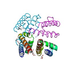 | | NavMs voltage-gated sodium channal pore and C-terminal domain soaked with Silver nitrate | | 分子名称: | HEGA-10, Ion transport protein, PENTAETHYLENE GLYCOL, ... | | 著者 | Naylor, C.E, Bagneris, C, Wallace, B.A. | | 登録日 | 2014-12-10 | | 公開日 | 2016-03-09 | | 最終更新日 | 2024-01-10 | | 実験手法 | X-RAY DIFFRACTION (2.62 Å) | | 主引用文献 | Molecular basis of ion permeability in a voltage-gated sodium channel.
Embo J., 35, 2016
|
|
6MEH
 | |
4X8Q
 | | X-ray crystal structure of AlkD2 from Streptococcus mutans | | 分子名称: | CHLORIDE ION, GLYCEROL, PHOSPHATE ION, ... | | 著者 | Mullins, E.A, Shi, R, Eichman, B.F. | | 登録日 | 2014-12-10 | | 公開日 | 2015-05-27 | | 最終更新日 | 2023-09-27 | | 実験手法 | X-RAY DIFFRACTION (1.729 Å) | | 主引用文献 | A New Family of HEAT-Like Repeat Proteins Lacking a Critical Substrate Recognition Motif Present in Related DNA Glycosylases.
Plos One, 10, 2015
|
|
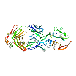
6LS9
 | | Crystal structure of bovine herpesvirus 1 glycoprotein D | | 分子名称: | 2-acetamido-2-deoxy-beta-D-glucopyranose, 2-acetamido-2-deoxy-beta-D-glucopyranose-(1-4)-2-acetamido-2-deoxy-beta-D-glucopyranose-(1-4)-2-acetamido-2-deoxy-beta-D-glucopyranose, Envelope glycoprotein D | | 著者 | Yue, D, Chen, Z.J, Yang, F.L, Ye, F, Lin, S, Cheng, Y.W, Wang, J.C, Chen, Z.M, Lin, X, Yang, J, Chen, H, Zhang, Z.L, You, Y, Sun, H.L, Wen, A, Wang, L.L, Zheng, Y, Cao, Y, Li, Y.H, Lu, G.W. | | 登録日 | 2020-01-17 | | 公開日 | 2020-06-17 | | 最終更新日 | 2023-11-29 | | 実験手法 | X-RAY DIFFRACTION (2.503 Å) | | 主引用文献 | Crystal structure of bovine herpesvirus 1 glycoprotein D bound to nectin-1 reveals the basis for its low-affinity binding to the receptor.
Sci Adv, 6, 2020
|
|
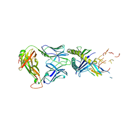
6MG4
 | | Structure of full-length human lambda-6A light chain JTO | | 分子名称: | JTO light chain | | 著者 | Morgan, G.J, Yan, N.L, Mortenson, D.E, Stanfield, R.L, Wilson, I.A, Kelly, J.W. | | 登録日 | 2018-09-12 | | 公開日 | 2019-04-10 | | 最終更新日 | 2023-10-11 | | 実験手法 | X-RAY DIFFRACTION (1.75 Å) | | 主引用文献 | Stabilization of amyloidogenic immunoglobulin light chains by small molecules.
Proc.Natl.Acad.Sci.USA, 116, 2019
|
|


