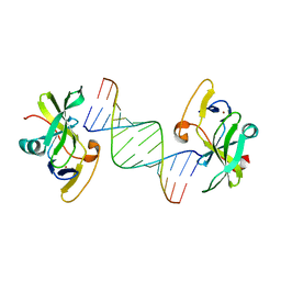
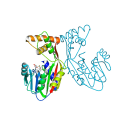
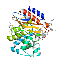
4DUM
 
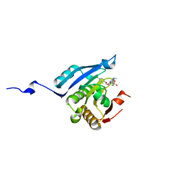 | | Co-crystal structure of eIF4E with inhibitor | | 分子名称: | (4-{7-[2-(4-chlorophenoxy)ethyl]-2-(methylamino)-6-oxo-6,7-dihydro-1H-purin-8-yl}phenyl)phosphonic acid, 1,2-ETHANEDIOL, Eukaryotic translation initiation factor 4E | | 著者 | Min, X, Johnstone, S, Walker, N, Wang, Z. | | 登録日 | 2012-02-22 | | 公開日 | 2012-04-11 | | 最終更新日 | 2024-02-28 | | 実験手法 | X-RAY DIFFRACTION (2.95 Å) | | 主引用文献 | Structure-Guided Design, Synthesis, and Evaluation of Guanine-Derived Inhibitors of the eIF4E mRNA-Cap Interaction.
J.Med.Chem., 55, 2012
|
|
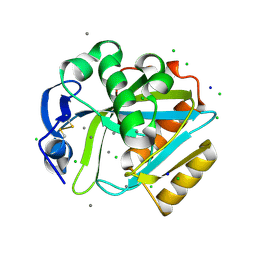
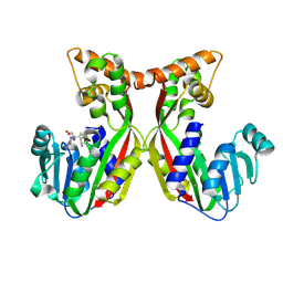
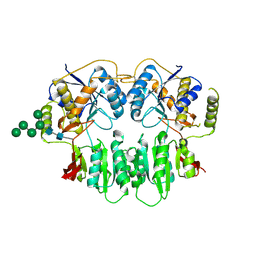
3K8S
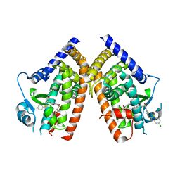 | | Crystal Structure of PPARg in complex with T2384 | | 分子名称: | 2-chloro-N-{3-chloro-4-[(5-chloro-1,3-benzothiazol-2-yl)sulfanyl]phenyl}-4-(trifluoromethyl)benzenesulfonamide, Peroxisome proliferator-activated receptor gamma | | 著者 | Wang, Z. | | 登録日 | 2009-10-14 | | 公開日 | 2009-11-03 | | 最終更新日 | 2023-09-06 | | 実験手法 | X-RAY DIFFRACTION (2.55 Å) | | 主引用文献 | T2384, a novel antidiabetic agent with unique peroxisome proliferator-activated receptor gamma binding properties
J.Biol.Chem., 283, 2008
|
|
7V2S
 | | Crystal structure of juvenile hormone acid methyltransferase JHAMT isoform3 from silkworm | | 分子名称: | Methyltranfer_dom domain-containing protein | | 著者 | Guo, P.C, Zhang, Y.S, Zhang, l, Xu, H.Y. | | 登録日 | 2021-08-09 | | 公開日 | 2022-08-10 | | 最終更新日 | 2023-11-15 | | 実験手法 | X-RAY DIFFRACTION (2.133 Å) | | 主引用文献 | Structural characterization and functional analysis of juvenile hormone acid methyltransferase JHAMT3 from the silkworm, Bombyx mori.
Insect Biochem.Mol.Biol., 151, 2022
|
|
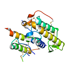
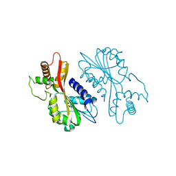
4DT6
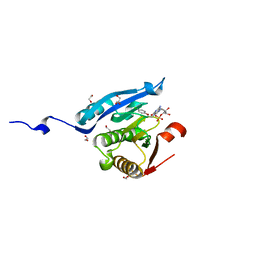 | | Co-crystal structure of eIF4E with inhibitor | | 分子名称: | 1,2-ETHANEDIOL, 7-[2-(4-chlorophenoxy)ethyl]guanosine 5'-(dihydrogen phosphate), Eukaryotic translation initiation factor 4E, ... | | 著者 | Min, X, Johnstone, S, Walker, N, Wang, Z. | | 登録日 | 2012-02-20 | | 公開日 | 2012-04-11 | | 最終更新日 | 2024-02-28 | | 実験手法 | X-RAY DIFFRACTION (2.6 Å) | | 主引用文献 | Structure-Guided Design, Synthesis, and Evaluation of Guanine-Derived Inhibitors of the eIF4E mRNA-Cap Interaction.
J.Med.Chem., 55, 2012
|
|
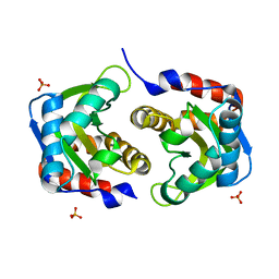
2DHO
 | | Crystal structure of human IPP isomerase I in space group P212121 | | 分子名称: | CHLORIDE ION, Isopentenyl-diphosphate delta-isomerase 1, MANGANESE (II) ION, ... | | 著者 | Zhang, C, Wei, Z, Gong, W. | | 登録日 | 2006-03-24 | | 公開日 | 2007-06-05 | | 最終更新日 | 2021-11-10 | | 実験手法 | X-RAY DIFFRACTION (1.6 Å) | | 主引用文献 | Crystal structures of human IPP isomerase: new insights into the catalytic mechanism
J.Mol.Biol., 366, 2007
|
|
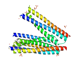
3LRR
 | |
3LEF
 | | Crystal structure of HIV epitope-scaffold 4E10_S0_1Z6NA_001 | | 分子名称: | 1,2-ETHANEDIOL, Uncharacterized protein 4E10_S0_1Z6NA_001 (T18) | | 著者 | Holmes, M.A. | | 登録日 | 2010-01-14 | | 公開日 | 2010-09-22 | | 最終更新日 | 2023-09-06 | | 実験手法 | X-RAY DIFFRACTION (2.3 Å) | | 主引用文献 | Computational Design of Epitope-Scaffolds Allows Induction of Antibodies Specific for a Poorly Immunogenic HIV Vaccine Epitope.
Structure, 18, 2010
|
|
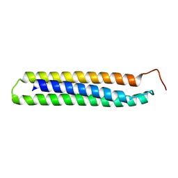
3LF6
 | |
3LG7
 | |
3LHP
 | |
3LF9
 | |
3LH2
 | |
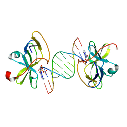
3LRN
 | | Crystal structure of human RIG-I CTD bound to a 14 bp GC 5' ppp dsRNA | | 分子名称: | Probable ATP-dependent RNA helicase DDX58, RNA (5'-R(*(GTP)P*GP*CP*GP*CP*GP*CP*GP*CP*GP*CP*GP*CP*C)-3'), ZINC ION | | 著者 | Li, P. | | 登録日 | 2010-02-11 | | 公開日 | 2010-06-02 | | 最終更新日 | 2024-04-03 | | 実験手法 | X-RAY DIFFRACTION (2.6 Å) | | 主引用文献 | The Structural Basis of 5' Triphosphate Double-Stranded RNA Recognition by RIG-I C-Terminal Domain.
Structure, 18, 2010
|
|
7VEO
 | | Crystal structure of juvenile hormone acid methyltransferase silkworm JHAMT isoform3 complex with S-Adenosyl-L-homocysteine | | 分子名称: | Methyltranfer_dom domain-containing protein, S-ADENOSYL-L-HOMOCYSTEINE | | 著者 | Guo, P.C, Zhang, Y.S, Zhang, l, Xu, H.Y. | | 登録日 | 2021-09-09 | | 公開日 | 2022-09-28 | | 最終更新日 | 2023-11-29 | | 実験手法 | X-RAY DIFFRACTION (2.53 Å) | | 主引用文献 | Structural characterization and functional analysis of juvenile hormone acid methyltransferase JHAMT3 from the silkworm, Bombyx mori.
Insect Biochem.Mol.Biol., 151, 2022
|
|
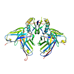
3R0L
 | | Crystal structure of crotoxin | | 分子名称: | ACETATE ION, CHLORIDE ION, Crotoxin chain A, ... | | 著者 | Saul, F.A, Faure, G. | | 登録日 | 2011-03-08 | | 公開日 | 2011-10-12 | | 最終更新日 | 2023-09-13 | | 実験手法 | X-RAY DIFFRACTION (1.35 Å) | | 主引用文献 | Crystal Structure of Crotoxin Reveals Key Residues Involved in the Stability and Toxicity of This Potent Heterodimeric Beta-Neurotoxin
J.Mol.Biol., 412, 2011
|
|
7EC0
 | | Crystal structure of juvenile hormone acid methyltransferase JHAMT in complex with S-Adenosyl homocysteine and methyl farnesoate | | 分子名称: | Juvenile hormone acid methyltransferase, Methyl farnesoate, S-ADENOSYL-L-HOMOCYSTEINE | | 著者 | Guo, P.C, Zhang, Y.S, Zhang, L, Xu, H.Y, Xia, Q.Y. | | 登録日 | 2021-03-11 | | 公開日 | 2021-09-29 | | 最終更新日 | 2023-11-29 | | 実験手法 | X-RAY DIFFRACTION (2.494 Å) | | 主引用文献 | Structural basis for juvenile hormone biosynthesis by the juvenile hormone acid methyltransferase.
J.Biol.Chem., 297, 2021
|
|
7EBX
 | | Crystal structure of juvenile hormone acid methyltransferase JHAMT in complex with S-adenosyl-L-homocysteine. | | 分子名称: | S-ADENOSYL-L-HOMOCYSTEINE, juvenile hormone acid methyltransferase | | 著者 | Guo, P.C, Zhang, Y.S, Zhang, L, Xu, H.Y, Xia, Q.Y. | | 登録日 | 2021-03-11 | | 公開日 | 2021-09-29 | | 最終更新日 | 2023-11-29 | | 実験手法 | X-RAY DIFFRACTION (2.89 Å) | | 主引用文献 | Structural basis for juvenile hormone biosynthesis by the juvenile hormone acid methyltransferase.
J.Biol.Chem., 297, 2021
|
|
7EBS
 | | Crystal structure of juvenile hormone acid methyltransferase JHAMT from silkworm | | 分子名称: | Juvenile hormone acid methyltransferase | | 著者 | Guo, P.C, Zhang, Y.S, Zhang, L, Xu, H.Y, Xia, Q.Y. | | 登録日 | 2021-03-11 | | 公開日 | 2021-09-29 | | 最終更新日 | 2022-02-23 | | 実験手法 | X-RAY DIFFRACTION (2.95 Å) | | 主引用文献 | Structural basis for juvenile hormone biosynthesis by the juvenile hormone acid methyltransferase.
J.Biol.Chem., 297, 2021
|
|
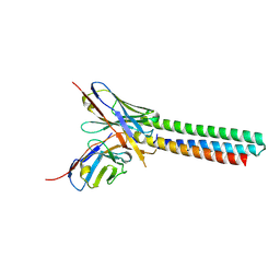
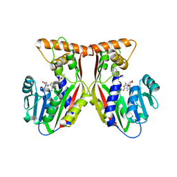
3SAJ
 | | Crystal Structure of glutamate receptor GluA1 Amino Terminal Domain | | 分子名称: | 2-acetamido-2-deoxy-beta-D-glucopyranose, 2-acetamido-2-deoxy-beta-D-glucopyranose-(1-4)-2-acetamido-2-deoxy-beta-D-glucopyranose, Glutamate receptor 1, ... | | 著者 | Jin, R, Zong, Y, Yao, G, Gu, S. | | 登録日 | 2011-06-02 | | 公開日 | 2011-06-22 | | 最終更新日 | 2020-07-29 | | 実験手法 | X-RAY DIFFRACTION (2.5 Å) | | 主引用文献 | Crystal structure of the glutamate receptor GluA1 N-terminal domain.
Biochem.J., 438, 2011
|
|
2FEK
 | | Structure of a protein tyrosine phosphatase | | 分子名称: | Low molecular weight protein-tyrosine-phosphatase wzb | | 著者 | Lescop, E, Jin, C. | | 登録日 | 2005-12-16 | | 公開日 | 2006-05-09 | | 最終更新日 | 2022-03-09 | | 実験手法 | SOLUTION NMR | | 主引用文献 | The solution structure of Escherichia coli Wzb reveals a novel substrate recognition mechanism of prokaryotic low molecular weight protein-tyrosine phosphatases
J.Biol.Chem., 281, 2006
|
|
3N8S
 | | Crystal Structure of BlaC-E166A covalently bound with Cefamandole | | 分子名称: | (2R)-2-[(1R)-1-{[(2R)-2-hydroxy-2-phenylacetyl]amino}-2-oxoethyl]-5-{[(1-methyl-1H-tetrazol-5-yl)sulfanyl]methyl}-3,6-dihydro-2H-1,3-thiazine-4-carboxylic acid, Beta-lactamase, PHOSPHATE ION | | 著者 | Tremblay, L.W, Blanchard, J.S. | | 登録日 | 2010-05-28 | | 公開日 | 2010-11-24 | | 最終更新日 | 2017-11-08 | | 実験手法 | X-RAY DIFFRACTION (2 Å) | | 主引用文献 | Structures of the Michaelis Complex (1.2 A) and the Covalent Acyl Intermediate (2.0 A) of Cefamandole Bound in the Active Sites of the Mycobacterium tuberculosis beta-Lactamase K73A and E166A Mutants.
Biochemistry, 49, 2010
|
|
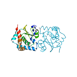
3NDP
 | | Crystal structure of human AK4(L171P) | | 分子名称: | Adenylate kinase isoenzyme 4, SULFATE ION | | 著者 | Liu, R, Wang, Y, Wei, Z, Gong, W. | | 登録日 | 2010-06-07 | | 公開日 | 2010-06-23 | | 最終更新日 | 2024-04-03 | | 実験手法 | X-RAY DIFFRACTION (2.3 Å) | | 主引用文献 | Crystal structure of human adenylate kinase 4 (L171P) suggests the role of hinge region in protein domain motion
Biochem.Biophys.Res.Commun., 379, 2009
|
|
3NY4
 | | Crystal Structure of BlaC-K73A bound with Cefamandole | | 分子名称: | (6R,7R)-7-{[(2R)-2-hydroxy-2-phenylacetyl]amino}-3-{[(1-methyl-1H-tetrazol-5-yl)sulfanyl]methyl}-8-oxo-5-thia-1-azabicyclo[4.2.0]oct-2-ene-2-carboxylic acid, Beta-lactamase, PHOSPHATE ION | | 著者 | Tremblay, L.W, Blanchard, J.S. | | 登録日 | 2010-07-14 | | 公開日 | 2010-11-24 | | 最終更新日 | 2024-02-21 | | 実験手法 | X-RAY DIFFRACTION (1.22 Å) | | 主引用文献 | Structures of the Michaelis Complex (1.2 A) and the Covalent Acyl Intermediate (2.0 A) of Cefamandole Bound in the Active Sites of the Mycobacterium tuberculosis beta-Lactamase K73A and E166A Mutants.
Biochemistry, 49, 2010
|
|
4HRV
 | |
3PAW
 | |


