5EQJ
 
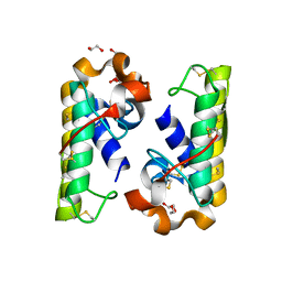
 | | Crystal structure of the two-subunit tRNA m1A58 methyltransferase from Saccharomyces cerevisiae | | 分子名称: | tRNA (adenine(58)-N(1))-methyltransferase catalytic subunit TRM61, tRNA (adenine(58)-N(1))-methyltransferase non-catalytic subunit TRM6 | | 著者 | Zhu, Y, Wang, M, Wang, C, Fan, X, Jiang, X, Teng, M, Li, X. | | 登録日 | 2015-11-13 | | 公開日 | 2016-09-14 | | 最終更新日 | 2024-03-20 | | 実験手法 | X-RAY DIFFRACTION (2.2 Å) | | 主引用文献 | Crystal structure of the two-subunit tRNA m(1)A58 methyltransferase TRM6-TRM61 from Saccharomyces cerevisiae.
Sci Rep, 6, 2016
|
|
3B4D
 | |
3B4M
 | |
3BS8
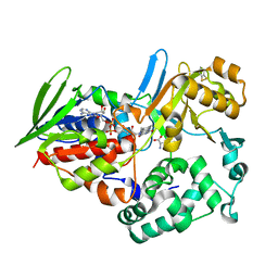
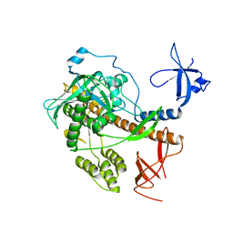
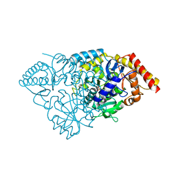 | | Crystal structure of Glutamate 1-Semialdehyde Aminotransferase complexed with pyridoxamine-5'-phosphate From Bacillus subtilis | | 分子名称: | 4'-DEOXY-4'-AMINOPYRIDOXAL-5'-PHOSPHATE, Glutamate-1-semialdehyde 2,1-aminomutase | | 著者 | Ge, H, Fan, J, Teng, M, Niu, L. | | 登録日 | 2007-12-22 | | 公開日 | 2008-12-23 | | 最終更新日 | 2023-11-01 | | 実験手法 | X-RAY DIFFRACTION (2.3 Å) | | 主引用文献 | Crystal structure of Glutamate1-semialdehyde aminotransferase from Bacillus subtilis with bound pyridoxamine-5'-phosphate
Biochem.Biophys.Res.Commun., 402, 2010
|
|
1ONJ
 | | Crystal structure of Atratoxin-b from Chinese cobra venom of Naja atra | | 分子名称: | Cobrotoxin b, SULFATE ION | | 著者 | Lou, X, Tu, X, Pan, G, Xu, C, Fan, R, Lu, W, Deng, W, Rao, P, Teng, M, Niu, L. | | 登録日 | 2003-02-28 | | 公開日 | 2004-02-28 | | 最終更新日 | 2017-10-11 | | 実験手法 | X-RAY DIFFRACTION (1.555 Å) | | 主引用文献 | Purification, N-terminal sequencing, crystallization and preliminary structural determination of atratoxin-b, a short-chain alpha-neurotoxin from Naja atra venom.
Acta Crystallogr.,Sect.D, 59, 2003
|
|
1REO
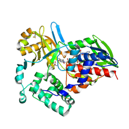
 | | L-amino acid oxidase from Agkistrodon halys pallas | | 分子名称: | 2-acetamido-2-deoxy-beta-D-glucopyranose, AHPLAAO, CITRIC ACID, ... | | 著者 | Zhang, H, Teng, M, Niu, L, Wang, Y, Wang, Y, Liu, Q, Huang, Q, Hao, Q, Dong, Y, Liu, P. | | 登録日 | 2003-11-07 | | 公開日 | 2004-05-04 | | 最終更新日 | 2023-10-25 | | 実験手法 | X-RAY DIFFRACTION (2.31 Å) | | 主引用文献 | Purification, partial characterization, crystallization and structural determination of AHP-LAAO, a novel L-amino-acid oxidase with cell apoptosis-inducing activity from Agkistrodon halys pallas venom.
Acta Crystallogr.,Sect.D, 60, 2004
|
|
1TDN
 | |
5DDZ
 | | Crystal structure of the RTA-c10-P2 complex | | 分子名称: | 60S acidic ribosomal protein P2, Ricin | | 著者 | Zhu, Y, Fan, X, Wang, C, Niu, L, Li, X, Teng, M. | | 登録日 | 2015-08-25 | | 公開日 | 2016-09-07 | | 最終更新日 | 2023-11-08 | | 実験手法 | X-RAY DIFFRACTION (1.5 Å) | | 主引用文献 | Structural insights into the interaction of the ribosomal P stalk protein P2 with a type II ribosome-inactivating protein ricin
Sci Rep, 6, 2016
|
|
1TDO
 | |
1TDK
 | |
5DPM
 | |
1VB0
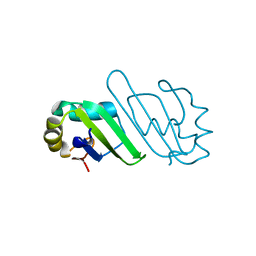
 | | Atomic resolution structure of atratoxin-b, one short-chain neurotoxin from Naja atra | | 分子名称: | 2-AMINO-2-HYDROXYMETHYL-PROPANE-1,3-DIOL, Cobrotoxin b, SULFATE ION | | 著者 | Lou, X, Liu, Q, Teng, M, Niu, L, Huang, Q, Hao, Q. | | 登録日 | 2004-02-20 | | 公開日 | 2004-12-21 | | 最終更新日 | 2023-10-25 | | 実験手法 | X-RAY DIFFRACTION (0.92 Å) | | 主引用文献 | The atomic resolution crystal structure of atratoxin determined by single wavelength anomalous diffraction phasing
J.Biol.Chem., 279, 2004
|
|
1V6P
 | | Crystal structure of Cobrotoxin | | 分子名称: | CHLORIDE ION, COPPER (II) ION, Cobrotoxin, ... | | 著者 | Lou, X, Tu, X, Wang, J, Teng, M, Niu, L, Liu, Q, Huang, Q, Hao, Q. | | 登録日 | 2003-12-03 | | 公開日 | 2004-12-21 | | 最終更新日 | 2023-12-27 | | 実験手法 | X-RAY DIFFRACTION (0.87 Å) | | 主引用文献 | The atomic resolution crystal structure of atratoxin determined by single wavelength anomalous diffraction phasing
J.Biol.Chem., 279, 2004
|
|
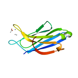
4OU6
 | | Crystal structure of DnaT84-153-dT10 ssDNA complex form 1 | | 分子名称: | DNA (5'-D(P*TP*TP*TP*TP*TP*TP*TP*TP*TP*T)-3'), Primosomal protein 1 | | 著者 | Liu, Z, Chen, P, Niu, L, Teng, M, Li, X. | | 登録日 | 2014-02-15 | | 公開日 | 2014-08-13 | | 最終更新日 | 2022-08-24 | | 実験手法 | X-RAY DIFFRACTION (1.96 Å) | | 主引用文献 | Crystal structure of DnaT84-153-dT10 ssDNA complex reveals a novel single-stranded DNA binding mode.
Nucleic Acids Res., 42, 2014
|
|
4OU7
 | | Crystal structure of DnaT84-153-dT10 ssDNA complex reveals a novel single-stranded DNA binding mode | | 分子名称: | DNA (5'-D(P*TP*TP*TP*TP*TP*TP*TP*TP*TP*T)-3'), Primosomal protein 1 | | 著者 | Liu, Z, Chen, P, Niu, L, Teng, M, Li, X. | | 登録日 | 2014-02-15 | | 公開日 | 2014-08-13 | | 最終更新日 | 2022-08-24 | | 実験手法 | X-RAY DIFFRACTION (2.83 Å) | | 主引用文献 | Crystal structure of DnaT84-153-dT10 ssDNA complex reveals a novel single-stranded DNA binding mode.
Nucleic Acids Res., 42, 2014
|
|
4Q77
 | | Crystal structure of Rot, a global regulator of virulence genes in Staphylococcus aureus | | 分子名称: | GLYCEROL, HTH-type transcriptional regulator rot | | 著者 | Zhu, Y, Fan, X, Li, X, Teng, M. | | 登録日 | 2014-04-24 | | 公開日 | 2014-09-17 | | 最終更新日 | 2024-03-20 | | 実験手法 | X-RAY DIFFRACTION (1.77 Å) | | 主引用文献 | Structure of Rot, a global regulator of virulence genes in Staphylococcus aureus.
Acta Crystallogr.,Sect.D, 70, 2014
|
|
4QWQ
 | | Crystal structure of the DNA-binding domain of the response regulator SaeR from Staphylococcus aureus | | 分子名称: | Response regulator SaeR | | 著者 | Fan, X, Zhu, Y, Zhang, X, Teng, M, Li, X. | | 登録日 | 2014-07-17 | | 公開日 | 2015-08-19 | | 最終更新日 | 2024-03-20 | | 実験手法 | X-RAY DIFFRACTION (2.501 Å) | | 主引用文献 | Structure of the DNA-binding domain of the response regulator SaeR from Staphylococcus aureus.
Acta Crystallogr.,Sect.D, 71, 2015
|
|
4RFP
 | |
4RO1
 | |
5H4Z
 | | Crystal structure of S202G mutant of human SYT-5 C2A domain | | 分子名称: | CALCIUM ION, CHLORIDE ION, Synaptotagmin-5 | | 著者 | Qiu, X, Ge, J, Yan, X, Gao, Y, Teng, M, Niu, L. | | 登録日 | 2016-11-02 | | 公開日 | 2016-11-30 | | 最終更新日 | 2023-11-08 | | 実験手法 | X-RAY DIFFRACTION (3.01 Å) | | 主引用文献 | Structural analysis of Ca(2+)-binding pocket of synaptotagmin 5 C2A domain
Int. J. Biol. Macromol., 95, 2017
|
|
5H4Y
 | | Crystal structure of human synaptotagmin 5 C2A domain | | 分子名称: | ACETATE ION, CALCIUM ION, Synaptotagmin-5 | | 著者 | Qiu, X, Gao, Y, Teng, M, Niu, L. | | 登録日 | 2016-11-02 | | 公開日 | 2016-11-30 | | 最終更新日 | 2024-03-20 | | 実験手法 | X-RAY DIFFRACTION (1.9 Å) | | 主引用文献 | Structural analysis of Ca(2+)-binding pocket of synaptotagmin 5 C2A domain
Int. J. Biol. Macromol., 95, 2017
|
|
5HXW
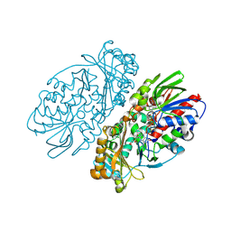
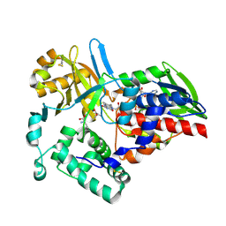
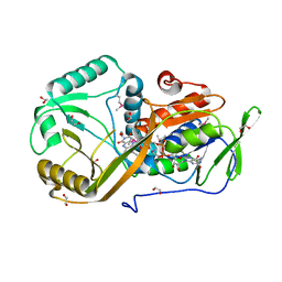
 | | L-amino acid deaminase from Proteus vulgaris | | 分子名称: | CETYL-TRIMETHYL-AMMONIUM, FLAVIN-ADENINE DINUCLEOTIDE, L-amino acid deaminase | | 著者 | Zhou, H, Ju, Y, Niu, L, Teng, M. | | 登録日 | 2016-01-31 | | 公開日 | 2016-08-03 | | 最終更新日 | 2023-11-08 | | 実験手法 | X-RAY DIFFRACTION (2.63 Å) | | 主引用文献 | Crystal structure of a membrane-bound l-amino acid deaminase from Proteus vulgaris
J.Struct.Biol., 195, 2016
|
|
5I39
 | | High resolution structure of L-amino acid deaminase from Proteus vulgaris with the deletion of the specific insertion sequence | | 分子名称: | 1,2-ETHANEDIOL, FLAVIN-ADENINE DINUCLEOTIDE, L-amino acid deaminase | | 著者 | Zhou, H, Ju, Y, Niu, L, Teng, M. | | 登録日 | 2016-02-10 | | 公開日 | 2016-08-03 | | 最終更新日 | 2016-08-24 | | 実験手法 | X-RAY DIFFRACTION (1.2 Å) | | 主引用文献 | Crystal structure of a membrane-bound l-amino acid deaminase from Proteus vulgaris
J.Struct.Biol., 195, 2016
|
|
3ONK
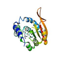
 | | yeast Ent3_ENTH domain | | 分子名称: | Epsin-3 | | 著者 | Wang, J, Fang, P, Niu, L, Teng, M. | | 登録日 | 2010-08-29 | | 公開日 | 2011-07-20 | | 最終更新日 | 2023-11-01 | | 実験手法 | X-RAY DIFFRACTION (2.09 Å) | | 主引用文献 | Epsin N-terminal homology domains bind on opposite sides of two SNAREs
Proc.Natl.Acad.Sci.USA, 108, 2011
|
|
3ONL
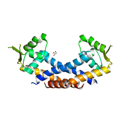
 | | yeast Ent3_ENTH-Vti1p_Habc complex structure | | 分子名称: | Epsin-3, t-SNARE VTI1 | | 著者 | Wang, J, Fang, P, Niu, L, Teng, M. | | 登録日 | 2010-08-29 | | 公開日 | 2011-07-20 | | 最終更新日 | 2023-11-01 | | 実験手法 | X-RAY DIFFRACTION (2.2 Å) | | 主引用文献 | Epsin N-terminal homology domains bind on opposite sides of two SNAREs
Proc.Natl.Acad.Sci.USA, 108, 2011
|
|


