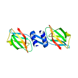
2R2J
 
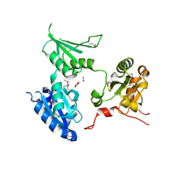 | | crystal structure of human ERp44 | | 分子名称: | FORMIC ACID, SUCCINIC ACID, Thioredoxin domain-containing protein 4 | | 著者 | Wang, L.K, Li, S.J, Sun, F, Wang, C.C. | | 登録日 | 2007-08-25 | | 公開日 | 2008-07-08 | | 最終更新日 | 2011-07-13 | | 実験手法 | X-RAY DIFFRACTION (2.6 Å) | | 主引用文献 | Crystal structure of human ERp44 shows a dynamic functional modulation by its carboxy-terminal tail.
Embo Rep., 2008
|
|
2ZGH
 | | Crystal Structure of active granzyme M bound to its product | | 分子名称: | Granzyme M, SSGKVPL, SULFATE ION | | 著者 | Wu, L.F, Wang, L, Hua, G.Q, Liu, K, Zhai, Y.J, Sun, F, Fan, Z.S. | | 登録日 | 2008-01-22 | | 公開日 | 2009-01-27 | | 最終更新日 | 2023-11-01 | | 実験手法 | X-RAY DIFFRACTION (2.17 Å) | | 主引用文献 | Structural basis for proteolytic specificity of the human apoptosis-inducing granzyme M
J.Immunol., 183, 2009
|
|
2ZGJ
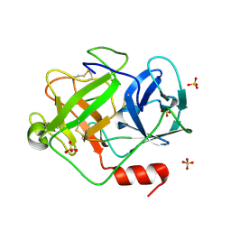 | | Crystal Structure of D86N-GzmM Complexed with Its Optimal Synthesized Substrate | | 分子名称: | Granzyme M, SSGKVPLS, SULFATE ION | | 著者 | Wu, L.F, Wang, L, Hua, G.Q, Liu, K, Zhai, Y.J, Sun, F, Fan, Z.S. | | 登録日 | 2008-01-22 | | 公開日 | 2009-01-27 | | 最終更新日 | 2023-11-01 | | 実験手法 | X-RAY DIFFRACTION (2.3 Å) | | 主引用文献 | Structural basis for proteolytic specificity of the human apoptosis-inducing granzyme M
J.Immunol., 183, 2009
|
|
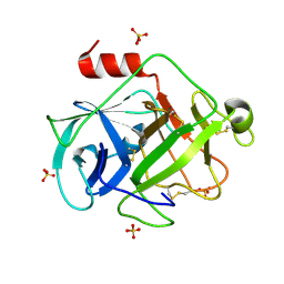
2ZKS
 | | Structural insights into the proteolytic machinery of apoptosis-inducing Granzyme M | | 分子名称: | Granzyme M, SULFATE ION, hGzmM inhibitor | | 著者 | Wu, L.F, Wang, L, Hua, G.Q, Liu, K, Yang, X, Zhai, Y.J, Sun, F, Fan, Z.S. | | 登録日 | 2008-03-28 | | 公開日 | 2009-03-31 | | 最終更新日 | 2024-04-03 | | 実験手法 | X-RAY DIFFRACTION (2.7 Å) | | 主引用文献 | Structural basis for proteolytic specificity of the human apoptosis-inducing granzyme M
J.Immunol., 183, 2009
|
|
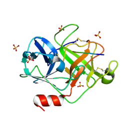
2ZGC
 | | Crystal Structure of Active Human Granzyme M | | 分子名称: | Granzyme M, SULFATE ION | | 著者 | Wu, L.F, Wang, L, Hua, G.Q, Liu, K, Zhai, Y.J, Sun, F, Fan, Z.S. | | 登録日 | 2008-01-21 | | 公開日 | 2009-01-27 | | 最終更新日 | 2023-11-01 | | 実験手法 | X-RAY DIFFRACTION (1.96 Å) | | 主引用文献 | Structural basis for proteolytic specificity of the human apoptosis-inducing granzyme M
J.Immunol., 183, 2009
|
|
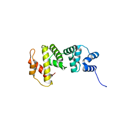
3A2A
 | | The structure of the carboxyl-terminal domain of the human voltage-gated proton channel Hv1 | | 分子名称: | CHLORIDE ION, Voltage-gated hydrogen channel 1 | | 著者 | Li, S.J, Unno, H, Zhou, Q, Zhao, Q, Zhai, Y, Sun, F. | | 登録日 | 2009-05-08 | | 公開日 | 2010-02-09 | | 最終更新日 | 2011-07-13 | | 実験手法 | X-RAY DIFFRACTION (2 Å) | | 主引用文献 | The role and structure of the carboxyl-terminal domain of the human voltage-gated proton channel Hv1.
J.Biol.Chem., 285, 2010
|
|
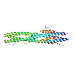
4I6P
 | | Crystal structure of Par3-NTD domain | | 分子名称: | Partitioning defective 3 homolog | | 著者 | Wang, W, Gao, F, Gong, W, Sun, F, Feng, W. | | 登録日 | 2012-11-29 | | 公開日 | 2013-07-17 | | 最終更新日 | 2023-09-20 | | 実験手法 | X-RAY DIFFRACTION (2.9 Å) | | 主引用文献 | Structural insights into the intrinsic self-assembly of par-3 N-terminal domain.
Structure, 21, 2013
|
|
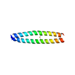
4CKH
 | | Helical reconstruction of ACAP1(BAR-PH domain) decorated membrane tubules by cryo-electron microscopy | | 分子名称: | ARF-GAP WITH COILED-COIL, ANK REPEAT AND PH DOMAIN-CONTAINING PROTEIN 1 | | 著者 | Pang, X.Y, Fan, J, Zhang, Y, Zhang, K, Gao, B.Q, Ma, J, Li, J, Deng, Y.C, Zhou, Q.J, Hsu, V, Sun, F. | | 登録日 | 2014-01-06 | | 公開日 | 2014-10-15 | | 最終更新日 | 2024-05-08 | | 実験手法 | ELECTRON MICROSCOPY (17 Å) | | 主引用文献 | A Ph Domain in Acap1 Possesses Key Features of the Bar Domain in Promoting Membrane Curvature.
Dev.Cell, 31, 2014
|
|
4CKG
 | | Helical reconstruction of ACAP1(BAR-PH domain) decorated membrane tubules by cryo-electron microscopy | | 分子名称: | ARF-GAP WITH COILED-COIL, ANK REPEAT AND PH DOMAIN-CONTAINING PROTEIN 1 | | 著者 | Pang, X.Y, Fan, J, Zhang, Y, Zhang, K, Gao, B.Q, Ma, J, Li, J, Deng, Y.C, Zhou, Q.J, Hsu, V, Sun, F. | | 登録日 | 2014-01-06 | | 公開日 | 2014-10-15 | | 最終更新日 | 2024-05-08 | | 実験手法 | ELECTRON MICROSCOPY (15 Å) | | 主引用文献 | A Ph Domain in Acap1 Possesses Key Features of the Bar Domain in Promoting Membrane Curvature.
Dev.Cell, 31, 2014
|
|
3J1P
 | |
5H3D
 | | Helical structure of membrane tubules decorated by ACAP1 (BARPH doamin) protein by cryo-electron microscopy and MD simulation | | 分子名称: | Arf-GAP with coiled-coil, ANK repeat and PH domain-containing protein 1 | | 著者 | Chan, C, Pang, X.Y, Zhang, Y, Sun, F, Fan, J. | | 登録日 | 2016-10-22 | | 公開日 | 2019-01-16 | | 最終更新日 | 2024-03-20 | | 実験手法 | ELECTRON MICROSCOPY (14 Å) | | 主引用文献 | ACAP1 assembles into an unusual protein lattice for membrane deformation through multiple stages.
Plos Comput.Biol., 15, 2019
|
|
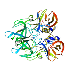
3F3H
 | | Crystal structure and anti-tumor activity of LZ-8 from the fungus Ganoderma lucidium | | 分子名称: | Immunomodulatory protein Ling Zhi-8 | | 著者 | Zhou, C.Z, Huang, L, He, Y.X, Bao, R, Sun, F, Liang, C, Liu, L. | | 登録日 | 2008-10-30 | | 公開日 | 2009-09-15 | | 最終更新日 | 2023-11-01 | | 実験手法 | X-RAY DIFFRACTION (2.1 Å) | | 主引用文献 | Crystal structure of LZ-8 from the medicinal fungus Ganoderma lucidium
Proteins, 75, 2009
|
|
5J8V
 | |
3J17
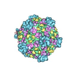 | | Structure of a transcribing cypovirus by cryo-electron microscopy | | 分子名称: | Structural protein VP3, Structural protein VP5, VP1 | | 著者 | Yang, C, Ji, G, Liu, H, Zhang, K, Liu, G, Sun, F, Zhu, P, Cheng, L. | | 登録日 | 2011-12-25 | | 公開日 | 2012-04-04 | | 最終更新日 | 2018-07-18 | | 実験手法 | ELECTRON MICROSCOPY (4.1 Å) | | 主引用文献 | Cryo-EM structure of a transcribing cypovirus.
Proc.Natl.Acad.Sci.USA, 109, 2012
|
|
3J1E
 | | Cryo-EM structure of 9-fold symmetric rATcpn-beta in apo state | | 分子名称: | Chaperonin beta subunit | | 著者 | Zhang, K, Wang, L, Liu, Y.X, Wang, X, Gao, B, Hu, Z.J, Ji, G, Chan, K.Y, Schulten, K, Dong, Z.Y, Sun, F. | | 登録日 | 2012-02-06 | | 公開日 | 2013-08-07 | | 最終更新日 | 2024-02-21 | | 実験手法 | ELECTRON MICROSCOPY (8.3 Å) | | 主引用文献 | Flexible interwoven termini determine the thermal stability of thermosomes.
Protein Cell, 4, 2013
|
|
3J1B
 | | Cryo-EM structure of 8-fold symmetric rATcpn-alpha in apo state | | 分子名称: | Chaperonin alpha subunit | | 著者 | Zhang, K, Wang, L, Liu, Y.X, Wang, X, Gao, B, Hu, Z.J, Ji, G, Chan, K.Y, Schulten, K, Dong, Z.Y, Sun, F. | | 登録日 | 2012-02-06 | | 公開日 | 2013-08-07 | | 最終更新日 | 2024-02-21 | | 実験手法 | ELECTRON MICROSCOPY (4.9 Å) | | 主引用文献 | Flexible interwoven termini determine the thermal stability of thermosomes.
Protein Cell, 4, 2013
|
|
3J1C
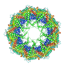 | | Cryo-EM structure of 9-fold symmetric rATcpn-alpha in apo state | | 分子名称: | Chaperonin alpha subunit | | 著者 | Zhang, K, Wang, L, Liu, Y.X, Wang, X, Gao, B, Hu, Z.J, Ji, G, Chan, K.Y, Schulten, K, Dong, Z.Y, Sun, F. | | 登録日 | 2012-02-06 | | 公開日 | 2013-08-07 | | 最終更新日 | 2024-02-21 | | 実験手法 | ELECTRON MICROSCOPY (9.1 Å) | | 主引用文献 | Flexible interwoven termini determine the thermal stability of thermosomes.
Protein Cell, 4, 2013
|
|
3K6J
 | | Crystal structure of the dehydrogenase part of multifuctional enzyme 1 from C.elegans | | 分子名称: | PHOSPHATE ION, Protein F01G10.3, confirmed by transcript evidence, ... | | 著者 | Ouyang, Z, Zhang, K, Zhai, Y, Lu, J, Sun, F. | | 登録日 | 2009-10-09 | | 公開日 | 2010-10-13 | | 最終更新日 | 2023-11-01 | | 実験手法 | X-RAY DIFFRACTION (2.2 Å) | | 主引用文献 | Crystal structure of the dehydrogenase part of multifuctional enzyme 1 from C.elegans
To be Published
|
|
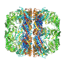
2AHM
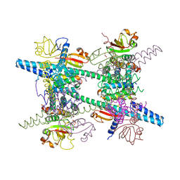 | | Crystal structure of SARS-CoV super complex of non-structural proteins: the hexadecamer | | 分子名称: | GLYCEROL, Replicase polyprotein 1ab, heavy chain, ... | | 著者 | Zhai, Y.J, Sun, F, Bartlam, M, Rao, Z. | | 登録日 | 2005-07-28 | | 公開日 | 2005-11-15 | | 最終更新日 | 2024-03-13 | | 実験手法 | X-RAY DIFFRACTION (2.4 Å) | | 主引用文献 | Insights into SARS-CoV transcription and replication from the structure of the nsp7-nsp8 hexadecamer
NAT.STRUCT.MOL.BIOL., 12, 2005
|
|
4OIY
 | | Crystal structure of Sec7p catalytic domain | | 分子名称: | MAGNESIUM ION, Protein transport protein SEC7 | | 著者 | Qiu, B, Zhang, K, Sun, F. | | 登録日 | 2014-01-20 | | 公開日 | 2014-07-02 | | 最終更新日 | 2023-11-08 | | 実験手法 | X-RAY DIFFRACTION (1.5 Å) | | 主引用文献 | C-terminal motif within Sec7 domain regulates guanine nucleotide exchange activity via tuning protein conformation
Biochem.Biophys.Res.Commun., 446, 2014
|
|
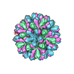
4EJR
 | | Crystal structure of major capsid protein S domain from rabbit hemorrhagic disease virus | | 分子名称: | Major capsid protein VP60 | | 著者 | Xu, F, Ma, J, Zhang, K, Wang, X, Sun, F. | | 登録日 | 2012-04-07 | | 公開日 | 2013-01-30 | | 最終更新日 | 2023-09-13 | | 実験手法 | X-RAY DIFFRACTION (2 Å) | | 主引用文献 | Atomic model of rabbit hemorrhagic disease virus by cryo-electron microscopy and crystallography.
Plos Pathog., 9, 2013
|
|
6LXT
 | | Structure of post fusion core of 2019-nCoV S2 subunit | | 分子名称: | Spike protein S2, TETRAETHYLENE GLYCOL, ZINC ION | | 著者 | Zhu, Y, Sun, F. | | 登録日 | 2020-02-11 | | 公開日 | 2020-02-26 | | 最終更新日 | 2023-11-29 | | 実験手法 | X-RAY DIFFRACTION (2.9 Å) | | 主引用文献 | Inhibition of SARS-CoV-2 (previously 2019-nCoV) infection by a highly potent pan-coronavirus fusion inhibitor targeting its spike protein that harbors a high capacity to mediate membrane fusion.
Cell Res., 30, 2020
|
|
4EGT
 | | Crystal structure of major capsid protein P domain from rabbit hemorrhagic disease virus | | 分子名称: | Major capsid protein VP60 | | 著者 | Wang, X, Xu, F, Zhang, K, Zhai, Y, Sun, F. | | 登録日 | 2012-04-01 | | 公開日 | 2013-01-30 | | 最終更新日 | 2023-09-13 | | 実験手法 | X-RAY DIFFRACTION (2 Å) | | 主引用文献 | Atomic model of rabbit hemorrhagic disease virus by cryo-electron microscopy and crystallography.
Plos Pathog., 9, 2013
|
|
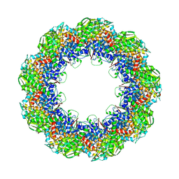
2GA6
 | | The crystal structure of SARS nsp10 without zinc ion as additive | | 分子名称: | ZINC ION, orf1a polyprotein | | 著者 | Su, D, Lou, Z, Sun, F, Zhai, Y, Yang, H, Rao, Z. | | 登録日 | 2006-03-08 | | 公開日 | 2006-08-15 | | 最終更新日 | 2023-10-25 | | 実験手法 | X-RAY DIFFRACTION (2.7 Å) | | 主引用文献 | Dodecamer Structure of Severe Acute Respiratory Syndrome Coronavirus Nonstructural Protein nsp10
J.Virol., 80, 2006
|
|
2G9T
 | | Crystal structure of the SARS coronavirus nsp10 at 2.1A | | 分子名称: | ZINC ION, orf1a polyprotein | | 著者 | Su, D, Lou, Z, Yang, H, Sun, F, Rao, Z. | | 登録日 | 2006-03-07 | | 公開日 | 2006-08-15 | | 最終更新日 | 2024-03-13 | | 実験手法 | X-RAY DIFFRACTION (2.1 Å) | | 主引用文献 | Dodecamer Structure of Severe Acute Respiratory Syndrome Coronavirus Nonstructural Protein nsp10
J.Virol., 80, 2006
|
|


