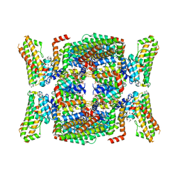
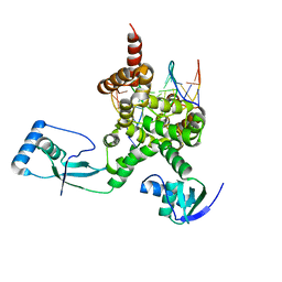
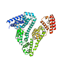
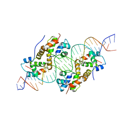
2UZP
 
 | | Crystal structure of the C2 domain of human protein kinase C gamma. | | 分子名称: | 1,2-ETHANEDIOL, CALCIUM ION, COBALT (II) ION, ... | | 著者 | Pike, A.C.W, Amos, A, Johansson, C, Sobott, F, Savitsky, P, Berridge, G, Fedorov, O, Umeano, C, Gorrec, F, Bunkoczi, G, Debreczeni, J, von Delft, F, Arrowsmith, C.H, Edwards, A, Weigelt, J, Sundstrom, M, Knapp, S. | | 登録日 | 2007-04-30 | | 公開日 | 2007-05-29 | | 最終更新日 | 2023-12-13 | | 実験手法 | X-RAY DIFFRACTION (2 Å) | | 主引用文献 | Crystal Structure of C2 Domain of Protein Kinase C Gamma
To be Published
|
|
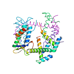
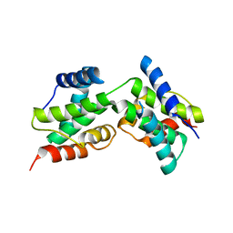
2J1L
 | | Crystal Structure of Human Rho-related GTP-binding protein RhoD | | 分子名称: | GUANOSINE-5'-DIPHOSPHATE, MAGNESIUM ION, RHO-RELATED GTP-BINDING PROTEIN RHOD | | 著者 | Pike, A.C.W, Johansson, C, Gileadi, C, Niesen, F.H, Sobott, F, Schoch, G, Elkins, J, Smee, C, Gorrec, F, Watt, S, Bray, J, Turnbull, A.P, von Delft, F, Arrowsmith, C, Edwards, A, Weigelt, J, Sundstrom, M, Doyle, D. | | 登録日 | 2006-08-14 | | 公開日 | 2006-09-18 | | 最終更新日 | 2023-12-13 | | 実験手法 | X-RAY DIFFRACTION (2.5 Å) | | 主引用文献 | Crystal Structure of Human Rho-Related GTP-Binding Protein Rhod
To be Published
|
|
2EXE
 | | Crystal structure of the phosphorylated CLK3 | | 分子名称: | Dual specificity protein kinase CLK3 | | 著者 | Papagrigoriou, E, Rellos, P, Das, S, Bullock, A, Ball, L.J, Turnbull, A, Savitsky, P, Fedorov, O, Johansson, C, Ugochukwu, E, Sobott, F, von Delft, F, Edwards, A, Sundstrom, M, Weigelt, J, Arrowsmith, C, Knapp, S, Structural Genomics Consortium (SGC) | | 登録日 | 2005-11-08 | | 公開日 | 2005-11-15 | | 最終更新日 | 2023-08-23 | | 実験手法 | X-RAY DIFFRACTION (2.35 Å) | | 主引用文献 | Crystal structure of the phosphorylated CLK3
TO BE PUBLISHED
|
|
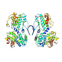
2JAM
 | | Crystal structure of human calmodulin-dependent protein kinase I G | | 分子名称: | 1,2-ETHANEDIOL, 5-[(E)-(5-CHLORO-2-OXO-1,2-DIHYDRO-3H-INDOL-3-YLIDENE)METHYL]-N-[2-(DIETHYLAMINO)ETHYL]-2,4-DIMETHYL-1H-PYRROLE-3-CARBOXAMIDE, CALCIUM ION, ... | | 著者 | Debreczeni, J.E, Bullock, A, Keates, T, Niesen, F.H, Salah, E, Shrestha, L, Smee, C, Sobott, F, Pike, A.C.W, Bunkoczi, G, von Delft, F, Turnbull, A, Weigelt, J, Arrowsmith, C.H, Edwards, A, Sundstrom, M, Knapp, S. | | 登録日 | 2006-11-29 | | 公開日 | 2007-03-13 | | 最終更新日 | 2023-12-13 | | 実験手法 | X-RAY DIFFRACTION (1.7 Å) | | 主引用文献 | Crystal Structure of Human Calmodulin-Dependent Protein Kinase I G
To be Published
|
|
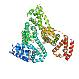
7B2L
 | | Structure of the endocytic adaptor complex AENTH | | 分子名称: | ANTH domain of Sla2, ENTH domain of epsin Ent1, [(2R)-2-octanoyloxy-3-[oxidanyl-[(1R,2R,3S,4R,5R,6S)-2,3,6-tris(oxidanyl)-4,5-diphosphonooxy-cyclohexyl]oxy-phosphoryl]oxy-propyl] octanoate | | 著者 | Klebl, D.P, Lizarrondo, J, Sobott, F, Garcia-Alai, M, Muench, S.P. | | 登録日 | 2020-11-27 | | 公開日 | 2021-05-05 | | 最終更新日 | 2021-06-02 | | 実験手法 | ELECTRON MICROSCOPY (3.9 Å) | | 主引用文献 | Structure of the endocytic adaptor complex reveals the basis for efficient membrane anchoring during clathrin-mediated endocytosis.
Nat Commun, 12, 2021
|
|
2CMW
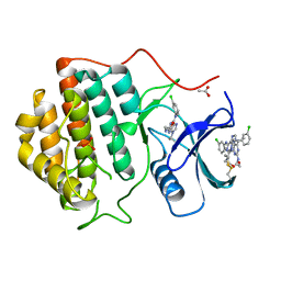 | | Structure of Human Casein kinase 1 gamma-1 in complex with 2-(2- Hydroxyethylamino)-6-(3-chloroanilino)-9-isopropylpurine | | 分子名称: | 2-(2-HYDROXYETHYLAMINO)-6-(3-CHLOROANILINO)-9-ISOPROPYLPURINE, ACETATE ION, CASEIN KINASE I ISOFORM GAMMA-1, ... | | 著者 | Bunkoczi, G, Rellos, P, Das, S, Savitsky, P, Niesen, F, Sobott, F, Fedorov, O, Pike, A.C.W, von Delft, F, Sundstrom, M, Arrowsmith, C, Edwards, A, Weigelt, J, Knapp, S. | | 登録日 | 2006-05-15 | | 公開日 | 2006-06-15 | | 最終更新日 | 2023-12-13 | | 実験手法 | X-RAY DIFFRACTION (1.75 Å) | | 主引用文献 | Structure of Human Casein Kinase 1 Gamma-1 in Complex with 2-(2-Hydroxyethylamino)-6-(3-Chloroanilino)-9-Isopropylpurine (Casp Target)
To be Published
|
|
2BUJ
 | | Crystal structure of the human Serine-threonine Kinase 16 in complex with staurosporine | | 分子名称: | CHLORIDE ION, SERINE/THREONINE-PROTEIN KINASE 16, STAUROSPORINE | | 著者 | Debreczeni, J.E, Eswaran, J, Bullock, A, Filippakopoulos, P, Kavanagh, K, Amos, A, Fedorov, O, Sobott, F, Ball, L.J, von Delft, F, Arrowsmith, C, Sundstrom, M, Edwards, A, Knapp, S. | | 登録日 | 2005-06-13 | | 公開日 | 2005-07-05 | | 最終更新日 | 2019-05-08 | | 実験手法 | X-RAY DIFFRACTION (2.6 Å) | | 主引用文献 | Crystal Structure of the Human Serine-Threonine Kinase 16 in Complex with Staurosporine
To be Published
|
|
2C47
 | | Structure of casein kinase 1 gamma 2 | | 分子名称: | (2R,3R,4S,5R)-2-(4-AMINO-5-IODO-7H-PYRROLO[2,3-D]PYRIMIDIN-7-YL)-5-(HYDROXYMETHYL)TETRAHYDROFURAN-3,4-DIOL, CASEIN KINASE 1 GAMMA 2 ISOFORM, MAGNESIUM ION | | 著者 | Bunkoczi, G, Rellos, P, Das, S, Ugochukwu, E, Fedorov, O, Sobott, F, Eswaran, J, Amos, A, Ball, L, von Delft, F, Bullock, A, Debreczeni, J, Turnbull, A, Sundstrom, M, Weigelt, J, Arrowsmith, C, Edwards, A, Knapp, S. | | 登録日 | 2005-10-16 | | 公開日 | 2005-11-07 | | 最終更新日 | 2023-12-13 | | 実験手法 | X-RAY DIFFRACTION (2.4 Å) | | 主引用文献 | The Structure of Casein Kinase 1 Gamma 2
To be Published
|
|
2CLS
 | | The crystal structure of the human RND1 GTPase in the active GTP bound state | | 分子名称: | GUANOSINE-5'-TRIPHOSPHATE, MAGNESIUM ION, RHO-RELATED GTP-BINDING PROTEIN RHO6 | | 著者 | Pike, A.C.W, Yang, X, Colebrook, S, Gileadi, O, Sobott, F, Bray, J, Wen Hwa, L, Marsden, B, Zhao, Y, Schoch, G, Elkins, J, Debreczeni, J.E, Turnbull, A.P, von Delft, F, Arrowsmith, C, Edwards, A, Weigelt, J, Sundstrom, M, Doyle, D. | | 登録日 | 2006-04-28 | | 公開日 | 2006-05-04 | | 最終更新日 | 2023-12-13 | | 実験手法 | X-RAY DIFFRACTION (2.31 Å) | | 主引用文献 | The Crystal Structure of the Human Rnd1 Gtpase in the Active GTP Bound State
To be Published
|
|
7R5A
 | |
6NW5
 | |
7OL9
 | |
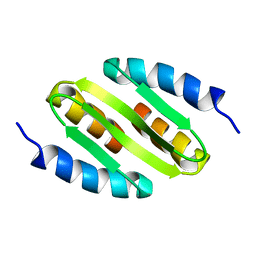
3DD9
 | | Structure of DocH66Y dimer | | 分子名称: | Death on curing protein | | 著者 | Garcia-Pino, A, Loris, R. | | 登録日 | 2008-06-05 | | 公開日 | 2009-06-09 | | 最終更新日 | 2023-11-01 | | 実験手法 | X-RAY DIFFRACTION (2.45 Å) | | 主引用文献 | The intrinsically disordered domain of the antitoxin Phd chaperones the toxin Doc against irreversible inactivation and misfolding
J. Biol. Chem., 289, 2014
|
|
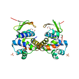
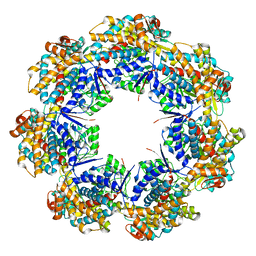
6SG4
 | | Structure of CDK2/cyclin A M246Q, S247EN | | 分子名称: | Cyclin-A2, Cyclin-dependent kinase 2 | | 著者 | Salamina, M, Basle, A, Massa, B, Noble, M.E.M, Endicott, J.A. | | 登録日 | 2019-08-02 | | 公開日 | 2021-01-27 | | 最終更新日 | 2024-01-24 | | 実験手法 | X-RAY DIFFRACTION (2.43 Å) | | 主引用文献 | Discriminative SKP2 Interactions with CDK-Cyclin Complexes Support a Cyclin A-Specific Role in p27KIP1 Degradation.
J.Mol.Biol., 433, 2021
|
|
2OC3
 | | Crystal Structure of the Catalytic Domain of Human Protein Tyrosine Phosphatase non-receptor Type 18 | | 分子名称: | Tyrosine-protein phosphatase non-receptor type 18 | | 著者 | Ugochukwu, E, Barr, A, Alfano, I, Gorrec, F, Umeano, C, Savitsky, P, Sobott, F, Eswaran, J, Papagrigoriou, E, Debreczeni, J.E, Turnbull, A, Bunkoczi, G, Sundstrom, M, Arrowsmith, C.H, Weigelt, J, Edwards, A, von Delft, F, Knapp, S, Structural Genomics Consortium (SGC) | | 登録日 | 2006-12-20 | | 公開日 | 2007-01-30 | | 最終更新日 | 2023-08-30 | | 実験手法 | X-RAY DIFFRACTION (1.5 Å) | | 主引用文献 | Large-scale structural analysis of the classical human protein tyrosine phosphatome.
Cell(Cambridge,Mass.), 136, 2009
|
|
2XDV
 | | Crystal Structure of the Catalytic Domain of FLJ14393 | | 分子名称: | 1,2-ETHANEDIOL, CADMIUM ION, MANGANESE (II) ION, ... | | 著者 | Krojer, T, Muniz, J.R.C, Ng, S.S, Pilka, E, Guo, K, Pike, A.C.W, Filippakopoulos, P, Knapp, S, Kavanagh, K.L, Gileadi, O, Bunkoczi, G, Yue, W.W, Niesen, F, Sobott, F, Fedorov, O, Savitsky, P, Kochan, G, Daniel, M, von Delft, F, Arrowsmith, C.H, Edwards, A.M, Weigelt, J, Bountra, C, Oppermann, U. | | 登録日 | 2010-05-07 | | 公開日 | 2010-05-26 | | 最終更新日 | 2018-02-21 | | 実験手法 | X-RAY DIFFRACTION (2.57 Å) | | 主引用文献 | Ribosomal oxygenases are structurally conserved from prokaryotes to humans.
Nature, 510, 2014
|
|
7AAI
 | | Crystal structure of Human serum albumin in complex with perfluorooctanoic acid (PFOA) at 2.10 Angstrom Resolution | | 分子名称: | (4S)-2-METHYL-2,4-PENTANEDIOL, 1,2-ETHANEDIOL, 3[N-MORPHOLINO]PROPANE SULFONIC ACID, ... | | 著者 | Maso, L, Liberi, S, Trande, M, Angelini, A, Cendron, L. | | 登録日 | 2020-09-04 | | 公開日 | 2021-02-24 | | 最終更新日 | 2024-01-31 | | 実験手法 | X-RAY DIFFRACTION (2.1 Å) | | 主引用文献 | Unveiling the binding mode of perfluorooctanoic acid to human serum albumin.
Protein Sci., 30, 2021
|
|
7AAE
 | | Crystal structure of Human serum albumin in complex with myristic acid at 2.27 Angstrom Resolution | | 分子名称: | (4S)-2-METHYL-2,4-PENTANEDIOL, Albumin, FORMIC ACID, ... | | 著者 | Maso, L, Liberi, S, Trande, M, Angelini, A, Cendron, L. | | 登録日 | 2020-09-04 | | 公開日 | 2021-02-24 | | 最終更新日 | 2024-01-31 | | 実験手法 | X-RAY DIFFRACTION (2.27 Å) | | 主引用文献 | Unveiling the binding mode of perfluorooctanoic acid to human serum albumin.
Protein Sci., 30, 2021
|
|
5NOC
 | |
6F8S
 | | Toxin-Antitoxin complex GraTA | | 分子名称: | DI(HYDROXYETHYL)ETHER, Putative Killer protein, SULFATE ION, ... | | 著者 | Talavera, A, Loris, R. | | 登録日 | 2017-12-13 | | 公開日 | 2018-07-11 | | 最終更新日 | 2019-03-13 | | 実験手法 | X-RAY DIFFRACTION (2.5 Å) | | 主引用文献 | A dual role in regulation and toxicity for the disordered N-terminus of the toxin GraT.
Nat Commun, 10, 2019
|
|
6F8H
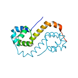 | | antitoxin GraA | | 分子名称: | XRE family transcriptional regulator | | 著者 | Talavera, A, Loris, R. | | 登録日 | 2017-12-13 | | 公開日 | 2019-01-30 | | 最終更新日 | 2019-03-13 | | 実験手法 | X-RAY DIFFRACTION (2.002 Å) | | 主引用文献 | A dual role in regulation and toxicity for the disordered N-terminus of the toxin GraT.
Nat Commun, 10, 2019
|
|
6FIX
 | |
7AZP
 | |
7B22
 | |
7B5P
 | |


