Loading
PDBj
MenuPDBj@FacebookPDBj@TwitterPDBj@YouTubewwPDB FoundationwwPDB
RCSB PDBPDBeBMRBAdv. SearchSearch help
Search by PDB author
8RUS
DownloadVisualize
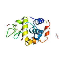
BU of 8rus by Molmil
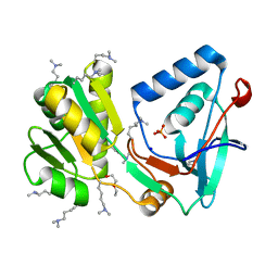
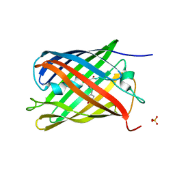
Hen egg-white lysozyme (HEWL) structure from EuXFEL FXE, multi-hit Droplet-on-Demand (DoD) injection, 9.3 keV photon energy, space group P432121
Descriptor: ACETATE ION, CHLORIDE ION, DI(HYDROXYETHYL)ETHER, ...
Authors:Perrett, S, van Thor, J.J.
Deposit date:2024-01-31
Release date:2024-07-24
Last modified:2024-10-09
Method:X-RAY DIFFRACTION (1.38 Å)
Cite:Kilohertz droplet-on-demand serial femtosecond crystallography at the European XFEL station FXE.
Struct Dyn., 11, 2024
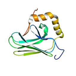
5GJJ
DownloadVisualize
BU of 5gjj by Molmil
Glutathionylated hHsp70 SBD
Descriptor: Heat shock 70 kDa protein 1A
Authors:Gong, W.B, Yang, J, Zhang, H, Perrett, S.
Deposit date:2016-06-30
Release date:2017-07-05
Last modified:2023-06-14
Method:SOLUTION NMR
Cite:Structure of glutathionylated hHsp70 SBD (385-641)
To Be Published
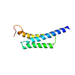
7DM4
DownloadVisualize
BU of 7dm4 by Molmil
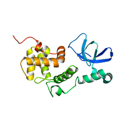
Solution structure of ARID4B Tudor domain
Descriptor: AT-rich interactive domain-containing protein 4B
Authors:Ren, J, Yao, H, Hu, W, Perrett, S, Feng, Y, Gong, W.
Deposit date:2020-12-02
Release date:2021-03-10
Last modified:2024-05-15
Method:SOLUTION NMR
Cite:Structural basis for the DNA-binding activity of human ARID4B Tudor domain.
J.Biol.Chem., 296, 2021
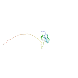
5Z8Q
DownloadVisualize
BU of 5z8q by Molmil
Solution structure of the SBDalpha domain of yeast Ssa1
Descriptor: Heat shock protein SSA1
Authors:Gong, W, Hu, W, Perrett, S.
Deposit date:2018-02-01
Release date:2018-09-26
Last modified:2024-05-15
Method:SOLUTION NMR
Cite:The C-terminal GGAP motif of Hsp70 mediates substrate recognition and stress response in yeast.
J. Biol. Chem., 293, 2018
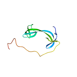
5Z8I
DownloadVisualize
BU of 5z8i by Molmil
Solution structure of the SBDbeta domain of yeast Ssa1
Descriptor: Heat shock protein SSA1
Authors:Gong, W, Hu, W, Wu, H, Perrett, S.
Deposit date:2018-01-31
Release date:2018-09-26
Last modified:2024-05-15
Method:SOLUTION NMR
Cite:The C-terminal GGAP motif of Hsp70 mediates substrate recognition and stress response in yeast.
J. Biol. Chem., 293, 2018
6L87
DownloadVisualize
BU of 6l87 by Molmil
Solution structure of the tandem PWWP-ARID domains of human RBBP1
Descriptor: AT-rich interactive domain-containing protein 4A
Authors:Gong, W.B, Perrett, S, Feng, Y.G.
Deposit date:2019-11-05
Release date:2021-01-06
Last modified:2024-05-15
Method:SOLUTION NMR
Cite:Structural insight into chromatin recognition by multiple domains of the tumor suppressor RBBP1.
J.Mol.Biol., 2021
1JON
DownloadVisualize
BU of 1jon by Molmil
GROEL (HSP60 CLASS) FRAGMENT COMPRISING RESIDUES 191-345
Descriptor: GROEL, HSP60 CLASS
Authors:Buckle, A.M, Fersht, A.R.
Deposit date:1996-05-30
Release date:1997-03-12
Last modified:2024-05-22
Method:X-RAY DIFFRACTION (2.5 Å)
Cite:Chaperone activity and structure of monomeric polypeptide binding domains of GroEL.
Proc.Natl.Acad.Sci.USA, 93, 1996
1VK1
DownloadVisualize
BU of 1vk1 by Molmil
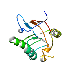
Conserved hypothetical protein from Pyrococcus furiosus Pfu-392566-001
Descriptor: Conserved hypothetical protein, DI(HYDROXYETHYL)ETHER, PHOSPHATE ION, ...
Authors:Shah, A, Liu, Z.J, Tempel, W, Chen, L, Lee, D, Yang, H, Chang, J, Zhao, M, Ng, J, Rose, J, Brereton, P.S, Izumi, M, Jenney Jr, F.E, Poole II, F.L, Shah, C, Sugar, F.J, Adams, M.W.W, Richardson, D.C, Richardson, J.S, Wang, B.C, Southeast Collaboratory for Structural Genomics (SECSG)
Deposit date:2004-04-13
Release date:2004-08-10
Last modified:2023-12-27
Method:X-RAY DIFFRACTION (1.2 Å)
Cite:(NZ)CH...O contacts assist crystallization of a ParB-like nuclease.
Bmc Struct.Biol., 7, 2007
6KGC
DownloadVisualize
BU of 6kgc by Molmil
Crystal structure of CaDoc0917(R49D)-CaCohA2 complex at pH 5.4
Descriptor: And cellulose-binding endoglucanase family 9 CelL ortholog dockerin domain, CALCIUM ION, Probably cellulosomal scaffolding protein, ...
Authors:Feng, Y, Yao, X.
Deposit date:2019-07-11
Release date:2020-07-08
Last modified:2023-11-22
Method:X-RAY DIFFRACTION (1.6 Å)
Cite:Discovery and mechanism of a pH-dependent dual-binding-site switch in the interaction of a pair of protein modules.
Sci Adv, 6, 2020
6KGD
DownloadVisualize
BU of 6kgd by Molmil
Crystal structure of CaDoc0917(R49D)-CaCohA2 complex at pH 8.0
Descriptor: And cellulose-binding endoglucanase family 9 CelL ortholog dockerin domain, CALCIUM ION, Probably cellulosomal scaffolding protein, ...
Authors:Feng, Y, Yao, X.
Deposit date:2019-07-11
Release date:2020-07-08
Last modified:2023-11-22
Method:X-RAY DIFFRACTION (1.65 Å)
Cite:Discovery and mechanism of a pH-dependent dual-binding-site switch in the interaction of a pair of protein modules.
Sci Adv, 6, 2020
6KGE
DownloadVisualize
BU of 6kge by Molmil
Crystal structure of CaDoc0917(R16D)-CaCohA2 complex at pH 5.5
Descriptor: And cellulose-binding endoglucanase family 9 CelL ortholog dockerin domain, CALCIUM ION, Probably cellulosomal scaffolding protein, ...
Authors:Feng, Y, Yao, X.
Deposit date:2019-07-11
Release date:2020-07-08
Last modified:2023-11-22
Method:X-RAY DIFFRACTION (2 Å)
Cite:Discovery and mechanism of a pH-dependent dual-binding-site switch in the interaction of a pair of protein modules.
Sci Adv, 6, 2020
6KG8
DownloadVisualize
BU of 6kg8 by Molmil
Solution structure of CaCohA2 from Clostridium acetobutylicum
Descriptor: Probably cellulosomal scaffolding protein, secreted cellulose-binding and cohesin domain
Authors:Feng, Y, Yao, X.
Deposit date:2019-07-11
Release date:2020-07-08
Last modified:2024-05-15
Method:SOLUTION NMR
Cite:Discovery and mechanism of a pH-dependent dual-binding-site switch in the interaction of a pair of protein modules.
Sci Adv, 6, 2020
6KG9
DownloadVisualize
BU of 6kg9 by Molmil
Solution structure of CaDoc0917 from Clostridium acetobutylicum
Descriptor: And cellulose-binding endoglucanase family 9 CelL ortholog dockerin domain, CALCIUM ION
Authors:Feng, Y, Yao, X.
Deposit date:2019-07-11
Release date:2020-07-08
Last modified:2024-05-01
Method:SOLUTION NMR
Cite:Discovery and mechanism of a pH-dependent dual-binding-site switch in the interaction of a pair of protein modules.
Sci Adv, 6, 2020
6KGF
DownloadVisualize
BU of 6kgf by Molmil
Crystal structure of CaDoc0917(R16D)-CaCohA2 complex at pH 8.2
Descriptor: And cellulose-binding endoglucanase family 9 CelL ortholog dockerin domain, CALCIUM ION, Probably cellulosomal scaffolding protein, ...
Authors:Feng, Y, Yao, X.
Deposit date:2019-07-11
Release date:2020-07-08
Last modified:2023-11-22
Method:X-RAY DIFFRACTION (2.3 Å)
Cite:Discovery and mechanism of a pH-dependent dual-binding-site switch in the interaction of a pair of protein modules.
Sci Adv, 6, 2020
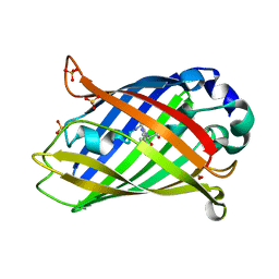
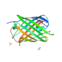
7QLL
DownloadVisualize
BU of 7qll by Molmil
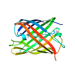
rsKiiro Thermal annealing at 290K of 200K Cis intermediate
Descriptor: GLYCEROL, SULFATE ION, rsKiiro
Authors:van Thor, J.J, Baxter, J.M.
Deposit date:2021-12-20
Release date:2023-07-05
Last modified:2023-11-15
Method:X-RAY DIFFRACTION (1.324 Å)
Cite:Optical control of ultrafast structural dynamics in a fluorescent protein.
Nat.Chem., 15, 2023
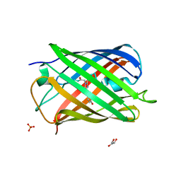
7QLI
DownloadVisualize
BU of 7qli by Molmil
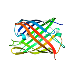
Cis structure of rsKiiro at 290 K
Descriptor: GLYCEROL, SULFATE ION, rsKiiro
Authors:van Thor, J.J, Baxter, J.M.
Deposit date:2021-12-20
Release date:2023-07-05
Last modified:2023-11-15
Method:X-RAY DIFFRACTION (1.155 Å)
Cite:Optical control of ultrafast structural dynamics in a fluorescent protein.
Nat.Chem., 15, 2023
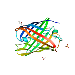
7QLK
DownloadVisualize
BU of 7qlk by Molmil
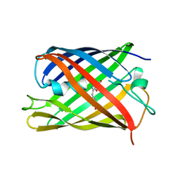
Cis structure intermediate of rsKiiro Illuminated at 200 K
Descriptor: GLYCEROL, SULFATE ION, rsKiiro
Authors:van Thor, J.J, Baxter, J.M.
Deposit date:2021-12-20
Release date:2023-07-05
Last modified:2023-11-15
Method:X-RAY DIFFRACTION (1.458 Å)
Cite:Optical control of ultrafast structural dynamics in a fluorescent protein.
Nat.Chem., 15, 2023
7QLJ
DownloadVisualize
BU of 7qlj by Molmil
Trans structure of rsKiiro Illuminated at 290 K
Descriptor: SULFATE ION, rsKiiro
Authors:van Thor, J.J, Baxter, J.M.
Deposit date:2021-12-20
Release date:2022-11-09
Last modified:2023-11-15
Method:X-RAY DIFFRACTION (1.02 Å)
Cite:Optical control of ultrafast structural dynamics in a fluorescent protein.
Nat.Chem., 15, 2023
7QLN
DownloadVisualize
BU of 7qln by Molmil
rsKiiro pump probe structure by TR-SFX
Descriptor: rsKiiro
Authors:van Thor, J.J.
Deposit date:2021-12-20
Release date:2023-10-18
Last modified:2023-11-15
Method:X-RAY DIFFRACTION (1.5 Å)
Cite:Optical control of ultrafast structural dynamics in a fluorescent protein.
Nat.Chem., 15, 2023
7QLO
DownloadVisualize
BU of 7qlo by Molmil
rsKiiro pump dump probe structure by TR-SFX
Descriptor: rsKiiro
Authors:van Thor, J.J.
Deposit date:2021-12-20
Release date:2023-10-18
Last modified:2023-11-15
Method:X-RAY DIFFRACTION (1.31 Å)
Cite:Optical control of ultrafast structural dynamics in a fluorescent protein.
Nat.Chem., 15, 2023
7QLM
DownloadVisualize
BU of 7qlm by Molmil
rsKiiro trans chromophore dark structure by SFX
Descriptor: rsKiiro
Authors:van Thor, J.J.
Deposit date:2021-12-20
Release date:2023-09-27
Last modified:2023-11-15
Method:X-RAY DIFFRACTION (1.3 Å)
Cite:Optical control of ultrafast structural dynamics in a fluorescent protein.
Nat.Chem., 15, 2023
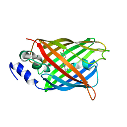
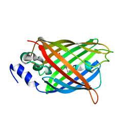
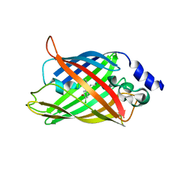
8A6G
DownloadVisualize
BU of 8a6g by Molmil
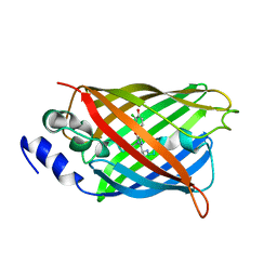
Room temperature rsEGFP2 with a chlorinated chromophore in the non-fluorescent OFF-state
Descriptor: Green fluorescent protein
Authors:Fadini, A, van Thor, J.
Deposit date:2022-06-17
Release date:2023-07-19
Last modified:2024-02-07
Method:X-RAY DIFFRACTION (1.63 Å)
Cite:Serial Femtosecond Crystallography Reveals that Photoactivation in a Fluorescent Protein Proceeds via the Hula Twist Mechanism.
J.Am.Chem.Soc., 145, 2023
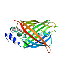
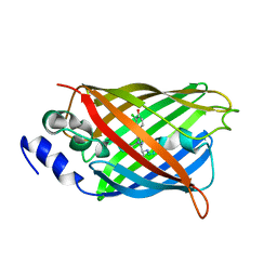
8A6R
DownloadVisualize
BU of 8a6r by Molmil
Room temperature rsEGFP2 with a chlorinated chromophore 100 ps after Photoexcitation
Descriptor: Green fluorescent protein
Authors:Fadini, A, van Thor, J.
Deposit date:2022-06-18
Release date:2023-07-19
Last modified:2024-02-07
Method:X-RAY DIFFRACTION (1.63 Å)
Cite:Serial Femtosecond Crystallography Reveals that Photoactivation in a Fluorescent Protein Proceeds via the Hula Twist Mechanism.
J.Am.Chem.Soc., 145, 2023
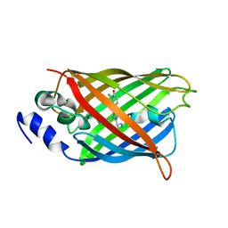
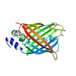
8A6O
DownloadVisualize
BU of 8a6o by Molmil
Room temperature rsEGFP2 with a chlorinated chromophore 600 fs after Photoexcitation
Descriptor: Green fluorescent protein
Authors:Fadini, A, van Thor, J.
Deposit date:2022-06-18
Release date:2023-07-19
Last modified:2024-02-07
Method:X-RAY DIFFRACTION (1.63 Å)
Cite:Serial Femtosecond Crystallography Reveals that Photoactivation in a Fluorescent Protein Proceeds via the Hula Twist Mechanism.
J.Am.Chem.Soc., 145, 2023
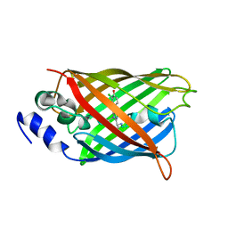
8A6S
DownloadVisualize
BU of 8a6s by Molmil
Room temperature rsEGFP2 with a chlorinated chromophore 1 microsecond after Photoexcitation
Descriptor: Green fluorescent protein
Authors:Fadini, A, van Thor, J.
Deposit date:2022-06-19
Release date:2023-07-19
Last modified:2024-02-07
Method:X-RAY DIFFRACTION (1.63 Å)
Cite:Serial Femtosecond Crystallography Reveals that Photoactivation in a Fluorescent Protein Proceeds via the Hula Twist Mechanism.
J.Am.Chem.Soc., 145, 2023


12>

227111

PDB entries from 2024-11-06

PDB statisticsPDBj update infoContact PDBjnumon