Loading
PDBj
MenuPDBj@FacebookPDBj@TwitterPDBj@YouTubewwPDB FoundationwwPDB
RCSB PDBPDBeBMRBAdv. SearchSearch help
Search by PDB author
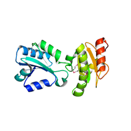
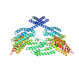
6MHM
DownloadVisualize
BU of 6mhm by Molmil
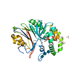
Crystal structure of human acid ceramidase in covalent complex with carmofur
Descriptor: 2-acetamido-2-deoxy-beta-D-glucopyranose, 2-acetamido-2-deoxy-beta-D-glucopyranose-(1-4)-2-acetamido-2-deoxy-beta-D-glucopyranose, 2-acetamido-2-deoxy-beta-D-glucopyranose-(1-4)-2-acetamido-2-deoxy-beta-D-glucopyranose-(1-4)-2-acetamido-2-deoxy-beta-D-glucopyranose, ...
Authors:Dementiev, A, Joachimiak, A, Doan, N.
Deposit date:2018-09-18
Release date:2019-01-23
Last modified:2023-10-11
Method:X-RAY DIFFRACTION (2.743 Å)
Cite:Molecular Mechanism of Inhibition of Acid Ceramidase by Carmofur.
J. Med. Chem., 62, 2019
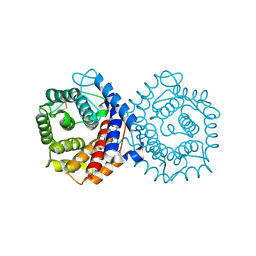
2H8L
DownloadVisualize
BU of 2h8l by Molmil
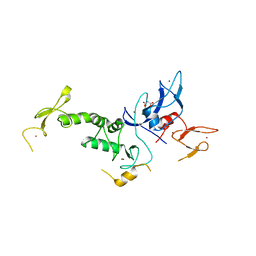
Crystal structure of the bb' fragment of ERp57
Descriptor: Protein disulfide-isomerase A3
Authors:Kozlov, G, Schrag, J.D, Cygler, M, Gehring, K.
Deposit date:2006-06-07
Release date:2006-08-29
Last modified:2011-07-13
Method:X-RAY DIFFRACTION (2 Å)
Cite:Crystal Structure of the bb' Domains of the Protein Disulfide Isomerase ERp57.
Structure, 14, 2006
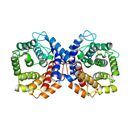
1QQF
DownloadVisualize
BU of 1qqf by Molmil
N-TERMINALLY TRUNCATED C3D,G FRAGMENT OF THE COMPLEMENT SYSTEM
Descriptor: PROTEIN (COMPLEMENT C3DG)
Authors:Zanotti, G, Bassetto, A, Battistutta, R, Stoppini, M, Berni, R.
Deposit date:1999-06-04
Release date:2000-07-31
Last modified:2015-01-21
Method:X-RAY DIFFRACTION (1.45 Å)
Cite:Structure at 1.44 A resolution of an N-terminally truncated form of the rat serum complement C3d fragment.
Biochim.Biophys.Acta, 1478, 2000
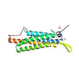
1QSJ
DownloadVisualize
BU of 1qsj by Molmil
N-TERMINALLY TRUNCATED C3DG FRAGMENT
Descriptor: COMPLEMENT C3 PRECURSOR
Authors:Zanotti, G, Bassetto, A, Battistutta, R, Stoppini, M, Folli, C, Berni, R.
Deposit date:1999-06-22
Release date:2000-07-31
Last modified:2021-11-03
Method:X-RAY DIFFRACTION (1.9 Å)
Cite:Structure at 1.44 A resolution of an N-terminally truncated form of the rat serum complement C3d fragment.
Biochim.Biophys.Acta, 1478, 2000
4K1O
DownloadVisualize
BU of 4k1o by Molmil
Crystal structure of the alphaN-catenin actin-binding domain
Descriptor: Catenin alpha-2, SULFATE ION
Authors:Ishiyama, N, Ikura, M.
Deposit date:2013-04-05
Release date:2013-05-01
Last modified:2024-02-28
Method:X-RAY DIFFRACTION (2.603 Å)
Cite:An autoinhibited structure of alpha-catenin and its implications for vinculin recruitment to adherens junctions.
J.Biol.Chem., 288, 2013
4K1N
DownloadVisualize
BU of 4k1n by Molmil
Crystal structure of full-length mouse alphaE-catenin
Descriptor: Catenin alpha-1
Authors:Ishiyama, N, Ikura, M.
Deposit date:2013-04-05
Release date:2013-05-01
Last modified:2023-09-20
Method:X-RAY DIFFRACTION (6.5 Å)
Cite:An autoinhibited structure of alpha-catenin and its implications for vinculin recruitment to adherens junctions.
J.Biol.Chem., 288, 2013
4K7D
DownloadVisualize
BU of 4k7d by Molmil
Crystal Structure of Parkin C-terminal RING domains
Descriptor: CHLORIDE ION, E3 ubiquitin-protein ligase parkin, MALONATE ION, ...
Authors:Sauve, V, Trempe, J.-F, Menade, M, Gehring, K.
Deposit date:2013-04-17
Release date:2013-05-15
Last modified:2024-02-28
Method:X-RAY DIFFRACTION (2.8 Å)
Cite:Structure of parkin reveals mechanisms for ubiquitin ligase activation.
Science, 340, 2013
3OEZ
DownloadVisualize
BU of 3oez by Molmil
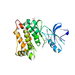
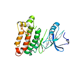
crystal structure of the L317I mutant of the chicken c-Src tyrosine kinase domain complexed with imatinib
Descriptor: 4-(4-METHYL-PIPERAZIN-1-YLMETHYL)-N-[4-METHYL-3-(4-PYRIDIN-3-YL-PYRIMIDIN-2-YLAMINO)-PHENYL]-BENZAMIDE, ACETATE ION, GLYCEROL, ...
Authors:Boubeva, R, Pernot, L, Perozzo, R, Scapozza, L.
Deposit date:2010-08-13
Release date:2011-08-17
Last modified:2023-09-06
Method:X-RAY DIFFRACTION (2.4 Å)
Cite:a single amino-acid dictates the dynamics of the switch between active and inactive C-Src conformation
To be Published
3OF0
DownloadVisualize
BU of 3of0 by Molmil
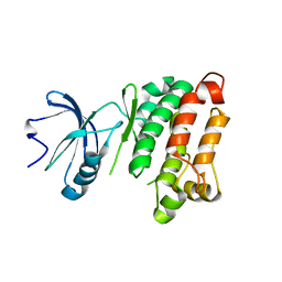
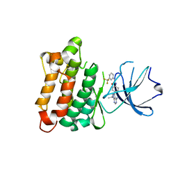
crystal structure of the L317I mutant of the chicken c-Src tyrosine kinase domain
Descriptor: Proto-oncogene tyrosine-protein kinase Src
Authors:Boubeva, R, Pernot, L, Perozzo, R, Scapozza, L.
Deposit date:2010-08-13
Release date:2011-08-17
Last modified:2023-09-06
Method:X-RAY DIFFRACTION (2.7 Å)
Cite:a single amino-acid dictates the dynamics of the switch between active and inactive C-Src conformation
To be Published
3QLG
DownloadVisualize
BU of 3qlg by Molmil
Crystal structure of the L317I mutant of the C-src tyrosine kinase domain complexed with dasatinib
Descriptor: N-(2-CHLORO-6-METHYLPHENYL)-2-({6-[4-(2-HYDROXYETHYL)PIPERAZIN-1-YL]-2-METHYLPYRIMIDIN-4-YL}AMINO)-1,3-THIAZOLE-5-CARBOXAMIDE, Proto-oncogene tyrosine-protein kinase Src
Authors:Boubeva, R, Pernot, L, Perozzo, R, Scapozza, L.
Deposit date:2011-02-02
Release date:2012-02-08
Last modified:2023-09-13
Method:X-RAY DIFFRACTION (2.75 Å)
Cite:A single amino-acid dictates the dynamics of the switch between active and inactive C-src conformation
To be Published
3QLF
DownloadVisualize
BU of 3qlf by Molmil
Crystal structure of the L317I mutant of the C-src tyrosine kinase domain complexed with pyrazolopyrimidine 5
Descriptor: 1-{4-[4-amino-1-(1-methylethyl)-1H-pyrazolo[3,4-d]pyrimidin-3-yl]phenyl}-3-[3-(trifluoromethyl)phenyl]urea, Proto-oncogene tyrosine-protein kinase Src
Authors:Boubeva, R, Pernot, L, Perozzo, R, Scapozza, L.
Deposit date:2011-02-02
Release date:2012-02-08
Last modified:2023-09-13
Method:X-RAY DIFFRACTION (2.75 Å)
Cite:A single amino-acid dictates the dynamics of the switch between active and inactive C-src conformation
To be Published

219869

數據於2024-05-15公開中

PDB statisticsPDBj update infoContact PDBjnumon