Loading
PDBj
MenuPDBj@FacebookPDBj@TwitterPDBj@YouTubewwPDB FoundationwwPDB
RCSB PDBPDBeBMRBAdv. SearchSearch help
Search by PDB author
8OLW
DownloadVisualize
BU of 8olw by Molmil
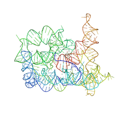
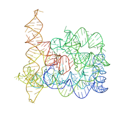
Structure of Oceanobacillus iheyensis group II intron before the first step of splicing in the presence of K+, Ca2+ and intronistat B
Descriptor: CALCIUM ION, Group IIC intron, POTASSIUM ION
Authors:Silvestri, I, Marcia, M.
Deposit date:2023-03-30
Release date:2024-06-19
Last modified:2024-07-03
Method:X-RAY DIFFRACTION (4 Å)
Cite:Targeting the conserved active site of splicing machines with specific and selective small molecule modulators.
Nat Commun, 15, 2024
8OLV
DownloadVisualize
BU of 8olv by Molmil
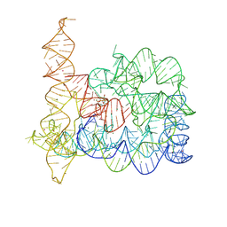
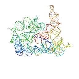
Structure of Oceanobacillus iheyensis group II intron in the presence of K+, Mg2+ and ARN25850
Descriptor: 2-[2,6-bis(bromanyl)-3,4,5-tris(oxidanyl)phenyl]carbonyl-~{N}-(2-pyrrolidin-1-ylethyl)-1-benzofuran-5-carboxamide, 4-(2-HYDROXYETHYL)-1-PIPERAZINE ETHANESULFONIC ACID, Group IIC intron, ...
Authors:Silvestri, I, Marcia, M.
Deposit date:2023-03-30
Release date:2024-06-19
Last modified:2024-07-03
Method:X-RAY DIFFRACTION (2.81 Å)
Cite:Targeting the conserved active site of splicing machines with specific and selective small molecule modulators.
Nat Commun, 15, 2024
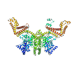
6YC5
DownloadVisualize
BU of 6yc5 by Molmil
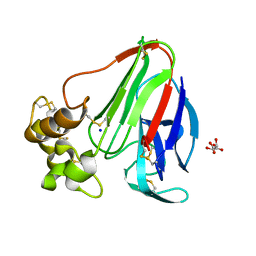
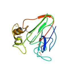
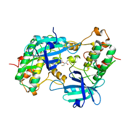
RT structure of Thaumatin obtained at 1.35 A resolution from crystal grown in a Kapton microchip.
Descriptor: L(+)-TARTARIC ACID, SODIUM ION, Thaumatin-1
Authors:Gavira, J.A, Martinez-Rodriguez, S.
Deposit date:2020-03-18
Release date:2020-08-12
Last modified:2024-01-24
Method:X-RAY DIFFRACTION (1.35 Å)
Cite:Attaining atomic resolution from in situ data collection at room temperature using counter-diffusion-based low-cost microchips.
Acta Crystallogr D Struct Biol, 76, 2020
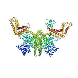
6YBI
DownloadVisualize
BU of 6ybi by Molmil
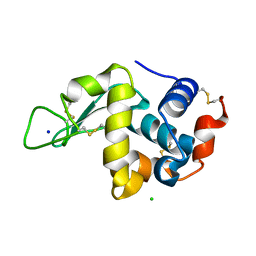
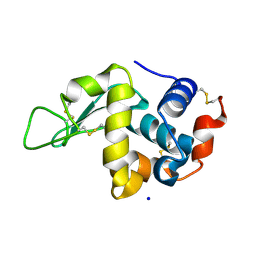
RT structure of HEW Lysozyme obtained at 1.12 A resolution from crystal grown in a Mylar microchip.
Descriptor: CHLORIDE ION, Lysozyme, SODIUM ION
Authors:Gavira, J.A, Martinez-Rodriguez, S.
Deposit date:2020-03-17
Release date:2020-08-12
Last modified:2024-01-24
Method:X-RAY DIFFRACTION (1.12 Å)
Cite:Attaining atomic resolution from in situ data collection at room temperature using counter-diffusion-based low-cost microchips.
Acta Crystallogr D Struct Biol, 76, 2020
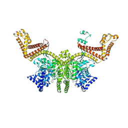
6YBO
DownloadVisualize
BU of 6ybo by Molmil
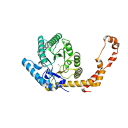
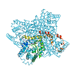
RT structure of Glucose Isomerase obtained at 1.06 A resolution from crystal grown in a Kapton microchip.
Descriptor: MAGNESIUM ION, SODIUM ION, Xylose isomerase
Authors:Gavira, J.A, Martinez-Rodriguez, S.
Deposit date:2020-03-17
Release date:2020-08-12
Last modified:2024-01-24
Method:X-RAY DIFFRACTION (1.06 Å)
Cite:Attaining atomic resolution from in situ data collection at room temperature using counter-diffusion-based low-cost microchips.
Acta Crystallogr D Struct Biol, 76, 2020
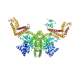
6YBX
DownloadVisualize
BU of 6ybx by Molmil
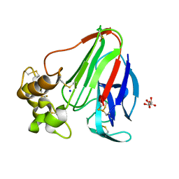
RT structure of Thaumatin obtained at 1.14 A resolution from crystal grown in a Mylar microchip.
Descriptor: L(+)-TARTARIC ACID, SODIUM ION, Thaumatin-1
Authors:Gavira, J.A, Martinez-Rodriguez, S.
Deposit date:2020-03-18
Release date:2020-08-12
Last modified:2024-01-24
Method:X-RAY DIFFRACTION (1.14 Å)
Cite:Attaining atomic resolution from in situ data collection at room temperature using counter-diffusion-based low-cost microchips.
Acta Crystallogr D Struct Biol, 76, 2020
6YBR
DownloadVisualize
BU of 6ybr by Molmil
RT structure of Glucose Isomerase obtained at 1.20 A resolution from crystal grown in a Mylar microchip.
Descriptor: MAGNESIUM ION, SODIUM ION, Xylose isomerase
Authors:Gavira, J.A, Martinez-Rodriguez, S.
Deposit date:2020-03-17
Release date:2020-08-12
Last modified:2024-01-24
Method:X-RAY DIFFRACTION (1.2 Å)
Cite:Attaining atomic resolution from in situ data collection at room temperature using counter-diffusion-based low-cost microchips.
Acta Crystallogr D Struct Biol, 76, 2020
6YBF
DownloadVisualize
BU of 6ybf by Molmil
RT structure of HEW Lysozyme obtained at 1.13 A resolution from crystal grown in a Kapton microchip.
Descriptor: CHLORIDE ION, Lysozyme, SODIUM ION
Authors:Gavira, J, Martinez-Rodriguez, S.
Deposit date:2020-03-17
Release date:2020-08-12
Last modified:2024-01-24
Method:X-RAY DIFFRACTION (1.13 Å)
Cite:Attaining atomic resolution from in situ data collection at room temperature using counter-diffusion-based low-cost microchips.
Acta Crystallogr D Struct Biol, 76, 2020
6ZHJ
DownloadVisualize
BU of 6zhj by Molmil
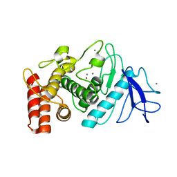
3D electron diffraction structure of thermolysin from Bacillus thermoproteolyticus
Descriptor: CALCIUM ION, Thermolysin, ZINC ION
Authors:Blum, T, Housset, D, Clabbers, M.T.B, van Genderen, E, Schoehn, G, Ling, W.L, Abrahams, J.P.
Deposit date:2020-06-23
Release date:2021-01-27
Last modified:2024-01-24
Method:ELECTRON CRYSTALLOGRAPHY (3.26 Å)
Cite:Statistically correcting dynamical electron scattering improves the refinement of protein nanocrystals, including charge refinement of coordinated metals.
Acta Crystallogr D Struct Biol, 77, 2021
6ZHN
DownloadVisualize
BU of 6zhn by Molmil
3D electron diffraction structure of thaumatin from Thaumatococcus daniellii
Descriptor: CHLORIDE ION, Thaumatin-1
Authors:Blum, T, Housset, D, Clabbers, M.T.B, van Genderen, E, Schoehn, G, Ling, W.L, Abrahams, J.P.
Deposit date:2020-06-23
Release date:2021-01-27
Last modified:2024-01-24
Method:ELECTRON CRYSTALLOGRAPHY (2.76 Å)
Cite:Statistically correcting dynamical electron scattering improves the refinement of protein nanocrystals, including charge refinement of coordinated metals.
Acta Crystallogr D Struct Biol, 77, 2021
1YRP
DownloadVisualize
BU of 1yrp by Molmil
Catalytic domain of human ZIP kinase phosphorylated at Thr265
Descriptor: 2,3-DIHYDROXY-1,4-DITHIOBUTANE, Death-associated protein kinase 3
Authors:Kursula, P, Vahokoski, J, Wilmanns, M.
Deposit date:2005-02-04
Release date:2006-06-20
Last modified:2016-05-18
Method:X-RAY DIFFRACTION (3.1 Å)
Cite:Death-Associated Protein Kinase Activity Is Regulated by Coupled Calcium/Calmodulin Binding to Two Distinct Sites
Structure, 2016
8Q4S
DownloadVisualize
BU of 8q4s by Molmil
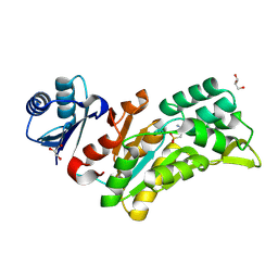
Crystal structure of phosphoserine phosphatase (SerB) from Brucella melitensis in complex with AP4 and magnesium.
Descriptor: (2S)-2-amino-4-phosphonobutanoic acid, GLYCEROL, MAGNESIUM ION, ...
Authors:Scaillet, T, Wouters, J.
Deposit date:2023-08-07
Release date:2024-08-21
Method:X-RAY DIFFRACTION (2.08 Å)
Cite:Crystal structure of phosphoserine phosphatase (SerB) from Brucella melitensis in complex with AP4 and magnesium.
To Be Published
4C0S
DownloadVisualize
BU of 4c0s by Molmil
Mammalian translation elongation factor eEF1A2
Descriptor: ELONGATION FACTOR 1-ALPHA 2, GUANOSINE-5'-DIPHOSPHATE, MAGNESIUM ION
Authors:Crepin, T, Shalak, V.F, Yaremchuk, A.D, Vlasenko, D.O, McCarthy, A.A, Negrutskii, B.S, Tukalo, M.A, El'skaya, A.V.
Deposit date:2013-08-07
Release date:2014-08-27
Last modified:2017-01-25
Method:X-RAY DIFFRACTION (2.703 Å)
Cite:Mammalian Translation Elongation Factor Eef1A2: X-Ray Structure and New Features of Gdp/GTP Exchange Mechanism in Higher Eukaryotes
Nucleic Acids Res., 42, 2014
8BHD
DownloadVisualize
BU of 8bhd by Molmil
N-terminal domain of Plasmodium berghei glutamyl-tRNA synthetase (Tbxo4 derivative crystal structure)
Descriptor: GLYCEROL, Glutamate--tRNA ligase, SULFATE ION, ...
Authors:Benas, P, Jaramillo Ponce, J.R, Legrand, P, Frugier, M, Sauter, C.
Deposit date:2022-10-31
Release date:2023-01-25
Last modified:2024-06-19
Method:X-RAY DIFFRACTION (3.17 Å)
Cite:Solution X-ray scattering highlights discrepancies in Plasmodium multi-aminoacyl-tRNA synthetase complexes.
Protein Sci., 32, 2023
4N0K
DownloadVisualize
BU of 4n0k by Molmil
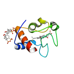
Atomic resolution crystal structure of a cytochrome c-calixarene complex
Descriptor: 25,26,27,28-tetrahydroxypentacyclo[19.3.1.1~3,7~.1~9,13~.1~15,19~]octacosa-1(25),3(28),4,6,9(27),10,12,15(26),16,18,21,23-dodecaene-5,11,17,23-tetrasulfonic acid, Cytochrome c iso-1, HEME C
Authors:McGovern, R.E, Pye, V.E, Crowley, P.B.
Deposit date:2013-10-02
Release date:2014-09-10
Last modified:2023-09-20
Method:X-RAY DIFFRACTION (1.05 Å)
Cite:A cytochrome c-calixarene structure at atomic resolution
To be Published
2FSF
DownloadVisualize
BU of 2fsf by Molmil
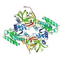
Escherichia coli SecA, the preprotein translocase dimeric ATPase
Descriptor: Preprotein translocase secA subunit
Authors:Papanikolau, Y, Petratos, K, Economou, A.
Deposit date:2006-01-23
Release date:2007-01-16
Last modified:2024-03-13
Method:X-RAY DIFFRACTION (2 Å)
Cite:Structure of dimeric SecA, the Escherichia coli preprotein translocase motor.
J.Mol.Biol., 366, 2007
2FSG
DownloadVisualize
BU of 2fsg by Molmil
Complex SecA:ATP from Escherichia coli
Descriptor: ADENOSINE-5'-TRIPHOSPHATE, Preprotein translocase secA subunit
Authors:Papanikolau, Y, Petratos, K, Economou, A.
Deposit date:2006-01-23
Release date:2007-01-16
Last modified:2011-07-13
Method:X-RAY DIFFRACTION (2.2 Å)
Cite:Structure of dimeric SecA, the Escherichia coli preprotein translocase motor.
J.Mol.Biol., 366, 2007
2FSH
DownloadVisualize
BU of 2fsh by Molmil
Complex SecA:AMP-PNP from Escherichia coli
Descriptor: PHOSPHOAMINOPHOSPHONIC ACID-ADENYLATE ESTER, Preprotein translocase secA subunit
Authors:Papanikolau, Y, Petratos, K, Economou, A.
Deposit date:2006-01-23
Release date:2007-01-16
Last modified:2023-10-25
Method:X-RAY DIFFRACTION (2 Å)
Cite:Structure of dimeric SecA, the Escherichia coli preprotein translocase motor.
J.Mol.Biol., 366, 2007
2FSI
DownloadVisualize
BU of 2fsi by Molmil
Complex SecA:ADP from Escherichia coli
Descriptor: ADENOSINE-5'-DIPHOSPHATE, Preprotein translocase secA subunit
Authors:Papanikolau, Y, Petratos, K, Economou, A.
Deposit date:2006-01-23
Release date:2007-01-16
Last modified:2023-10-25
Method:X-RAY DIFFRACTION (2.11 Å)
Cite:Structure of dimeric SecA, the Escherichia coli preprotein translocase motor.
J.Mol.Biol., 366, 2007
8RUM
DownloadVisualize
BU of 8rum by Molmil
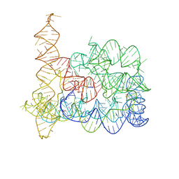
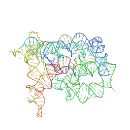
Structure of Oceanobacillus iheyensis group II intron in the presence of Li+, Mg2+, and intronistat B
Descriptor: Domains 1-5, MAGNESIUM ION, ~{N}-(2-pyrrolidin-1-ylethyl)-2-[3,4,5-tris(oxidanyl)phenyl]carbonyl-1-benzofuran-5-carboxamide
Authors:Silvestri, I, Marcia, M.
Deposit date:2024-01-31
Release date:2024-06-19
Last modified:2024-07-03
Method:X-RAY DIFFRACTION (3.62 Å)
Cite:Targeting the conserved active site of splicing machines with specific and selective small molecule modulators.
Nat Commun, 15, 2024
8RUI
DownloadVisualize
BU of 8rui by Molmil
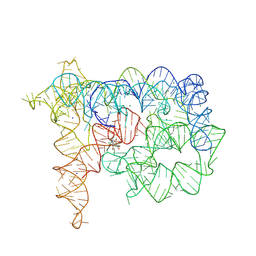
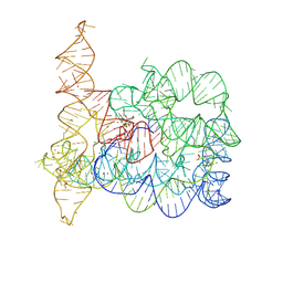
Structure of Oceanobacillus iheyensis group II intron in the presence of K+, Mg2+, 5'-exon, and intronistat B after 1h soaking
Descriptor: Domains 1-5, MAGNESIUM ION, POTASSIUM ION, ...
Authors:Silvestri, I, Marcia, M.
Deposit date:2024-01-30
Release date:2024-06-19
Last modified:2024-07-03
Method:X-RAY DIFFRACTION (3.31 Å)
Cite:Targeting the conserved active site of splicing machines with specific and selective small molecule modulators.
Nat Commun, 15, 2024
8RUH
DownloadVisualize
BU of 8ruh by Molmil
Structure of Oceanobacillus iheyensis group II intron in the presence of K+, Mg2+ and 5'-exon
Descriptor: 4-(2-HYDROXYETHYL)-1-PIPERAZINE ETHANESULFONIC ACID, 5'-exon, Domains 1-5, ...
Authors:Silvestri, I, Marcia, M.
Deposit date:2024-01-30
Release date:2024-06-19
Last modified:2024-07-03
Method:X-RAY DIFFRACTION (4.55 Å)
Cite:Targeting the conserved active site of splicing machines with specific and selective small molecule modulators.
Nat Commun, 15, 2024
8RUK
DownloadVisualize
BU of 8ruk by Molmil
Structure of Oceanobacillus iheyensis group II intron in the presence of Na+, Mg2+, and ARN25850
Descriptor: 2-[2,6-bis(bromanyl)-3,4,5-tris(oxidanyl)phenyl]carbonyl-~{N}-(2-pyrrolidin-1-ylethyl)-1-benzofuran-5-carboxamide, Domains 1-5, MAGNESIUM ION, ...
Authors:Silvestri, I, Marcia, M.
Deposit date:2024-01-31
Release date:2024-06-19
Last modified:2024-07-03
Method:X-RAY DIFFRACTION (4.81 Å)
Cite:Targeting the conserved active site of splicing machines with specific and selective small molecule modulators.
Nat Commun, 15, 2024
8RUJ
DownloadVisualize
BU of 8ruj by Molmil
Structure of Oceanobacillus iheyensis group II intron in the presence of K+, Mg2+, 5'-exon, and ARN25850 after 1h soaking
Descriptor: 2-[2,6-bis(bromanyl)-3,4,5-tris(oxidanyl)phenyl]carbonyl-~{N}-(2-pyrrolidin-1-ylethyl)-1-benzofuran-5-carboxamide, Domains 1-5, MAGNESIUM ION, ...
Authors:Silvestri, I, Marcia, M.
Deposit date:2024-01-30
Release date:2024-06-19
Last modified:2024-07-03
Method:X-RAY DIFFRACTION (3.12 Å)
Cite:Targeting the conserved active site of splicing machines with specific and selective small molecule modulators.
Nat Commun, 15, 2024
8RUL
DownloadVisualize
BU of 8rul by Molmil
Structure of Oceanobacillus iheyensis group II intron in the presence of Li+ and Mg2+
Descriptor: 4-(2-HYDROXYETHYL)-1-PIPERAZINE ETHANESULFONIC ACID, Domains 1-5, MAGNESIUM ION
Authors:Silvestri, I, Marcia, M.
Deposit date:2024-01-31
Release date:2024-06-19
Last modified:2024-07-03
Method:X-RAY DIFFRACTION (3.62 Å)
Cite:Targeting the conserved active site of splicing machines with specific and selective small molecule modulators.
Nat Commun, 15, 2024

225158

PDB entries from 2024-09-18

PDB statisticsPDBj update infoContact PDBjnumon