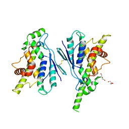
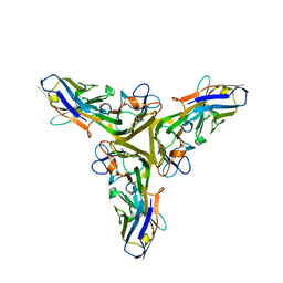
2GN7
 
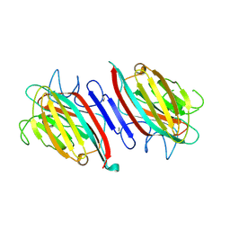 | | Metal-free (apo) P. angolensis seed lectin in complex with Man-alpha(1-3)Man-alpha(1-6)Man | | 分子名称: | alpha-D-mannopyranose, beta-D-mannopyranose-(1-3)-[alpha-D-mannopyranose-(1-6)]alpha-D-mannopyranose, lectin | | 著者 | Garcia-Pino, A, Buts, L, Wyns, L, Loris, R. | | 登録日 | 2006-04-09 | | 公開日 | 2006-07-25 | | 最終更新日 | 2020-07-29 | | 実験手法 | X-RAY DIFFRACTION (2.9 Å) | | 主引用文献 | Interplay Between Metal Binding and cis/trans Isomerization in Legume Lectins: Structural and Thermodynamic Study of P. angolensis Lectin.
J.Mol.Biol., 361, 2006
|
|
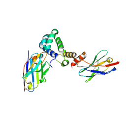
2GMP
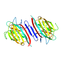 | | Metal-free (apo) P. angolensis seed lectin in complex with GlcNAC-beta(1-2)Man | | 分子名称: | 2-acetamido-2-deoxy-beta-D-glucopyranose-(1-2)-alpha-D-mannopyranose, SULFATE ION, lectin | | 著者 | Garcia-Pino, A, Buts, L, Wyns, L, Loris, R. | | 登録日 | 2006-04-07 | | 公開日 | 2006-07-25 | | 最終更新日 | 2020-07-29 | | 実験手法 | X-RAY DIFFRACTION (2.5 Å) | | 主引用文献 | Interplay Between Metal Binding and cis/trans Isomerization in Legume Lectins: Structural and Thermodynamic Study of P. angolensis Lectin.
J.Mol.Biol., 361, 2006
|
|
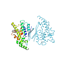
2GNT
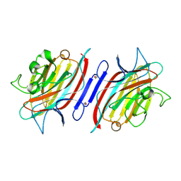 | | EDTA treated P. angolensis lectin (PAL) remetallized with calcium (1 hour treatment) | | 分子名称: | CALCIUM ION, lectin | | 著者 | Garcia-Pino, A, Buts, L, Wyns, L, Loris, R. | | 登録日 | 2006-04-11 | | 公開日 | 2006-07-25 | | 最終更新日 | 2019-12-25 | | 実験手法 | X-RAY DIFFRACTION (2.02 Å) | | 主引用文献 | Interplay Between Metal Binding and cis/trans Isomerization in Legume Lectins: Structural and Thermodynamic Study of P. angolensis Lectin.
J.Mol.Biol., 361, 2006
|
|
2GNB
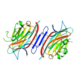 | | EDTA-treated (2 weeks) P. angolensis lectin | | 分子名称: | alpha-D-mannopyranose, lectin | | 著者 | Garcia-Pino, A, Buts, L, Wyns, L, Loris, R. | | 登録日 | 2006-04-09 | | 公開日 | 2006-07-25 | | 最終更新日 | 2020-07-29 | | 実験手法 | X-RAY DIFFRACTION (2.27 Å) | | 主引用文献 | Interplay Between Metal Binding and cis/trans Isomerization in Legume Lectins: Structural and Thermodynamic Study of P. angolensis Lectin.
J.Mol.Biol., 361, 2006
|
|
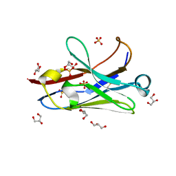
6GSP
 | | RIBONUCLEASE T1/3'-GMP, 15 WEEKS | | 分子名称: | CALCIUM ION, GUANOSINE-3'-MONOPHOSPHATE, RIBONUCLEASE T1 | | 著者 | Zegers, I, Wyns, L. | | 登録日 | 1997-12-09 | | 公開日 | 1998-03-18 | | 最終更新日 | 2023-08-09 | | 実験手法 | X-RAY DIFFRACTION (2.2 Å) | | 主引用文献 | Hydrolysis of a slow cyclic thiophosphate substrate of RNase T1 analyzed by time-resolved crystallography.
Nat.Struct.Biol., 5, 1998
|
|
7GSP
 | | RIBONUCLEASE T1/2',3'-CGPS, NON-PRODUCTIVE | | 分子名称: | GUANOSINE-2',3'-CYCLOPHOSPHOROTHIOATE, PHOSPHATE ION, RIBONUCLEASE T1 | | 著者 | Zegers, I, Wyns, L. | | 登録日 | 1997-12-10 | | 公開日 | 1998-03-18 | | 最終更新日 | 2023-08-09 | | 実験手法 | X-RAY DIFFRACTION (2 Å) | | 主引用文献 | Hydrolysis of a slow cyclic thiophosphate substrate of RNase T1 analyzed by time-resolved crystallography.
Nat.Struct.Biol., 5, 1998
|
|
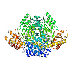
3N5F
 | | Crystal Structure of L-N-carbamoylase from Geobacillus stearothermophilus CECT43 | | 分子名称: | CACODYLATE ION, COBALT (II) ION, ISOPROPYL ALCOHOL, ... | | 著者 | Garcia-Pino, A, Martinez-Rodriguez, S, Gavira, J.A. | | 登録日 | 2010-05-25 | | 公開日 | 2011-05-25 | | 最終更新日 | 2023-09-06 | | 実験手法 | X-RAY DIFFRACTION (2.75 Å) | | 主引用文献 | Mutational and structural analysis of L-N-carbamoylase reveals new insights into a peptidase m20/m25/m40 family member.
J.Bacteriol., 194, 2012
|
|
7Q6K
 | |
7Q6J
 | |
7R1Z
 | |
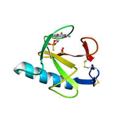
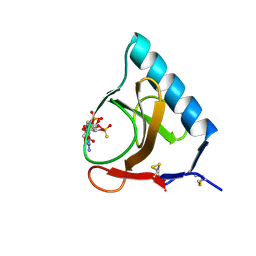
7R20
 | | Anti-Arc nanobody E5 | | 分子名称: | Anti-Arc nanobody E5, GLYCEROL, SULFATE ION | | 著者 | Markusson, S, Kursula, P. | | 登録日 | 2022-02-03 | | 公開日 | 2022-06-01 | | 最終更新日 | 2024-01-31 | | 実験手法 | X-RAY DIFFRACTION (1.42 Å) | | 主引用文献 | High-affinity anti-Arc nanobodies provide tools for structural and functional studies.
Plos One, 17, 2022
|
|
7YWD
 | |
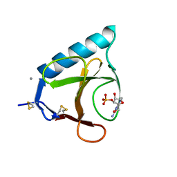
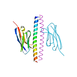
6YTU
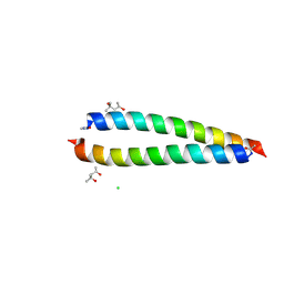 | | Atomic-resolution structure of the coiled-coil dimerisation domain of human Arc | | 分子名称: | (4S)-2-METHYL-2,4-PENTANEDIOL, Activity-regulated cytoskeleton-associated protein, CHLORIDE ION | | 著者 | Hallin, E.I, Touma, C, Bramham, C.R, Kursula, P. | | 登録日 | 2020-04-24 | | 公開日 | 2021-03-03 | | 最終更新日 | 2021-05-12 | | 実験手法 | X-RAY DIFFRACTION (0.95 Å) | | 主引用文献 | Arc self-association and formation of virus-like capsids are mediated by an N-terminal helical coil motif.
Febs J., 288, 2021
|
|
7A4Y
 | |
7A4T
 | |
7A50
 | |
7A48
 | |
7A4D
 | |
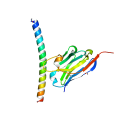
5M2J
 | | Complex between human TNF alpha and Llama VHH2 | | 分子名称: | Anti-(ED-B) scFV, Tumor necrosis factor | | 著者 | Cambillau, C, Spinelli, S, Desmyter, A, de Haard, H. | | 登録日 | 2016-10-13 | | 公開日 | 2017-08-30 | | 最終更新日 | 2024-01-17 | | 実験手法 | X-RAY DIFFRACTION (1.9 Å) | | 主引用文献 | Bivalent Llama Single-Domain Antibody Fragments against Tumor Necrosis Factor Have Picomolar Potencies due to Intramolecular Interactions.
Front Immunol, 8, 2017
|
|
5M2M
 | | Complex between human TNF alpha and Llama VHH3 | | 分子名称: | Llama nanobody VHH3, Tumor necrosis factor | | 著者 | Cambillau, C, Spinelli, S, Desmyter, A, de Haard, H. | | 登録日 | 2016-10-13 | | 公開日 | 2017-08-30 | | 最終更新日 | 2024-01-17 | | 実験手法 | X-RAY DIFFRACTION (2.3 Å) | | 主引用文献 | Bivalent Llama Single-Domain Antibody Fragments against Tumor Necrosis Factor Have Picomolar Potencies due to Intramolecular Interactions.
Front Immunol, 8, 2017
|
|
5M2I
 | | Structure of human Tumor Necrosis Factor (TNF) in complex with the Llama VHH1 | | 分子名称: | Tumor necrosis factor, VHH1 | | 著者 | Cambillau, C, Spinelli, S, Desmyter, A, de Haard, H. | | 登録日 | 2016-10-13 | | 公開日 | 2017-08-30 | | 最終更新日 | 2024-01-17 | | 実験手法 | X-RAY DIFFRACTION (2.15 Å) | | 主引用文献 | Bivalent Llama Single-Domain Antibody Fragments against Tumor Necrosis Factor Have Picomolar Potencies due to Intramolecular Interactions.
Front Immunol, 8, 2017
|
|
4GSP
 | | RIBONUCLEASE T1 COMPLEXED WITH 2',3'-CGPS + 3'-GMP, 7 DAYS | | 分子名称: | CALCIUM ION, GUANOSINE-2',3'-CYCLOPHOSPHOROTHIOATE, GUANOSINE-3'-MONOPHOSPHATE, ... | | 著者 | Zegers, I, Wyns, L. | | 登録日 | 1997-12-02 | | 公開日 | 1998-08-12 | | 最終更新日 | 2023-08-09 | | 実験手法 | X-RAY DIFFRACTION (1.65 Å) | | 主引用文献 | Hydrolysis of a slow cyclic thiophosphate substrate of RNase T1 analyzed by time-resolved crystallography.
Nat.Struct.Biol., 5, 1998
|
|
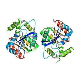
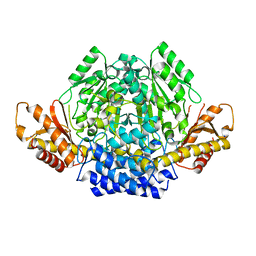
6ZEK
 | | Crystal structure of mouse CSAD | | 分子名称: | CHLORIDE ION, COBALT (II) ION, Cysteine sulfinic acid decarboxylase, ... | | 著者 | Mahootchi, E, Raasakka, A, Haavik, J, Kursula, P. | | 登録日 | 2020-06-16 | | 公開日 | 2021-04-28 | | 最終更新日 | 2024-01-24 | | 実験手法 | X-RAY DIFFRACTION (2.1 Å) | | 主引用文献 | Structure and substrate specificity determinants of the taurine biosynthetic enzyme cysteine sulphinic acid decarboxylase.
J.Struct.Biol., 213, 2021
|
|
6ZMM
 | |
7A0A
 | |


