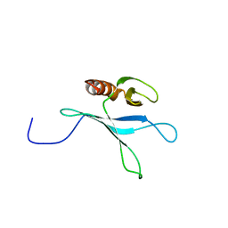
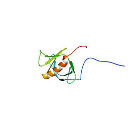
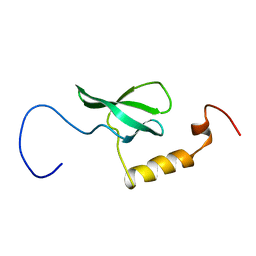
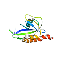
2D9U
 
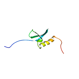 | | Solution structure of the Chromo domain of chromobox homolog 2 from human | | Descriptor: | Chromobox protein homolog 2 (isoform 2) | | Authors: | Li, H, Saito, K, Koshiba, S, Inoue, M, Kigawa, T, Yokoyama, S, RIKEN Structural Genomics/Proteomics Initiative (RSGI) | | Deposit date: | 2005-12-13 | | Release date: | 2007-01-02 | | Last modified: | 2022-03-09 | | Method: | SOLUTION NMR | | Cite: | Solution structure of the Chromo domain of chromobox homolog 2 from human
To be Published
|
|
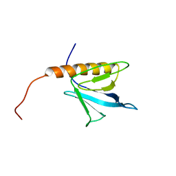
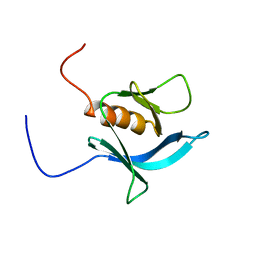
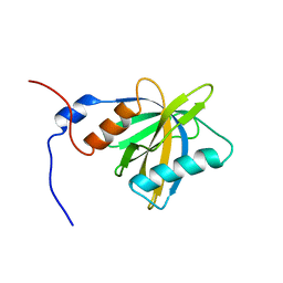
2D9V
 | | Solution structure of the PH domain of Pleckstrin homology domain-containing protein family B member 1 from mouse | | Descriptor: | Pleckstrin homology domain-containing protein family B member 1 | | Authors: | Li, H, Tomizawa, T, Koshiba, S, Inoue, M, Kigawa, T, Yokoyama, S, RIKEN Structural Genomics/Proteomics Initiative (RSGI) | | Deposit date: | 2005-12-13 | | Release date: | 2006-06-13 | | Last modified: | 2022-03-09 | | Method: | SOLUTION NMR | | Cite: | Solution structure of the PH domain of Pleckstrin homology domain-containing protein family B member 1 from mouse
To be Published
|
|
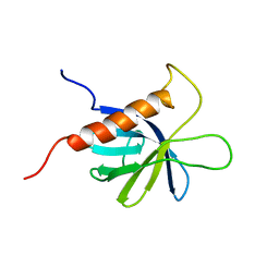
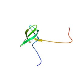
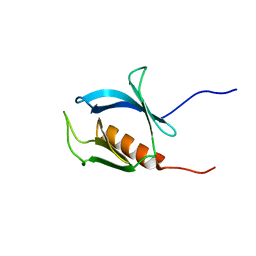
2D9X
 | | Solution structure of the PH domain of Oxysterol binding protein-related protein 11 from human | | Descriptor: | Oxysterol binding protein-related protein 11 | | Authors: | Li, H, Sato, M, Koshiba, S, Inoue, M, Kigawa, T, Yokoyama, S, RIKEN Structural Genomics/Proteomics Initiative (RSGI) | | Deposit date: | 2005-12-13 | | Release date: | 2006-06-13 | | Last modified: | 2022-03-09 | | Method: | SOLUTION NMR | | Cite: | Solution structure of the PH domain of Oxysterol binding protein-related protein 11 from human
To be Published
|
|
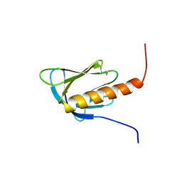
2D9Z
 | | Solution structure of the PH domain of Protein kinase C, nu type from human | | Descriptor: | Protein kinase C, nu type | | Authors: | Li, H, Tomizawa, T, Koshiba, S, Inoue, M, Kigawa, T, Yokoyama, S, RIKEN Structural Genomics/Proteomics Initiative (RSGI) | | Deposit date: | 2005-12-13 | | Release date: | 2007-01-02 | | Last modified: | 2022-03-09 | | Method: | SOLUTION NMR | | Cite: | Solution structure of the PH domain of Protein kinase C, nu type from human
To be Published
|
|
2D9W
 | | Solution structure of the PH domain of Docking protein 2 from human | | Descriptor: | Docking protein 2 | | Authors: | Li, H, Tomizawa, T, Koshiba, S, Inoue, M, Kigawa, T, Yokoyama, S, RIKEN Structural Genomics/Proteomics Initiative (RSGI) | | Deposit date: | 2005-12-13 | | Release date: | 2006-06-13 | | Last modified: | 2022-03-09 | | Method: | SOLUTION NMR | | Cite: | Solution structure of the PH domain of Docking protein 2 from human
To be Published
|
|
2D9Y
 | | Solution structure of the PH domain of PEPP-3 from human | | Descriptor: | Pleckstrin homology domain-containing protein family A member 6 | | Authors: | Li, H, Tochio, N, Koshiba, S, Inoue, M, Kigawa, T, Yokoyama, S, RIKEN Structural Genomics/Proteomics Initiative (RSGI) | | Deposit date: | 2005-12-13 | | Release date: | 2006-06-13 | | Last modified: | 2022-03-09 | | Method: | SOLUTION NMR | | Cite: | Solution structure of the PH domain of PEPP-3 from human
To be Published
|
|
2DA0
 | | Solution structure of the PH domain of PIP2-dependent ARF1 GTPase-activating protein from human | | Descriptor: | 130-kDa phosphatidylinositol 4,5-biphosphate-dependent ARF1 GTPase-activating protein | | Authors: | Li, H, Tomizawa, T, Koshiba, S, Inoue, M, Kigawa, T, Yokoyama, S, RIKEN Structural Genomics/Proteomics Initiative (RSGI) | | Deposit date: | 2005-12-13 | | Release date: | 2006-06-13 | | Last modified: | 2022-03-09 | | Method: | SOLUTION NMR | | Cite: | Solution structure of the PH domain of PIP2-dependent ARF1 GTPase-activating protein from human
To be Published
|
|
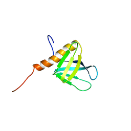
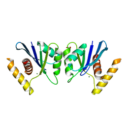
2D9T
 | | Solution structure of the Tudor domain of Tudor domain containing protein 3 from mouse | | Descriptor: | Tudor domain-containing protein 3 | | Authors: | Li, H, Tomizawa, T, Koshiba, S, Inoue, M, Kigawa, T, Yokoyama, S, RIKEN Structural Genomics/Proteomics Initiative (RSGI) | | Deposit date: | 2005-12-13 | | Release date: | 2006-06-13 | | Last modified: | 2022-03-09 | | Method: | SOLUTION NMR | | Cite: | Solution structure of the Tudor domain of Tudor domain containing protein 3 from mouse
To be Published
|
|
2DKP
 | | Solution structure of the PH domain of pleckstrin homology domain-containing protein family A member 5 from human | | Descriptor: | Pleckstrin homology domain-containing family A member 5 | | Authors: | Li, H, Tomizawa, T, Koshiba, S, Inoue, M, Kigawa, T, Yokoyama, S, RIKEN Structural Genomics/Proteomics Initiative (RSGI) | | Deposit date: | 2006-04-13 | | Release date: | 2006-10-13 | | Last modified: | 2022-03-09 | | Method: | SOLUTION NMR | | Cite: | Solution structure of the PH domain of pleckstrin homology domain-containing protein family A member 5 from human
To be Published
|
|
2DKQ
 | | Solution structure of the PTB domain of KIAA1075 protein from human | | Descriptor: | KIAA1075 protein | | Authors: | Li, H, Tochio, N, Koshiba, S, Inoue, M, Kigawa, T, Yokoyama, S, RIKEN Structural Genomics/Proteomics Initiative (RSGI) | | Deposit date: | 2006-04-13 | | Release date: | 2006-10-13 | | Last modified: | 2022-03-09 | | Method: | SOLUTION NMR | | Cite: | Solution structure of the PTB domain of KIAA1075 protein from human
To be Published
|
|
2DHI
 | | Solution structure of the PH domain of Evectin-2 from mouse | | Descriptor: | Pleckstrin homology domain-containing family B member 2 | | Authors: | Li, H, Yoneyama, M, Koshiba, S, Inoue, M, Kigawa, T, Yokoyama, S, RIKEN Structural Genomics/Proteomics Initiative (RSGI) | | Deposit date: | 2006-03-24 | | Release date: | 2006-09-24 | | Last modified: | 2022-03-09 | | Method: | SOLUTION NMR | | Cite: | Solution structure of the PH domain of Evectin-2 from mouse
To be Published
|
|
2DN6
 | | Solution structure of the PH domain of KIAA0640 protein from human | | Descriptor: | KIAA0640 protein | | Authors: | Li, H, Tochio, N, Koshiba, S, Inoue, M, Kigawa, T, Yokoyama, S, RIKEN Structural Genomics/Proteomics Initiative (RSGI) | | Deposit date: | 2006-04-25 | | Release date: | 2006-10-25 | | Last modified: | 2022-03-09 | | Method: | SOLUTION NMR | | Cite: | Solution structure of the PH domain of KIAA0640 protein from human
To be Published
|
|
2DHJ
 | | Solution structure of the PH domain of Rho GTPase activating protein 21 from human | | Descriptor: | Rho GTPase activating protein 21 | | Authors: | Li, H, Tochio, N, Koshiba, S, Inoue, M, Kigawa, T, Yokoyama, S, RIKEN Structural Genomics/Proteomics Initiative (RSGI) | | Deposit date: | 2006-03-24 | | Release date: | 2006-09-24 | | Last modified: | 2022-03-09 | | Method: | SOLUTION NMR | | Cite: | Solution structure of the PH domain of Rho GTPase activating protein 21 from human
To be Published
|
|
2DHK
 | | Solution structure of the PH domain of TBC1 domain family member 2 protein from human | | Descriptor: | TBC1 domain family member 2 | | Authors: | Li, H, Tochio, N, Koshiba, S, Harada, T, Watanabe, S, Kigawa, T, Yokoyama, S, RIKEN Structural Genomics/Proteomics Initiative (RSGI) | | Deposit date: | 2006-03-24 | | Release date: | 2006-09-24 | | Last modified: | 2022-03-09 | | Method: | SOLUTION NMR | | Cite: | Solution structure of the PH domain of TBC1 domain family member 2 protein from human
To be Published
|
|
2E6J
 | | Solution structure of the C-terminal PapD-like domain from human HYDIN protein | | Descriptor: | HYDIN protein | | Authors: | Li, H, Sato, M, Koshiba, S, Watanabe, S, Harada, T, Kigawa, T, Yokoyama, S, RIKEN Structural Genomics/Proteomics Initiative (RSGI) | | Deposit date: | 2006-12-27 | | Release date: | 2007-07-03 | | Last modified: | 2022-03-09 | | Method: | SOLUTION NMR | | Cite: | Solution structure of the C-terminal PapD-like domain from human HYDIN protein
To be Published
|
|
2E6I
 | | Solution structure of the BTK motif of tyrosine-protein kinase ITK from human | | Descriptor: | Tyrosine-protein kinase ITK/TSK, ZINC ION | | Authors: | Li, H, Tochio, N, Koshiba, S, Watanabe, S, Harada, T, Kigawa, T, Yokoyama, S, RIKEN Structural Genomics/Proteomics Initiative (RSGI) | | Deposit date: | 2006-12-27 | | Release date: | 2007-07-03 | | Last modified: | 2022-03-09 | | Method: | SOLUTION NMR | | Cite: | Solution structure of the BTK motif of tyrosine-protein kinase ITK from human
To be Published
|
|
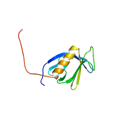
2EFI
 | | Solution structure of the chromo domain of Mortality factor 4-like protein 1 from human | | Descriptor: | Mortality factor 4-like protein 1 | | Authors: | Li, H, Sato, M, Tochio, N, Tomizawa, T, Koshiba, S, Harada, T, Watanabe, S, Kigawa, T, Yokoyama, S, RIKEN Structural Genomics/Proteomics Initiative (RSGI) | | Deposit date: | 2007-02-22 | | Release date: | 2007-08-28 | | Last modified: | 2022-03-09 | | Method: | SOLUTION NMR | | Cite: | Solution structure of the chromo domain of Mortality factor 4-like protein 1 from human
To be Published
|
|
2F6N
 | |
2F6J
 | |
2FSA
 | |
7CJI
 | | Photosystem II structure in the S1 state | | Descriptor: | 1,2-DI-O-ACYL-3-O-[6-DEOXY-6-SULFO-ALPHA-D-GLUCOPYRANOSYL]-SN-GLYCEROL, 1,2-DIPALMITOYL-PHOSPHATIDYL-GLYCEROLE, 1,2-DISTEAROYL-MONOGALACTOSYL-DIGLYCERIDE, ... | | Authors: | Li, H, Shen, J.-R, Suga, M. | | Deposit date: | 2020-07-11 | | Release date: | 2021-04-28 | | Last modified: | 2023-11-29 | | Method: | X-RAY DIFFRACTION (2.35 Å) | | Cite: | Capturing structural changes of the S 1 to S 2 transition of photosystem II using time-resolved serial femtosecond crystallography.
Iucrj, 8, 2021
|
|
7CJJ
 | | Photosystem II structure in the S2 state | | Descriptor: | 1,2-DI-O-ACYL-3-O-[6-DEOXY-6-SULFO-ALPHA-D-GLUCOPYRANOSYL]-SN-GLYCEROL, 1,2-DIPALMITOYL-PHOSPHATIDYL-GLYCEROLE, 1,2-DISTEAROYL-MONOGALACTOSYL-DIGLYCERIDE, ... | | Authors: | Li, H, Shen, J.-R, Suga, M. | | Deposit date: | 2020-07-11 | | Release date: | 2021-04-28 | | Last modified: | 2024-04-03 | | Method: | X-RAY DIFFRACTION (2.4 Å) | | Cite: | Capturing structural changes of the S 1 to S 2 transition of photosystem II using time-resolved serial femtosecond crystallography.
Iucrj, 8, 2021
|
|
7COU
 | | Structure of cyanobacterial photosystem II in the dark S1 state | | Descriptor: | 1,2-DI-O-ACYL-3-O-[6-DEOXY-6-SULFO-ALPHA-D-GLUCOPYRANOSYL]-SN-GLYCEROL, 1,2-DIPALMITOYL-PHOSPHATIDYL-GLYCEROLE, 1,2-DISTEAROYL-MONOGALACTOSYL-DIGLYCERIDE, ... | | Authors: | Li, H, Shen, J.-R, Suga, M. | | Deposit date: | 2020-08-05 | | Release date: | 2021-04-28 | | Last modified: | 2023-11-29 | | Method: | X-RAY DIFFRACTION (2.25 Å) | | Cite: | Capturing structural changes of the S 1 to S 2 transition of photosystem II using time-resolved serial femtosecond crystallography.
Iucrj, 8, 2021
|
|
2KDW
 | |
5E6F
 | | Canarypox virus resolvase | | Descriptor: | CNPV261 Holliday junction resolvase protein, D(-)-TARTARIC ACID, MAGNESIUM ION | | Authors: | Li, H, Hwang, Y, Perry, K, Bushman, F.D, Van Duyne, G.D. | | Deposit date: | 2015-10-09 | | Release date: | 2016-03-30 | | Last modified: | 2023-09-27 | | Method: | X-RAY DIFFRACTION (2.6 Å) | | Cite: | Structure and Metal Binding Properties of a Poxvirus Resolvase.
J.Biol.Chem., 291, 2016
|
|


