1JN7
 
 | |
1GNF
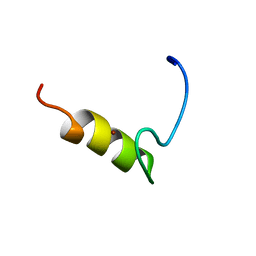
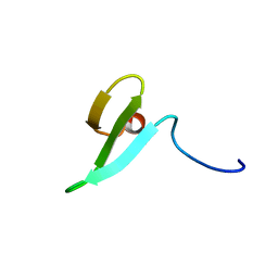
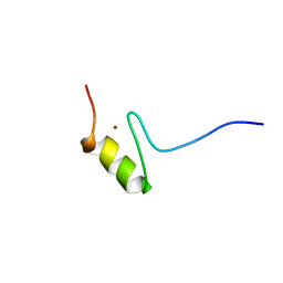
 | | SOLUTION STRUCTURE OF THE N-TERMINAL ZINC FINGER OF MURINE GATA-1, NMR, 25 STRUCTURES | | 分子名称: | TRANSCRIPTION FACTOR GATA-1, ZINC ION | | 著者 | Kowalski, K, Czolij, R, King, G.F, Crossley, M, Mackay, J.P. | | 登録日 | 1998-10-12 | | 公開日 | 1999-06-08 | | 最終更新日 | 2024-05-01 | | 実験手法 | SOLUTION NMR | | 主引用文献 | The solution structure of the N-terminal zinc finger of GATA-1 reveals a specific binding face for the transcriptional co-factor FOG.
J.Biomol.NMR, 13, 1999
|
|
1WR4
 | |
1WLX
 | |
1WR7
 | |
1WR3
 | |
1WMV
 | |
1FV5
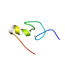
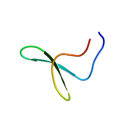
 | | SOLUTION STRUCTURE OF THE FIRST ZINC FINGER FROM THE DROSOPHILA U-SHAPED TRANSCRIPTION FACTOR | | 分子名称: | FIRST ZINC FINGER OF U-SHAPED, ZINC ION | | 著者 | Liew, C.K, Kowalski, K, Fox, A.H, Newton, A, Sharpe, B.K, Crossley, M, Mackay, J.P. | | 登録日 | 2000-09-18 | | 公開日 | 2000-10-04 | | 最終更新日 | 2022-02-23 | | 実験手法 | SOLUTION NMR | | 主引用文献 | Solution structures of two CCHC zinc fingers from the FOG family protein U-shaped that mediate protein-protein interactions.
Structure Fold.Des., 8, 2000
|
|
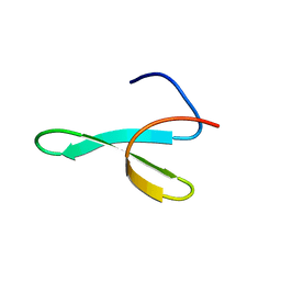
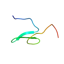
1FU9
 | | SOLUTION STRUCTURE OF THE NINTH ZINC-FINGER DOMAIN OF THE U-SHAPED TRANSCRIPTION FACTOR | | 分子名称: | U-SHAPED TRANSCRIPTIONAL COFACTOR, ZINC ION | | 著者 | Liew, C.K, Kowalski, K, Fox, A.H, Newton, A, Sharpe, B.K, Crossley, M, Mackay, J.P. | | 登録日 | 2000-09-14 | | 公開日 | 2000-10-04 | | 最終更新日 | 2022-02-23 | | 実験手法 | SOLUTION NMR | | 主引用文献 | Solution structures of two CCHC zinc fingers from the FOG family protein U-shaped that mediate protein-protein interactions.
Structure Fold.Des., 8, 2000
|
|
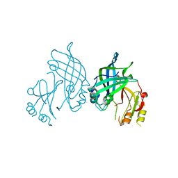
4XXR
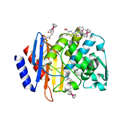 | | Atomic Resolution X-Ray Crystal Structure of a Ruthenocene Conjugated Beta-Lactam Antibiotic in Complex with CTX-M-14 E166A Beta-Lactamase | | 分子名称: | CTX-M-14 Class A Beta-Lactamase, POTASSIUM ION, [(1,2,3,4,5-eta)-1-(4-{[(4-carboxy-5,5-dimethyl-1,3-thiazolidin-2-yl)methyl]amino}-4-oxobutanoyl)cyclopentadienyl][(1,2,3,4,5-eta)-cyclopentadienyl]ruthenium, ... | | 著者 | Lewandowski, E.M, Chen, Y. | | 登録日 | 2015-01-30 | | 公開日 | 2015-03-18 | | 最終更新日 | 2023-09-27 | | 実験手法 | X-RAY DIFFRACTION (1.18 Å) | | 主引用文献 | Antibacterial properties and atomic resolution X-ray complex crystal structure of a ruthenocene conjugated beta-lactam antibiotic.
Chem.Commun.(Camb.), 51, 2015
|
|
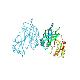
6VNU
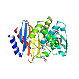 | | X-ray Crystal Structure of Ruthenocenyl-7-Aminocephalosporanic Acid Covalent Acyl-Enzyme Complex with CTX-M-14 E166A Beta-Lactamase | | 分子名称: | Beta-lactamase, POTASSIUM ION, [(1,2,3,4,5-eta)-1-(4-{[carboxy(4-carboxy-5-methylidene-5,6-dihydro-2H-1,3-thiazin-2-yl)methyl]amino}-4-oxobutanoyl)cyclopentadienyl][(1,2,3,4,5-eta)-cyclopentadienyl]ruthenium, ... | | 著者 | Lewandowski, E.M, Jacobs, L.M.C, Chen, Y. | | 登録日 | 2020-01-29 | | 公開日 | 2020-04-01 | | 最終更新日 | 2023-10-11 | | 実験手法 | X-RAY DIFFRACTION (1.47 Å) | | 主引用文献 | Metallocenyl 7-ACA Conjugates: Antibacterial Activity Studies and Atomic-Resolution X-ray Crystal Structure with CTX-M beta-Lactamase.
Chembiochem, 21, 2020
|
|
6GXJ
 | | X-ray structure of DiRu-1-encapsulated Apoferritin | | 分子名称: | CADMIUM ION, CHLORIDE ION, Ferritin light chain, ... | | 著者 | Pica, A, Ferraro, G, Merlino, A. | | 登録日 | 2018-06-27 | | 公開日 | 2019-02-06 | | 最終更新日 | 2024-01-17 | | 実験手法 | X-RAY DIFFRACTION (1.43 Å) | | 主引用文献 | Encapsulation of the Dinuclear Trithiolato-Bridged Arene Ruthenium Complex Diruthenium-1 in an Apoferritin Nanocage: Structure and Cytotoxicity.
ChemMedChem, 14, 2019
|
|
5TOP
 | |
5TOY
 | |
5UJO
 | |
5VLE
 | |
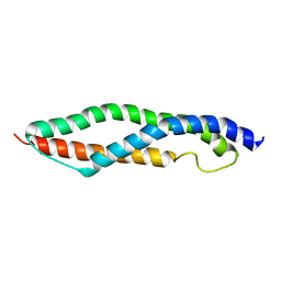
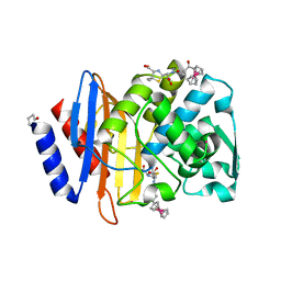
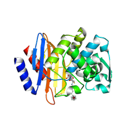
8AYU
 | | Crystal structure of SUDV VP40 L117A mutant | | 分子名称: | Matrix protein VP40 | | 著者 | Werner, A.-D, Steinchen, W, Werel, L, Kowalski, K, Essen, L.-O, Becker, S. | | 登録日 | 2022-09-03 | | 公開日 | 2023-09-13 | | 最終更新日 | 2024-04-24 | | 実験手法 | X-RAY DIFFRACTION (2 Å) | | 主引用文献 | Crystal structure of SUDV VP40 L117A mutant
To Be Published
|
|
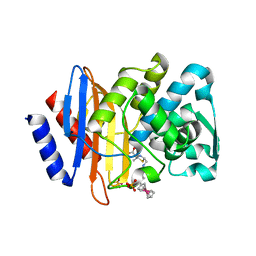
8AYT
 | | Crystal structure of SUDV VP40 W95A mutant | | 分子名称: | Matrix protein VP40 | | 著者 | Werner, A.-D, Steinchen, W, Werel, L, Kowalski, K, Essen, L.-O, Becker, S. | | 登録日 | 2022-09-03 | | 公開日 | 2023-09-13 | | 最終更新日 | 2024-04-24 | | 実験手法 | X-RAY DIFFRACTION (1.9 Å) | | 主引用文献 | Crystal structure of SUDV VP40 W95A mutant
To Be Published
|
|


