Loading
PDBj
MenuPDBj@FacebookPDBj@TwitterPDBj@YouTubewwPDB FoundationwwPDB
RCSB PDBPDBeBMRBAdv. SearchSearch help
Search by PDB author
5N12
DownloadVisualize
BU of 5n12 by Molmil
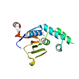
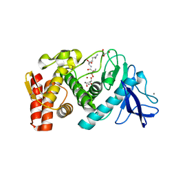
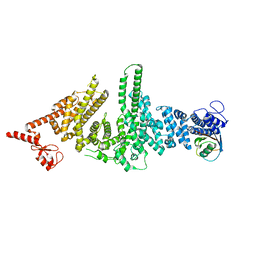
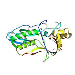
Crystal structure of TCE treated rPPEP-1
Descriptor: 2,2,2-tris-chloroethanol, 2-AMINO-2-HYDROXYMETHYL-PROPANE-1,3-DIOL, Pro-Pro endopeptidase, ...
Authors:Pichlo, C, Schacherl, M, Baumann, U.
Deposit date:2017-02-04
Release date:2018-05-30
Last modified:2024-01-17
Method:X-RAY DIFFRACTION (1.38 Å)
Cite:Improved protein-crystal identification by using 2,2,2-trichloroethanol as a fluorescence enhancer.
Acta Crystallogr F Struct Biol Commun, 74, 2018
2N7D
DownloadVisualize
BU of 2n7d by Molmil
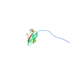
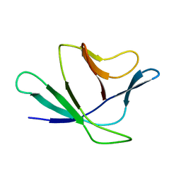
Solution structure of the UBL domain of human Ddi2
Descriptor: Protein DDI1 homolog 2
Authors:Siva, M, Grantz Saskova, K, Veverka, V.
Deposit date:2015-09-08
Release date:2016-07-20
Last modified:2023-06-14
Method:SOLUTION NMR
Cite:Human DNA-Damage-Inducible 2 Protein Is Structurally and Functionally Distinct from Its Yeast Ortholog.
Sci Rep, 6, 2016
6W9O
DownloadVisualize
BU of 6w9o by Molmil
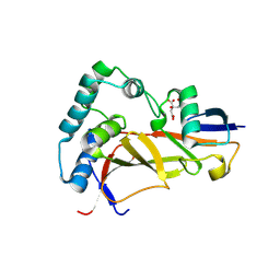
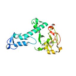
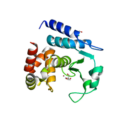
Crystal structure of an OTU deubiquitinase from Wolbachia pipientis wMel
Descriptor: ACETATE ION, OTU domain-containing protein wMelOTU
Authors:Schubert, A.F, Pruneda, J.N, Komander, D.
Deposit date:2020-03-23
Release date:2020-07-01
Last modified:2023-10-18
Method:X-RAY DIFFRACTION (1.47 Å)
Cite:Identification and characterization of diverse OTU deubiquitinases in bacteria.
Embo J., 39, 2020
6W9R
DownloadVisualize
BU of 6w9r by Molmil
Crystal structure of an OTU deubiquitinase from Wolbachia pipientis wMel bound to ubiquitin
Descriptor: CITRATE ANION, OTU domain-containing protein wMelOTU, Ubiquitin
Authors:Schubert, A.F, Pruneda, J.N, Komander, D.
Deposit date:2020-03-23
Release date:2020-07-01
Last modified:2023-10-18
Method:X-RAY DIFFRACTION (1.82 Å)
Cite:Identification and characterization of diverse OTU deubiquitinases in bacteria.
Embo J., 39, 2020
6W9S
DownloadVisualize
BU of 6w9s by Molmil
Crystal structure of an OTU deubiquitinase from Escherichia albertii bound to ubiquitin
Descriptor: FORMIC ACID, OTU domain-containing protein EschOTU, Ubiquitin
Authors:Schubert, A.F, Pruneda, J.N, Komander, D.
Deposit date:2020-03-23
Release date:2020-07-01
Last modified:2020-08-12
Method:X-RAY DIFFRACTION (2.1 Å)
Cite:Identification and characterization of diverse OTU deubiquitinases in bacteria.
Embo J., 39, 2020
6FSM
DownloadVisualize
BU of 6fsm by Molmil
Crystal structure of TCE-treated Thermolysin
Descriptor: CALCIUM ION, GLYCEROL, LYSINE, ...
Authors:Pichlo, C, Baumann, U.
Deposit date:2018-02-19
Release date:2018-05-02
Last modified:2024-01-17
Method:X-RAY DIFFRACTION (1.39 Å)
Cite:Improved protein-crystal identification by using 2,2,2-trichloroethanol as a fluorescence enhancer.
Acta Crystallogr F Struct Biol Commun, 74, 2018
6FSJ
DownloadVisualize
BU of 6fsj by Molmil
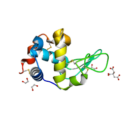
Crystal structure of TCE-treated Lysozyme
Descriptor: 1,2-ETHANEDIOL, CITRIC ACID, Lysozyme C
Authors:Pichlo, C, Baumann, U.
Deposit date:2018-02-19
Release date:2018-05-02
Last modified:2024-01-17
Method:X-RAY DIFFRACTION (1.2 Å)
Cite:Improved protein-crystal identification by using 2,2,2-trichloroethanol as a fluorescence enhancer.
Acta Crystallogr F Struct Biol Commun, 74, 2018
5O6C
DownloadVisualize
BU of 5o6c by Molmil
Crystal Structure of a threonine-selective RCR E3 ligase
Descriptor: E3 ubiquitin-protein ligase MYCBP2, ZINC ION
Authors:Pao, K.-C, Rafie, K.Z, van Aalten, D, Virdee, S.
Deposit date:2017-06-06
Release date:2018-04-18
Last modified:2018-05-02
Method:X-RAY DIFFRACTION (1.75 Å)
Cite:Activity-based E3 ligase profiling uncovers an E3 ligase with esterification activity.
Nature, 556, 2018
3M63
DownloadVisualize
BU of 3m63 by Molmil
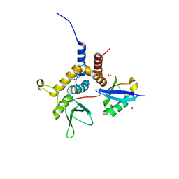
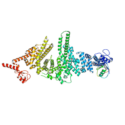
Crystal structure of Ufd2 in complex with the ubiquitin-like (UBL) domain of Dsk2
Descriptor: PENTAETHYLENE GLYCOL, POTASSIUM ION, Ubiquitin conjugation factor E4, ...
Authors:Haenzelmann, P, Schindelin, H.
Deposit date:2010-03-15
Release date:2010-04-28
Last modified:2023-09-06
Method:X-RAY DIFFRACTION (2.4 Å)
Cite:The yeast E4 ubiquitin ligase Ufd2 interacts with the ubiquitin-like domains of Rad23 and Dsk2 via a novel and distinct ubiquitin-like binding domain.
J.Biol.Chem., 285, 2010
3M62
DownloadVisualize
BU of 3m62 by Molmil
Crystal structure of Ufd2 in complex with the ubiquitin-like (UBL) domain of Rad23
Descriptor: PENTAETHYLENE GLYCOL, POTASSIUM ION, UV excision repair protein RAD23, ...
Authors:Haenzelmann, P, Schindelin, H.
Deposit date:2010-03-15
Release date:2010-04-28
Last modified:2023-09-06
Method:X-RAY DIFFRACTION (2.4 Å)
Cite:The yeast E4 ubiquitin ligase Ufd2 interacts with the ubiquitin-like domains of Rad23 and Dsk2 via a novel and distinct ubiquitin-like binding domain.
J.Biol.Chem., 285, 2010
2Z4D
DownloadVisualize
BU of 2z4d by Molmil
NMR Structures of Yeast Proteasome Component Rpn13
Descriptor: 26S proteasome regulatory subunit RPN13
Authors:Zhang, N, Walters, K.J.
Deposit date:2007-06-15
Release date:2008-05-20
Last modified:2022-03-16
Method:SOLUTION NMR
Cite:Proteasome subunit Rpn13 is a novel ubiquitin receptor.
Nature, 453, 2008
6NYT
DownloadVisualize
BU of 6nyt by Molmil
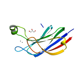
Munc13-1 C2B-domain, calcium bound
Descriptor: CALCIUM ION, CHLORIDE ION, GLYCEROL, ...
Authors:Tomchick, D.R, Rizo, J, Machius, M, Lu, J.
Deposit date:2019-02-12
Release date:2019-02-20
Last modified:2023-10-11
Method:X-RAY DIFFRACTION (1.369 Å)
Cite:Munc13 C2B domain is an activity-dependent Ca2+ regulator of synaptic exocytosis.
Nat. Struct. Mol. Biol., 17, 2010
6NYC
DownloadVisualize
BU of 6nyc by Molmil
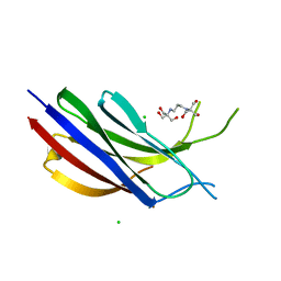
Munc13-1 C2B-domain, calcium free
Descriptor: 2-[3-(2-HYDROXY-1,1-DIHYDROXYMETHYL-ETHYLAMINO)-PROPYLAMINO]-2-HYDROXYMETHYL-PROPANE-1,3-DIOL, CHLORIDE ION, Munc13-1
Authors:Tomchick, D.R, Rizo, J, Machius, M, Lu, J.
Deposit date:2019-02-11
Release date:2019-02-20
Last modified:2023-10-11
Method:X-RAY DIFFRACTION (1.893 Å)
Cite:Munc13 C2B domain is an activity-dependent Ca2+ regulator of synaptic exocytosis.
Nat. Struct. Mol. Biol., 17, 2010
6R99
DownloadVisualize
BU of 6r99 by Molmil
Crystal Structure of Human CLN protein 5 (Ceroid Lipofuscinosis Neuronal Protein 5)
Descriptor: 2-acetamido-2-deoxy-beta-D-glucopyranose, 2-acetamido-2-deoxy-beta-D-glucopyranose-(1-4)-2-acetamido-2-deoxy-beta-D-glucopyranose, Ceroid-lipofuscinosis neuronal protein 5, ...
Authors:Luebben, A.V, Sheldrick, G.M.
Deposit date:2019-04-03
Release date:2020-04-22
Last modified:2022-09-28
Method:X-RAY DIFFRACTION (2.7 Å)
Cite:Cln5 represents a new type of cysteine-based S -depalmitoylase linked to neurodegeneration.
Sci Adv, 8, 2022
1DP7
DownloadVisualize
BU of 1dp7 by Molmil
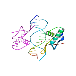
COCRYSTAL STRUCTURE OF RFX-DBD IN COMPLEX WITH ITS COGNATE X-BOX BINDING SITE
Descriptor: 1,2-ETHANEDIOL, DI(HYDROXYETHYL)ETHER, DNA (5'-D(*CP*GP*(BRU)P*TP*AP*CP*CP*AP*(BRU)P*GP*GP*TP*AP*AP*CP*G)-3'), ...
Authors:Gajiwala, K.S, Chen, H, Cornille, F, Roques, B.P, Reith, W, Mach, B, Burley, S.K.
Deposit date:1999-12-23
Release date:2000-03-06
Last modified:2024-02-07
Method:X-RAY DIFFRACTION (1.5 Å)
Cite:Structure of the winged-helix protein hRFX1 reveals a new mode of DNA binding.
Nature, 403, 2000
<12


219140

PDB entries from 2024-05-01

PDB statisticsPDBj update infoContact PDBjnumon