Loading
PDBj
MenuPDBj@FacebookPDBj@TwitterPDBj@YouTubewwPDB FoundationwwPDB
RCSB PDBPDBeBMRBAdv. SearchSearch help
Search by PDB author
1KBA
DownloadVisualize
BU of 1kba by Molmil
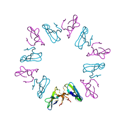
CRYSTAL STRUCTURE OF KAPPA-BUNGAROTOXIN AT 2.3-ANGSTROM RESOLUTION
Descriptor: KAPPA-BUNGAROTOXIN
Authors:Dewan, J.C, Grant, G.A, Sacchettini, J.C.
Deposit date:1994-07-13
Release date:1994-12-20
Last modified:2024-05-29
Method:X-RAY DIFFRACTION (2.3 Å)
Cite:Crystal structure of kappa-bungarotoxin at 2.3-A resolution.
Biochemistry, 33, 1994
4RQO
DownloadVisualize
BU of 4rqo by Molmil
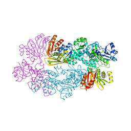
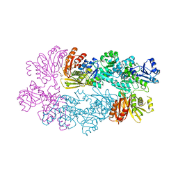
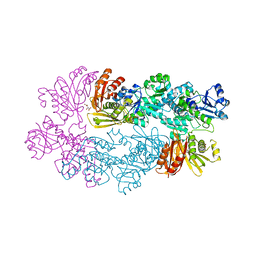
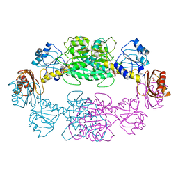
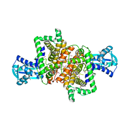
Crystal structure of L-Serine Dehydratase from Legionella pneumophila
Descriptor: CHLORIDE ION, IRON/SULFUR CLUSTER, L-serine dehydratase
Authors:Thoden, J.B, Holden, H.M, Grant, G.A.
Deposit date:2014-11-04
Release date:2014-11-19
Last modified:2014-12-24
Method:X-RAY DIFFRACTION (2.25 Å)
Cite:Structure of l-Serine Dehydratase from Legionella pneumophila: Novel Use of the C-Terminal Cysteine as an Intrinsic Competitive Inhibitor.
Biochemistry, 53, 2014
1YGY
DownloadVisualize
BU of 1ygy by Molmil
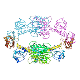
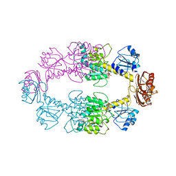
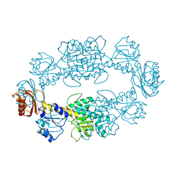
Crystal Structure of D-3-Phosphoglycerate dehydrogenase From Mycobacterium tuberculosis
Descriptor: D-3-phosphoglycerate dehydrogenase, L(+)-TARTARIC ACID
Authors:Dey, S, Grant, G.A, Sacchettini, J.C, TB Structural Genomics Consortium (TBSGC)
Deposit date:2005-01-05
Release date:2005-01-25
Last modified:2019-07-10
Method:X-RAY DIFFRACTION (2.3 Å)
Cite:Crystal Structure of Mycobacterium tuberculosis D-3-Phosphoglycerate Dehydrogenase: EXTREME ASYMMETRY IN A TETRAMER OF IDENTICAL SUBUNITS
J.Biol.Chem., 280, 2005
1PSD
DownloadVisualize
BU of 1psd by Molmil
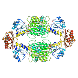
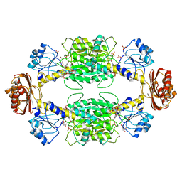
THE ALLOSTERIC LIGAND SITE IN THE VMAX-TYPE COOPERATIVE ENZYME PHOSPHOGLYCERATE DEHYDROGENASE
Descriptor: D-3-PHOSPHOGLYCERATE DEHYDROGENASE (PHOSPHOGLYCERATE DEHYDROGENASE), NICOTINAMIDE-ADENINE-DINUCLEOTIDE, SERINE
Authors:Schuller, D.J, Grant, G.A, Banaszak, L.J.
Deposit date:1995-05-02
Release date:1995-07-31
Last modified:2024-02-14
Method:X-RAY DIFFRACTION (2.75 Å)
Cite:The allosteric ligand site in the Vmax-type cooperative enzyme phosphoglycerate dehydrogenase.
Nat.Struct.Biol., 2, 1995
1SC6
DownloadVisualize
BU of 1sc6 by Molmil
Crystal Structure of W139G D-3-Phosphoglycerate dehydrogenase complexed with NAD+
Descriptor: D-3-phosphoglycerate dehydrogenase, NICOTINAMIDE-ADENINE-DINUCLEOTIDE
Authors:Bell, J.K, Grant, G.A, Banaszak, L.J.
Deposit date:2004-02-11
Release date:2005-02-22
Last modified:2021-10-27
Method:X-RAY DIFFRACTION (2.09 Å)
Cite:Multiconformational states in phosphoglycerate dehydrogenase
Biochemistry, 43, 2004
1YBA
DownloadVisualize
BU of 1yba by Molmil
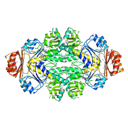
The active form of phosphoglycerate dehydrogenase
Descriptor: 2-OXOGLUTARIC ACID, D-3-phosphoglycerate dehydrogenase, NICOTINAMIDE-ADENINE-DINUCLEOTIDE, ...
Authors:Thompson, J.R, Banaszak, L.J.
Deposit date:2004-12-20
Release date:2005-04-26
Last modified:2024-04-03
Method:X-RAY DIFFRACTION (2.24 Å)
Cite:Vmax Regulation through Domain and Subunit Changes. The Active Form of Phosphoglycerate Dehydrogenase
Biochemistry, 44, 2005
3DC2
DownloadVisualize
BU of 3dc2 by Molmil
Crystal structure of serine bound D-3-phosphoglycerate dehydrogenase from Mycobacterium tuberculosis
Descriptor: D-3-phosphoglycerate dehydrogenase, L(+)-TARTARIC ACID, SERINE
Authors:Dey, S, Sacchettini, J.C.
Deposit date:2008-06-03
Release date:2008-07-22
Last modified:2023-11-01
Method:X-RAY DIFFRACTION (2.7 Å)
Cite:Structural analysis of substrate and effector binding in Mycobacterium tuberculosis D-3-phosphoglycerate dehydrogenase
Biochemistry, 47, 2008
3DDN
DownloadVisualize
BU of 3ddn by Molmil
Crystal structure of hydroxypyruvic acid phosphate bound D-3-phosphoglycerate dehydrogenase in mycobacterium tuberculosis
Descriptor: 2-oxo-3-(phosphonooxy)propanoic acid, D-3-phosphoglycerate dehydrogenase, L(+)-TARTARIC ACID
Authors:Dey, S, Sacchettini, J.C.
Deposit date:2008-06-05
Release date:2008-07-22
Last modified:2023-11-01
Method:X-RAY DIFFRACTION (2.4 Å)
Cite:Structural analysis of substrate and effector binding in Mycobacterium tuberculosis D-3-phosphoglycerate dehydrogenase
Biochemistry, 47, 2008
2P9G
DownloadVisualize
BU of 2p9g by Molmil
Crystal structure of serine bound G336V,G337V double mutant of E.coli phosphoglycerate dehydrogenase
Descriptor: 1,4-DIHYDRONICOTINAMIDE ADENINE DINUCLEOTIDE, D-3-phosphoglycerate dehydrogenase, SERINE
Authors:Dey, S, Sacchettini, J.C.
Deposit date:2007-03-25
Release date:2007-04-24
Last modified:2023-08-30
Method:X-RAY DIFFRACTION (2.8 Å)
Cite:The Effect of Hinge Mutations on Effector Binding and Domain Rotation in Escherichia coli D-3-Phosphoglycerate Dehydrogenase.
J.Biol.Chem., 282, 2007
2P9E
DownloadVisualize
BU of 2p9e by Molmil
Crystal Structure of G336V mutant of E.coli phosphoglycerate dehydrogenase
Descriptor: 1,4-DIHYDRONICOTINAMIDE ADENINE DINUCLEOTIDE, CITRIC ACID, D-3-phosphoglycerate dehydrogenase, ...
Authors:Dey, S, Sacchettini, J.C.
Deposit date:2007-03-25
Release date:2007-04-24
Last modified:2023-08-30
Method:X-RAY DIFFRACTION (2.6 Å)
Cite:The Effect of Hinge Mutations on Effector Binding and Domain Rotation in Escherichia coli D-3-Phosphoglycerate Dehydrogenase
J.Biol.Chem., 282, 2007
2P9C
DownloadVisualize
BU of 2p9c by Molmil
Crystal structure of serine bound G336V mutant of E.coli phosphoglycerate dehydrogenase
Descriptor: 1,4-DIHYDRONICOTINAMIDE ADENINE DINUCLEOTIDE, D-3-phosphoglycerate dehydrogenase, SERINE
Authors:Dey, S, Sacchettini, J.C.
Deposit date:2007-03-24
Release date:2007-04-24
Last modified:2023-08-30
Method:X-RAY DIFFRACTION (2.46 Å)
Cite:The Effect of Hinge Mutations on Effector Binding and Domain Rotation in Escherichia coli D-3-Phosphoglycerate Dehydrogenase
J.Biol.Chem., 282, 2007
2PA3
DownloadVisualize
BU of 2pa3 by Molmil
crystal structure of serine bound G336V mutant of E.coli phosphoglycerate dehydrogenase
Descriptor: 1,4-DIHYDRONICOTINAMIDE ADENINE DINUCLEOTIDE, D-3-phosphoglycerate dehydrogenase, SERINE
Authors:Dey, S, Sacchettini, J.C.
Deposit date:2007-03-27
Release date:2007-04-24
Last modified:2023-08-30
Method:X-RAY DIFFRACTION (2.74 Å)
Cite:The Effect of Hinge Mutations on Effector Binding and Domain Rotation in Escherichia coli D-3-Phosphoglycerate Dehydrogenase.
J.Biol.Chem., 282, 2007
1ZID
DownloadVisualize
BU of 1zid by Molmil
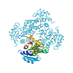
LONG FATTY ACID CHAIN ENOYL-ACP REDUCTASE (INHA) IN COMPLEX WITH AN ISONICOTINIC-ACYL-NADH INHIBITOR
Descriptor: ENOYL-[ACYL-CARRIER-PROTEIN] REDUCTASE, ISONICOTINIC-ACETYL-NICOTINAMIDE-ADENINE DINUCLEOTIDE
Authors:Rozwarski, D.A, Jacobs Jr, W.R, Sacchettini, J.C, TB Structural Genomics Consortium (TBSGC)
Deposit date:1997-03-25
Release date:1998-03-25
Last modified:2024-05-22
Method:X-RAY DIFFRACTION (2.7 Å)
Cite:Modification of the NADH of the isoniazid target (InhA) from Mycobacterium tuberculosis.
Science, 279, 1998

222036

PDB entries from 2024-07-03

PDB statisticsPDBj update infoContact PDBjnumon