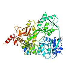
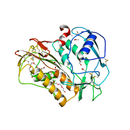
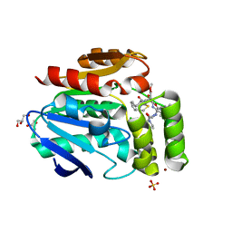
Loading
PDBj
MenuPDBj@FacebookPDBj@TwitterPDBj@YouTubewwPDB FoundationwwPDB
RCSB PDBPDBeBMRBAdv. SearchSearch help
Search by PDB author
6P0J
DownloadVisualize
BU of 6p0j by Molmil
Crystal structure of GDP-bound human RalA
Descriptor: CALCIUM ION, GUANOSINE-5'-DIPHOSPHATE, Ras-related protein Ral-A
Authors:Bum-Erdene, K, Gonzalez-Gutierrez, G, Liu, D, Meroueh, S.O.
Deposit date:2019-05-17
Release date:2020-03-04
Last modified:2023-10-11
Method:X-RAY DIFFRACTION (1.31 Å)
Cite:Small-molecule covalent bond formation at tyrosine creates a binding site and inhibits activation of Ral GTPases.
Proc.Natl.Acad.Sci.USA, 117, 2020
6PHJ
DownloadVisualize
BU of 6phj by Molmil
Crystal structure of native glucagon in space group P213 at 1.99 A resolution
Descriptor: Glucagon
Authors:Mroz, P.A, Gonzalez-Gutierrez, G, DiMarchi, R.D.
Deposit date:2019-06-25
Release date:2020-07-01
Last modified:2023-10-11
Method:X-RAY DIFFRACTION (1.99 Å)
Cite:High resolution X-ray structure of glucagon and selected stereo-inversed analogs in novel crystallographic packing arrangement.
To Be Published
6PHO
DownloadVisualize
BU of 6pho by Molmil
Crystal structure of glucagon analog with selenomethionine substitutions at position 1 and 27 in space group I41 at 1.42 A resolution
Descriptor: Glucagon
Authors:Mroz, P.A, Gonzalez-Gutierrez, G, DiMarchi, R.D.
Deposit date:2019-06-25
Release date:2020-07-01
Method:X-RAY DIFFRACTION (1.42 Å)
Cite:High resolution X-ray structure of glucagon and selected stereo-inversed analogs in novel crystallographic packing arrangement.
To Be Published
6PHK
DownloadVisualize
BU of 6phk by Molmil
Crystal structure of glucagon analog with mono-stereoinversion at position 21 (D-Asp21) in space group I41 at 1.18 A resolution
Descriptor: Glucagon
Authors:Mroz, P.A, Gonzalez-Gutierrez, G, DiMarchi, R.D.
Deposit date:2019-06-25
Release date:2020-07-01
Last modified:2023-10-11
Method:X-RAY DIFFRACTION (1.18 Å)
Cite:High resolution X-ray structure of glucagon and selected stereo-inversed analogs in novel crystallographic packing arrangement.
To Be Published
6PHP
DownloadVisualize
BU of 6php by Molmil
Crystal structure of glucagon analog with 4-bromo-phenylalanine substitutions at position 6 and 22 in space group I41 at 1.65 A resolution
Descriptor: Glucagon
Authors:Mroz, P.A, Gonzalez-Gutierrez, G, DiMarchi, R.D.
Deposit date:2019-06-25
Release date:2020-07-01
Last modified:2023-11-15
Method:X-RAY DIFFRACTION (1.65 Å)
Cite:High resolution X-ray structure of glucagon and selected stereo-inversed analogs in novel crystallographic packing arrangement.
To Be Published
6SH7
DownloadVisualize
BU of 6sh7 by Molmil
Crystal structure of the human DEAH-helicase DHX15 in complex with the NKRF G-patch
Descriptor: NF-kappa-B-repressing factor, Pre-mRNA-splicing factor ATP-dependent RNA helicase DHX15
Authors:Jonas, S, Studer, M.K, Ivanovic, L.
Deposit date:2019-08-05
Release date:2020-04-22
Last modified:2024-01-24
Method:X-RAY DIFFRACTION (2.21 Å)
Cite:Structural basis for DEAH-helicase activation by G-patch proteins.
Proc.Natl.Acad.Sci.USA, 117, 2020
6PQ7
DownloadVisualize
BU of 6pq7 by Molmil
Structure of the iMango-III fluorescent aptamer at room temperature.
Descriptor: MAGNESIUM ION, POTASSIUM ION, RNA (37-MER), ...
Authors:Trachman III, R.J, Ferre-D'Amare, A.R.
Deposit date:2019-07-08
Release date:2019-07-31
Last modified:2023-10-11
Method:X-RAY DIFFRACTION (3 Å)
Cite:Co-crystal structure of the iMango-III fluorescent RNA aptamer using an X-ray free-electron laser.
Acta Crystallogr.,Sect.F, 75, 2019
6PHN
DownloadVisualize
BU of 6phn by Molmil
Crystal structure of glucagon analog composed of D-amino acids with mono-stereoinversion at position 23 (L-Val23) in space group I41 at 1.33 A resolution
Descriptor: D-glucagon L-Val23, SODIUM ION
Authors:Mroz, P.A, Gonzalez-Gutierrez, G, DiMarchi, R.D.
Deposit date:2019-06-25
Release date:2020-07-01
Last modified:2023-11-15
Method:X-RAY DIFFRACTION (1.33 Å)
Cite:High resolution X-ray structure of glucagon and selected stereo-inversed analogs in novel crystallographic packing arrangement.
To Be Published
6PHI
DownloadVisualize
BU of 6phi by Molmil
Crystal structure of native glucagon in space group I41 at 1.1 A resolution
Descriptor: CHLORIDE ION, Glucagon
Authors:Mroz, P.A, Gonzalez-Gutierrez, G, DiMarchi, R.D.
Deposit date:2019-06-25
Release date:2020-07-01
Last modified:2023-10-11
Method:X-RAY DIFFRACTION (1.1 Å)
Cite:High resolution X-ray structure of glucagon and selected stereo-inversed analogs in novel crystallographic packing arrangement.
To Be Published
6PHM
DownloadVisualize
BU of 6phm by Molmil
Crystal structure of glucagon analog fully composed of D-amino acids in space group I41 at 1.1 A resolution
Descriptor: D-glucagon, SULFATE ION
Authors:Mroz, P.A, Gonzalez-Gutierrez, G, DiMarchi, R.D.
Deposit date:2019-06-25
Release date:2020-07-01
Last modified:2023-11-15
Method:X-RAY DIFFRACTION (1.1 Å)
Cite:High resolution X-ray structure of glucagon and selected stereo-inversed analogs in novel crystallographic packing arrangement.
To Be Published
6P7M
DownloadVisualize
BU of 6p7m by Molmil
Cryo-EM structure of LbCas12a-crRNA: AcrVA4 (1:2 complex)
Descriptor: Cas12a, MAGNESIUM ION, anti-CRISPR VA4, ...
Authors:Knott, G.J, Liu, J.J, Doudna, J.A.
Deposit date:2019-06-06
Release date:2019-08-21
Last modified:2024-03-20
Method:ELECTRON MICROSCOPY (3 Å)
Cite:Structural basis for AcrVA4 inhibition of specific CRISPR-Cas12a.
Elife, 8, 2019
6PHL
DownloadVisualize
BU of 6phl by Molmil
Crystal structure of glucagon analog with mono-stereoinversion at position 23 (D-Val23) in space group I41 at 1.44 A resolution
Descriptor: Glucagon
Authors:Mroz, P.A, Gonzalez-Gutierrez, G, DiMarchi, R.D.
Deposit date:2019-06-25
Release date:2020-07-01
Last modified:2023-10-11
Method:X-RAY DIFFRACTION (1.443 Å)
Cite:High resolution X-ray structure of glucagon and selected stereo-inversed analogs in novel crystallographic packing arrangement.
To Be Published
6PHQ
DownloadVisualize
BU of 6phq by Molmil
Crystal structure of glucagon analog fully composed of D-amino acids with 4-bromo-D-phenylalanine substitutions at position 6 and 22 in space group I41 at 1.1 A resolution
Descriptor: D-glucagon D-BrPhe 6,22
Authors:Mroz, P.A, Gonzalez-Gutierrez, G, DiMarchi, R.D.
Deposit date:2019-06-25
Release date:2020-07-01
Last modified:2023-11-15
Method:X-RAY DIFFRACTION (1.32 Å)
Cite:High resolution X-ray structure of glucagon and selected stereo-inversed analogs in novel crystallographic packing arrangement.
To Be Published
6P7N
DownloadVisualize
BU of 6p7n by Molmil
Cryo-EM structure of LbCas12a-crRNA: AcrVA4 (2:2 complex)
Descriptor: Cas12a, MAGNESIUM ION, anti-CRISPR VA4, ...
Authors:Knott, G.J, Liu, J.J, Doudna, J.A.
Deposit date:2019-06-06
Release date:2019-08-21
Last modified:2023-08-16
Method:ELECTRON MICROSCOPY (4.9 Å)
Cite:Structural basis for AcrVA4 inhibition of specific CRISPR-Cas12a.
Elife, 8, 2019
6TDB
DownloadVisualize
BU of 6tdb by Molmil
Neuropilin2-b1 domain in a complex with the C-terminal VEGFB167 peptide
Descriptor: 1,2-ETHANEDIOL, ACETYL GROUP, C-terminal VEGFB167 peptide, ...
Authors:Eldrid, C, Yu, L, Yelland, T, Fotinou, C, Djordjevic, S.
Deposit date:2019-11-08
Release date:2020-11-18
Last modified:2024-01-24
Method:X-RAY DIFFRACTION (2.45 Å)
Cite:NRP2-b1 domain in a complex with the C-terminal VEGFB167 peptide
To Be Published
6T6X
DownloadVisualize
BU of 6t6x by Molmil
Structure of the Bottromycin epimerase BotH in complex with substrate
Descriptor: (4~{R})-2-[(1~{R})-1-[[(2~{S})-2-[[(2~{S})-3-methyl-2-[[(4~{Z},6~{S},9~{S},12~{S})-2,8,11-tris(oxidanylidene)-6,9-di(propan-2-yl)-1,4,7,10-tetrazabicyclo[10.3.0]pentadec-4-en-5-yl]amino]butanoyl]amino]-3-phenyl-propanoyl]amino]-3-oxidanyl-3-oxidanylidene-propyl]-4,5-dihydro-1,3-thiazole-4-carboxylic acid, BotH
Authors:Koehnke, J, Sikandar, A.
Deposit date:2019-10-20
Release date:2020-07-15
Last modified:2024-01-24
Method:X-RAY DIFFRACTION (1.25 Å)
Cite:The bottromycin epimerase BotH defines a group of atypical alpha / beta-hydrolase-fold enzymes.
Nat.Chem.Biol., 16, 2020
6TOC
DownloadVisualize
BU of 6toc by Molmil
Crystal structure of the oligomerisation domain of the transcription factor PHOSPHATE STARVATION RESPONSE 1 from Arabidopsis (crystal form 3).
Descriptor: Protein PHOSPHATE STARVATION RESPONSE 1
Authors:Hothorn, M.
Deposit date:2019-12-11
Release date:2021-01-13
Last modified:2024-01-24
Method:X-RAY DIFFRACTION (1.853 Å)
Cite:Inositol pyrophosphates promote the interaction of SPX domains with the coiled-coil motif of PHR transcription factors to regulate plant phosphate homeostasis.
Nat Commun, 12, 2021
6QIX
DownloadVisualize
BU of 6qix by Molmil
The crystal structure of Trichuris muris p43
Descriptor: 2-acetamido-2-deoxy-beta-D-glucopyranose, ACETATE ION, CALCIUM ION, ...
Authors:Levy, C.W.
Deposit date:2019-01-21
Release date:2019-06-12
Last modified:2020-07-29
Method:X-RAY DIFFRACTION (1.65 Å)
Cite:The major secreted protein of the whipworm parasite tethers to matrix and inhibits interleukin-13 function.
Nat Commun, 10, 2019
6T6H
DownloadVisualize
BU of 6t6h by Molmil
Apo structure of the Bottromycin epimerase BotH
Descriptor: BotH, SODIUM ION, SULFATE ION
Authors:Koehnke, J, Sikandar, A.
Deposit date:2019-10-18
Release date:2020-07-15
Last modified:2020-08-26
Method:X-RAY DIFFRACTION (1.18 Å)
Cite:The bottromycin epimerase BotH defines a group of atypical alpha / beta-hydrolase-fold enzymes.
Nat.Chem.Biol., 16, 2020
6T6Z
DownloadVisualize
BU of 6t6z by Molmil
Structure of the Bottromycin epimerase BotH in complex with a bottromycin A2 derivative
Descriptor: BotH, Bottromycin A2 derivative
Authors:Koehnke, J, Sikandar, A.
Deposit date:2019-10-20
Release date:2020-07-15
Last modified:2024-01-24
Method:X-RAY DIFFRACTION (1.7 Å)
Cite:The bottromycin epimerase BotH defines a group of atypical alpha / beta-hydrolase-fold enzymes.
Nat.Chem.Biol., 16, 2020
6T6Y
DownloadVisualize
BU of 6t6y by Molmil
Structure of the Bottromycin epimerase BotH in complex with Bottromycin A2
Descriptor: BotH, Bottromycin A2
Authors:Koehnke, J, Sikandar, A.
Deposit date:2019-10-20
Release date:2020-07-15
Last modified:2024-01-24
Method:X-RAY DIFFRACTION (1.4 Å)
Cite:The bottromycin epimerase BotH defines a group of atypical alpha / beta-hydrolase-fold enzymes.
Nat.Chem.Biol., 16, 2020
6T70
DownloadVisualize
BU of 6t70 by Molmil
Structure of the Bottromycin epimerase BotH in complex with Bottromycin A2 derivative
Descriptor: BotH, Bottromycin A2 derivative, CHLORIDE ION, ...
Authors:Koehnke, J, Sikandar, A.
Deposit date:2019-10-20
Release date:2020-07-15
Last modified:2024-01-24
Method:X-RAY DIFFRACTION (1.58 Å)
Cite:The bottromycin epimerase BotH defines a group of atypical alpha / beta-hydrolase-fold enzymes.
Nat.Chem.Biol., 16, 2020
6TX4
DownloadVisualize
BU of 6tx4 by Molmil
CRYSTAL STRUCTURE OF HUMAN FKBP51 FK1 DOMAIN A19T MUTANT IN COMPLEX WITH 2-PYRIDONE
Descriptor: 1~{H}-pyridin-2-one, CHLORIDE ION, Peptidyl-prolyl cis-trans isomerase FKBP5, ...
Authors:Fiegen, D, Draxler, S.W.
Deposit date:2020-01-13
Release date:2020-05-27
Last modified:2024-01-24
Method:X-RAY DIFFRACTION (1.06 Å)
Cite:Hybrid Screening Approach for Very Small Fragments: X-ray and Computational Screening on FKBP51.
J.Med.Chem., 63, 2020
7SQM
DownloadVisualize
BU of 7sqm by Molmil
Discovery and Preclinical Pharmacology of INE963, A Potent and Fast-Acting Blood-Stage Antimalarial with a High Barrier to Resistance and Potential for Single-Dose Cure in Uncomplicated Malaria
Descriptor: 1-[(4S)-5-(2,4-difluorophenyl)imidazo[2,1-b][1,3,4]thiadiazol-2-yl]-4-methylpiperidin-4-amine, GLYCEROL, Serine/threonine-protein kinase haspin
Authors:Shu, W, Yokokawa, F.
Deposit date:2021-11-05
Release date:2021-12-29
Last modified:2023-10-18
Method:X-RAY DIFFRACTION (1.78 Å)
Cite:Failure of artesunate-mefloquine combination therapy for uncompli-cated Plasmodium falciparum malaria in southern Cambodia.
Malar. J., 2009, 8, 10, 2009
7T7U
DownloadVisualize
BU of 7t7u by Molmil
Light Harvesting complex phycocyanin PC 630, from the cryptophyte Chroomonas sp. M1627
Descriptor: DiCys-(15,16)-Dihydrobiliverdin, GLYCEROL, PHYCOCYANOBILIN, ...
Authors:Michie, K.A, Harrop, S.J, Rathbone, H.W, Wilk, K.E, Curmi, P.M.G.
Deposit date:2021-12-15
Release date:2023-02-15
Last modified:2023-10-25
Method:X-RAY DIFFRACTION (1.8 Å)
Cite:Molecular structures reveal the origin of spectral variation in cryptophyte light harvesting antenna proteins.
Protein Sci., 32, 2023

218853

PDB entries from 2024-04-24

PDB statisticsPDBj update infoContact PDBjnumon