Loading
PDBj
MenuPDBj@FacebookPDBj@TwitterPDBj@YouTubewwPDB FoundationwwPDB
RCSB PDBPDBeBMRBAdv. SearchSearch help
Search by PDB author
1VR0
DownloadVisualize
BU of 1vr0 by Molmil
Crystal structure of putative 2-phosphosulfolactate phosphatase (15026306) from Clostridium acetobutylicum at 2.6 A resolution
Descriptor: (2R)-3-SULFOLACTIC ACID, MAGNESIUM ION, Probable 2-phosphosulfolactate phosphatase
Authors:Joint Center for Structural Genomics (JCSG)
Deposit date:2005-01-26
Release date:2005-02-15
Last modified:2023-01-25
Method:X-RAY DIFFRACTION (2.49 Å)
Cite:Crystal structure of 2-phosphosulfolactate phosphatase (ComB) from Clostridium acetobutylicum at 2.6 A resolution reveals a new fold with a novel active site.
Proteins, 65, 2006
1VQ3
DownloadVisualize
BU of 1vq3 by Molmil
Crystal structure of Phosphoribosylformylglycinamidine synthase, purS subunit (EC 6.3.5.3) (TM1244) from Thermotoga maritima at 1.90 A resolution
Descriptor: Phosphoribosylformylglycinamidine synthase, purS subunit
Authors:Joint Center for Structural Genomics (JCSG)
Deposit date:2004-12-15
Release date:2004-12-28
Last modified:2023-09-20
Method:X-RAY DIFFRACTION (1.9 Å)
Cite:Crystal structure of phosphoribosylformyl-glycinamidine synthase II, PurS subunit (TM1244) from Thermotoga maritima at 1.90 A resolution.
Proteins, 65, 2006
1VR8
DownloadVisualize
BU of 1vr8 by Molmil
Crystal structure of GTP binding regulator (TM1622) from Thermotoga Maritima at 1.75 A resolution
Descriptor: AZIDE ION, GLYCEROL, GTP binding regulator
Authors:Joint Center for Structural Genomics (JCSG)
Deposit date:2005-02-16
Release date:2005-03-15
Last modified:2023-01-25
Method:X-RAY DIFFRACTION (1.75 Å)
Cite:Crystal structure of an ORFan protein (TM1622) from Thermotoga maritima at 1.75 A resolution reveals a fold similar to the Ran-binding protein Mog1p.
Proteins, 65, 2006
1ZCZ
DownloadVisualize
BU of 1zcz by Molmil
Crystal structure of Phosphoribosylaminoimidazolecarboxamide formyltransferase / IMP cyclohydrolase (TM1249) from THERMOTOGA MARITIMA at 1.88 A resolution
Descriptor: Bifunctional purine biosynthesis protein purH, POTASSIUM ION, TETRAETHYLENE GLYCOL
Authors:Joint Center for Structural Genomics (JCSG)
Deposit date:2005-04-13
Release date:2005-04-26
Last modified:2023-09-20
Method:X-RAY DIFFRACTION (1.88 Å)
Cite:Crystal structure of AICAR transformylase IMP cyclohydrolase (TM1249) from Thermotoga maritima at 1.88 A resolution.
Proteins, 71, 2008
5V7Q
DownloadVisualize
BU of 5v7q by Molmil
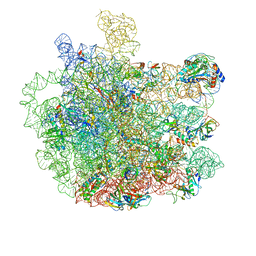
Cryo-EM structure of the large ribosomal subunit from Mycobacterium tuberculosis bound with a potent linezolid analog
Descriptor: 23S rRNA, 50S ribosomal protein L13, 50S ribosomal protein L14, ...
Authors:Yang, K, Chang, J.-Y, Cui, Z, Zhang, J.
Deposit date:2017-03-20
Release date:2017-09-20
Last modified:2024-03-13
Method:ELECTRON MICROSCOPY (3.7 Å)
Cite:Structural insights into species-specific features of the ribosome from the human pathogen Mycobacterium tuberculosis.
Nucleic Acids Res., 45, 2017
2A6A
DownloadVisualize
BU of 2a6a by Molmil
Crystal structure of Glycoprotein endopeptidase (tm0874) from THERMOTOGA MARITIMA at 2.50 A resolution
Descriptor: UNKNOWN LIGAND, hypothetical protein TM0874
Authors:Joint Center for Structural Genomics (JCSG)
Deposit date:2005-07-02
Release date:2005-07-19
Last modified:2023-01-25
Method:X-RAY DIFFRACTION (2.5 Å)
Cite:Structure of an essential bacterial protein YeaZ (TM0874) from Thermotoga maritima at 2.5 A resolution.
Acta Crystallogr.,Sect.F, 66, 2010
2ETS
DownloadVisualize
BU of 2ets by Molmil
CRYSTAL STRUCTURE OF A BACTERIAL DOMAIN OF UNKNOWN FUNCTION FROM DUF1798 FAMILY (MW1337) FROM STAPHYLOCOCCUS AUREUS SUBSP. AUREUS AT 2.25 A RESOLUTION
Descriptor: CHLORIDE ION, PHOSPHATE ION, hypothetical protein
Authors:Joint Center for Structural Genomics (JCSG)
Deposit date:2005-10-27
Release date:2005-11-08
Last modified:2023-01-25
Method:X-RAY DIFFRACTION (2.25 Å)
Cite:Crystal structures of MW1337R and lin2004: representatives of a novel protein family that adopt a four-helical bundle fold.
Proteins, 71, 2008
2F46
DownloadVisualize
BU of 2f46 by Molmil
Crystal structure of a putative phosphatase (nma1982) from neisseria meningitidis z2491 at 1.41 A resolution
Descriptor: CHLORIDE ION, UNKNOWN LIGAND, hypothetical protein
Authors:Joint Center for Structural Genomics (JCSG)
Deposit date:2005-11-22
Release date:2006-02-07
Last modified:2023-01-25
Method:X-RAY DIFFRACTION (1.41 Å)
Cite:Crystal structure of NMA1982 from Neisseria meningitidis at 1.5 A resolution provides a structural scaffold for nonclassical, eukaryotic-like phosphatases.
Proteins, 69, 2007
2FG0
DownloadVisualize
BU of 2fg0 by Molmil
Crystal structure of a putative gamma-d-glutamyl-l-diamino acid endopeptidase (npun_r0659) from nostoc punctiforme pcc 73102 at 1.79 A resolution
Descriptor: COG0791: Cell wall-associated hydrolases (invasion-associated proteins), GLYCEROL
Authors:Joint Center for Structural Genomics (JCSG)
Deposit date:2005-12-20
Release date:2006-01-10
Last modified:2023-01-25
Method:X-RAY DIFFRACTION (1.79 Å)
Cite:Structural Basis of Murein Peptide Specificity of a gamma-D-Glutamyl-L-Diamino Acid Endopeptidase.
Structure, 17, 2009
2EVR
DownloadVisualize
BU of 2evr by Molmil
CRYSTAL STRUCTURE OF A PUTATIVE GAMMA-D-GLUTAMYL-L-DIAMINO ACID ENDOPEPTIDASE (NPUN_R0659) FROM NOSTOC PUNCTIFORME PCC 73102 AT 1.60 A RESOLUTION
Descriptor: 1,2-ETHANEDIOL, CHLORIDE ION, COG0791: Cell wall-associated hydrolases (invasion-associated proteins), ...
Authors:Joint Center for Structural Genomics (JCSG)
Deposit date:2005-10-31
Release date:2005-11-22
Last modified:2023-01-25
Method:X-RAY DIFFRACTION (1.6 Å)
Cite:Structural Basis of Murein Peptide Specificity of a gamma-D-Glutamyl-L-Diamino Acid Endopeptidase.
Structure, 17, 2009
2FEA
DownloadVisualize
BU of 2fea by Molmil
Crystal structure of MtnX phosphatase from Bacillus Subtilis at 2.00 A resolution
Descriptor: 1,2-ETHANEDIOL, 2-hydroxy-3-keto-5-methylthiopentenyl-1-phosphate phosphatase, MAGNESIUM ION, ...
Authors:Joint Center for Structural Genomics (JCSG)
Deposit date:2005-12-15
Release date:2005-12-27
Last modified:2023-01-25
Method:X-RAY DIFFRACTION (2 Å)
Cite:Crystal structure of MtnX phosphatase from Bacillus subtilis at 2.0 A resolution provides a structural basis for bipartite phosphomonoester hydrolysis of 2-hydroxy-3-keto-5-methylthiopentenyl-1-phosphate.
Proteins, 69, 2007
2FNA
DownloadVisualize
BU of 2fna by Molmil
Crystal structure of an archaeal aaa+ atpase (sso1545) from sulfolobus solfataricus p2 at 2.00 A resolution
Descriptor: 1,2-ETHANEDIOL, ADENOSINE-5'-DIPHOSPHATE, Conserved hypothetical protein, ...
Authors:Joint Center for Structural Genomics (JCSG)
Deposit date:2006-01-10
Release date:2006-02-07
Last modified:2023-01-25
Method:X-RAY DIFFRACTION (2 Å)
Cite:Crystal structure of a novel archaeal AAA+ ATPase SSO1545 from Sulfolobus solfataricus.
Proteins, 74, 2009
2G36
DownloadVisualize
BU of 2g36 by Molmil
Crystal structure of Tryptophanyl-tRNA synthetase (EC 6.1.1.2) (Tryptophan-tRNA ligase)(TrpRS) (tm0492) from THERMOTOGA MARITIMA at 2.50 A resolution
Descriptor: IRON/SULFUR CLUSTER, TRYPTOPHAN, Tryptophanyl-tRNA synthetase
Authors:Joint Center for Structural Genomics (JCSG)
Deposit date:2006-02-17
Release date:2006-03-28
Last modified:2024-04-03
Method:X-RAY DIFFRACTION (2.5 Å)
Cite:Structure of a tryptophanyl-tRNA synthetase containing an iron-sulfur cluster.
Acta Crystallogr.,Sect.F, 66, 2010
2FNO
DownloadVisualize
BU of 2fno by Molmil
Crystal structure of a glutathione s-transferase (atu5508) from agrobacterium tumefaciens str. c58 at 2.00 A resolution
Descriptor: AGR_pAT_752p, THIOCYANATE ION
Authors:Joint Center for Structural Genomics (JCSG)
Deposit date:2006-01-11
Release date:2006-02-14
Last modified:2023-01-25
Method:X-RAY DIFFRACTION (2 Å)
Cite:Comparative structural analysis of a novel glutathioneS-transferase (ATU5508) from Agrobacterium tumefaciens at 2.0 A resolution.
Proteins, 65, 2006
3IRB
DownloadVisualize
BU of 3irb by Molmil
Crystal structure of protein with unknown function from DUF35 family (13815350) from SULFOLOBUS SOLFATARICUS at 1.80 A resolution
Descriptor: ACETIC ACID, SULFATE ION, ZINC ION, ...
Authors:Joint Center for Structural Genomics (JCSG)
Deposit date:2009-08-21
Release date:2009-09-01
Last modified:2023-02-01
Method:X-RAY DIFFRACTION (1.8 Å)
Cite:The structure of SSO2064, the first representative of Pfam family PF01796, reveals a novel two-domain zinc-ribbon OB-fold architecture with a potential acyl-CoA-binding role.
Acta Crystallogr.,Sect.F, 66, 2010
3L5O
DownloadVisualize
BU of 3l5o by Molmil
Crystal structure of protein with unknown function from DUF364 family (ZP_00559375.1) from Desulfitobacterium hafniense DCB-2 at 2.01 A resolution
Descriptor: 1,2-ETHANEDIOL, CHLORIDE ION, IMIDAZOLE, ...
Authors:Joint Center for Structural Genomics, Joint Center for Structural Genomics (JCSG)
Deposit date:2009-12-22
Release date:2010-02-02
Last modified:2023-02-01
Method:X-RAY DIFFRACTION (2.01 Å)
Cite:Structure of the first representative of Pfam family PF04016 (DUF364) reveals enolase and Rossmann-like folds that combine to form a unique active site with a possible role in heavy-metal chelation.
Acta Crystallogr.,Sect.F, 66, 2010
1VK9
DownloadVisualize
BU of 1vk9 by Molmil
CRYSTAL STRUCTURE OF A DUF1893 family protein (TM1506) FROM THERMOTOGA MARITIMA AT 2.70 A RESOLUTION
Descriptor: UNKNOWN LIGAND, ZINC ION, conserved hypothetical protein TM1506
Authors:Joint Center for Structural Genomics (JCSG)
Deposit date:2004-05-06
Release date:2004-05-18
Last modified:2023-01-25
Method:X-RAY DIFFRACTION (2.7 Å)
Cite:Crystal structure of an ADP-ribosylated protein with a cytidine deaminase-like fold, but unknown function (TM1506), from Thermotoga maritima at 2.70 A resolution.
Proteins, 71, 2008
3H50
DownloadVisualize
BU of 3h50 by Molmil
CRYSTAL STRUCTURE OF A TETRACENOMYCIN POLYKETIDE SYNTHESIS PROTEIN (TCMJ) FROM XANTHOMONAS CAMPESTRIS PV. CAMPESTRIS AT 1.60 A RESOLUTION
Descriptor: ACETATE ION, Tetracenomycin polyketide synthesis protein, ZINC ION
Authors:Joint Center for Structural Genomics (JCSG)
Deposit date:2009-04-21
Release date:2009-05-05
Last modified:2023-02-01
Method:X-RAY DIFFRACTION (1.6 Å)
Cite:Conformational changes associated with the binding of zinc acetate at the putative active site of XcTcmJ, a cupin from Xanthomonas campestris pv. campestris.
Acta Crystallogr.,Sect.F, 66, 2010
3HBZ
DownloadVisualize
BU of 3hbz by Molmil
Crystal structure of a putative glycoside hydrolase (bt_2081) from bacteroides thetaiotaomicron vpi-5482 at 2.05 A resolution
Descriptor: ACETATE ION, CACODYLATE ION, CALCIUM ION, ...
Authors:Joint Center for Structural Genomics (JCSG)
Deposit date:2009-05-05
Release date:2009-05-26
Last modified:2023-02-01
Method:X-RAY DIFFRACTION (2.05 Å)
Cite:Structure of Bacteroides thetaiotaomicron BT2081 at 2.05 A resolution: the first structural representative of a new protein family that may play a role in carbohydrate metabolism.
Acta Crystallogr.,Sect.F, 66, 2010
3HSA
DownloadVisualize
BU of 3hsa by Molmil
Crystal structure of pleckstrin homology domain (YP_926556.1) from SHEWANELLA AMAZONENSIS SB2B at 1.99 A resolution
Descriptor: DI(HYDROXYETHYL)ETHER, GLYCEROL, pleckstrin homology domain
Authors:Joint Center for Structural Genomics (JCSG)
Deposit date:2009-06-10
Release date:2009-06-23
Last modified:2023-02-01
Method:X-RAY DIFFRACTION (1.99 Å)
Cite:Bacterial pleckstrin homology domains: a prokaryotic origin for the PH domain.
J.Mol.Biol., 396, 2010
3K5J
DownloadVisualize
BU of 3k5j by Molmil
Crystal structure of Putative SUFU (suppressor of fused protein) homolog (YP_208451.1) from Neisseria gonorrhoeae FA 1090 at 1.40 A resolution
Descriptor: GLYCEROL, SULFATE ION, Suppressor of fused family protein
Authors:Joint Center for Structural Genomics (JCSG)
Deposit date:2009-10-07
Release date:2010-01-26
Last modified:2019-07-17
Method:X-RAY DIFFRACTION (1.4 Å)
Cite:The crystal structure of a bacterial Sufu-like protein defines a novel group of bacterial proteins that are similar to the N-terminal domain of human Sufu.
Protein Sci., 19, 2010
3KK7
DownloadVisualize
BU of 3kk7 by Molmil
Crystal structure of Putative cell invasion protein with MAC/Perforin domain (NP_812351.1) from BACTERIODES THETAIOTAOMICRON VPI-5482 at 2.46 A resolution
Descriptor: (4S)-2-METHYL-2,4-PENTANEDIOL, 1,2-ETHANEDIOL, CHLORIDE ION, ...
Authors:Joint Center for Structural Genomics (JCSG)
Deposit date:2009-11-04
Release date:2009-11-24
Last modified:2023-02-01
Method:X-RAY DIFFRACTION (2.46 Å)
Cite:Structure of a membrane-attack complex/perforin (MACPF) family protein from the human gut symbiont Bacteroides thetaiotaomicron.
Acta Crystallogr.,Sect.F, 66, 2010
7SIU
DownloadVisualize
BU of 7siu by Molmil
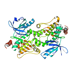
Crystal structure of HPK1 (MAP4K1) complex with inhibitor A-745
Descriptor: 6-[(5R)-5-benzamidocyclohex-1-en-1-yl]-3-[(1-methyl-1H-pyrazol-4-yl)amino]pyrazine-2-carboxamide, Mitogen-activated protein kinase kinase kinase kinase 1, SULFATE ION
Authors:Longenecker, K.L, Korepanova, A, Qiu, W.
Deposit date:2021-10-14
Release date:2022-03-02
Last modified:2023-10-18
Method:X-RAY DIFFRACTION (1.786 Å)
Cite:The HPK1 Inhibitor A-745 Verifies the Potential of Modulating T Cell Kinase Signaling for Immunotherapy.
Acs Chem.Biol., 17, 2022
3GF8
DownloadVisualize
BU of 3gf8 by Molmil
Crystal structure of putative polysaccharide binding proteins (DUF1812) (NP_809975.1) from BACTEROIDES THETAIOTAOMICRON VPI-5482 at 2.20 A resolution
Descriptor: 1,2-ETHANEDIOL, putative polysaccharide binding proteins (DUF1812)
Authors:Joint Center for Structural Genomics (JCSG)
Deposit date:2009-02-26
Release date:2009-03-24
Last modified:2023-02-01
Method:X-RAY DIFFRACTION (2.2 Å)
Cite:A conserved fold for fimbrial components revealed by the crystal structure of a putative fimbrial assembly protein (BT1062) from Bacteroides thetaiotaomicron at 2.2 A resolution
Acta Crystallogr.,Sect.F, 66, 2010
3B77
DownloadVisualize
BU of 3b77 by Molmil
Crystal structure of a ph domain containing bacterial protein (exig_2160) from exiguobacterium sibiricum 255-15 at 2.42 A resolution
Descriptor: Uncharacterized protein
Authors:Joint Center for Structural Genomics (JCSG)
Deposit date:2007-10-30
Release date:2007-11-13
Last modified:2023-01-25
Method:X-RAY DIFFRACTION (2.42 Å)
Cite:Bacterial pleckstrin homology domains: a prokaryotic origin for the PH domain.
J.Mol.Biol., 396, 2010

220472

數據於2024-05-29公開中

PDB statisticsPDBj update infoContact PDBjnumon