3SJC
 
 | |
3SJA
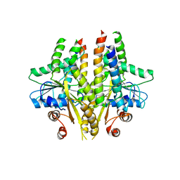
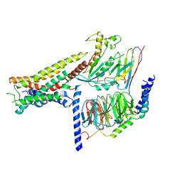
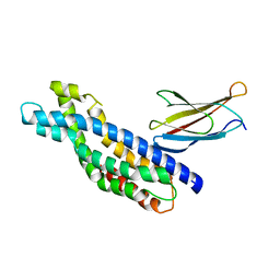
 | | Crystal structure of S. cerevisiae Get3 in the open state in complex with Get1 cytosolic domain | | 分子名称: | ATPase GET3, Golgi to ER traffic protein 1, PHOSPHATE ION, ... | | 著者 | Reitz, S, Wild, K, Sinning, I. | | 登録日 | 2011-06-21 | | 公開日 | 2011-07-06 | | 最終更新日 | 2024-02-28 | | 実験手法 | X-RAY DIFFRACTION (3 Å) | | 主引用文献 | Structural basis for tail-anchored membrane protein biogenesis by the Get3-receptor complex.
Science, 333, 2011
|
|
3SJD
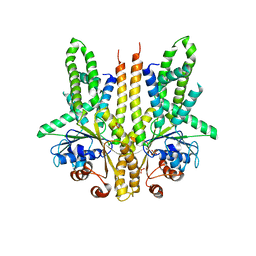
 | | Crystal structure of S. cerevisiae Get3 with bound ADP-Mg2+ in complex with Get2 cytosolic domain | | 分子名称: | ADENOSINE-5'-DIPHOSPHATE, ATPase GET3, Golgi to ER traffic protein 2, ... | | 著者 | Reitz, S, Wild, K, Sinning, I. | | 登録日 | 2011-06-21 | | 公開日 | 2011-07-13 | | 最終更新日 | 2024-02-28 | | 実験手法 | X-RAY DIFFRACTION (4.6 Å) | | 主引用文献 | Structural basis for tail-anchored membrane protein biogenesis by the Get3-receptor complex.
Science, 333, 2011
|
|
7OVC
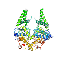
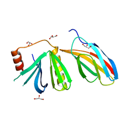
 | | Structure of the human UFC1 protein in complex with the UBA5 C-terminal UFC1-binding motif. | | 分子名称: | Ubiquitin-fold modifier-conjugating enzyme 1, Ubiquitin-like modifier-activating enzyme 5 | | 著者 | Wesch, W, Loehr, F, Rogova, N, Doetsch, V, Rogov, V.V. | | 登録日 | 2021-06-14 | | 公開日 | 2021-08-04 | | 最終更新日 | 2023-06-14 | | 実験手法 | SOLUTION NMR | | 主引用文献 | A Concerted Action of UBA5 C-Terminal Unstructured Regions Is Important for Transfer of Activated UFM1 to UFC1.
Int J Mol Sci, 22, 2021
|
|
8POK
 | | Cryo-EM structure of cell-free synthesized human histamine H2 receptor coupled to heterotrimeric Gs protein in lipid environment | | 分子名称: | Guanine nucleotide-binding protein G(I)/G(S)/G(O) subunit gamma-2, Guanine nucleotide-binding protein G(I)/G(S)/G(T) subunit beta-1, HISTAMINE, ... | | 著者 | Schnelle, K, Koeck, Z, Persechino, M, Umbach, S, Schihada, H, Januliene, D, Parey, K, Pockes, S, Kolb, P, Doetsch, V, Moeller, A, Hilger, D, Bernhard, F. | | 登録日 | 2023-07-05 | | 公開日 | 2024-03-06 | | 最終更新日 | 2024-03-13 | | 実験手法 | ELECTRON MICROSCOPY (3.4 Å) | | 主引用文献 | Cryo-EM structure of cell-free synthesized human histamine 2 receptor/G s complex in nanodisc environment.
Nat Commun, 15, 2024
|
|
4BPD
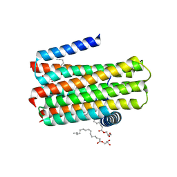 | | Structure determination of an integral membrane kinase | | 分子名称: | (2S)-2,3-DIHYDROXYPROPYL(7Z)-PENTADEC-7-ENOATE, DIACYLGLYCEROL KINASE, ZINC ION | | 著者 | Li, D, Boland, C, Caffrey, M. | | 登録日 | 2013-05-24 | | 公開日 | 2014-05-07 | | 最終更新日 | 2023-12-20 | | 実験手法 | X-RAY DIFFRACTION (3.3 Å) | | 主引用文献 | Cell-Free Expression and in Meso Crystallisation of an Integral Membrane Kinase for Structure Determination.
Cell.Mol.Life Sci., 71, 2014
|
|
4D2E
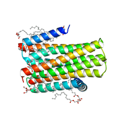 | | Crystal structure of an integral membrane kinase - v2.3 | | 分子名称: | (2R)-2,3-DIHYDROXYPROPYL(7Z)-PENTADEC-7-ENOATE, (2S)-2,3-DIHYDROXYPROPYL(7Z)-PENTADEC-7-ENOATE, CITRATE ANION, ... | | 著者 | Li, D, Boland, C, Caffrey, M. | | 登録日 | 2014-05-09 | | 公開日 | 2014-07-23 | | 最終更新日 | 2023-12-20 | | 実験手法 | X-RAY DIFFRACTION (2.28 Å) | | 主引用文献 | Cell-Free Expression and in Meso Crystallisation of an Integral Membrane Kinase for Structure Determination.
Cell.Mol.Life Sci., 71, 2014
|
|
4YMH
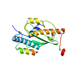
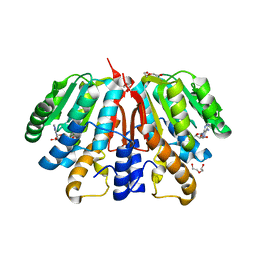 | | Crystal structure of SAH-bound Podospora anserina methyltransferase PaMTH1 | | 分子名称: | DI(HYDROXYETHYL)ETHER, Putative SAM-dependent O-methyltranferase, S-ADENOSYL-L-HOMOCYSTEINE | | 著者 | Kudlinzki, D, Linhard, V.L, Chatterjee, D, Saxena, K, Sreeramulu, S, Schwalbe, H. | | 登録日 | 2015-03-06 | | 公開日 | 2015-05-27 | | 最終更新日 | 2024-01-10 | | 実験手法 | X-RAY DIFFRACTION (1.876 Å) | | 主引用文献 | Structure and Biophysical Characterization of the S-Adenosylmethionine-dependent O-Methyltransferase PaMTH1, a Putative Enzyme Accumulating during Senescence of Podospora anserina.
J.Biol.Chem., 290, 2015
|
|
5N7E
 | | Crystal structure of the Dbl-homology domain of Bcr-Abl in complex with monobody Mb(Bcr-DH_4). | | 分子名称: | Breakpoint cluster region protein, Mb(Bcr-DH_4) | | 著者 | Reckel, S, Reynaud, A, Pojer, F, Hantschel, O. | | 登録日 | 2017-02-20 | | 公開日 | 2017-12-27 | | 最終更新日 | 2024-01-17 | | 実験手法 | X-RAY DIFFRACTION (1.647 Å) | | 主引用文献 | Structural and functional dissection of the DH and PH domains of oncogenic Bcr-Abl tyrosine kinase.
Nat Commun, 8, 2017
|
|
5OC7
 | | Crystal structure of the pleckstrin-homology domain of Bcr-Abl in complex with monobody Mb(Bcr-PH_4). | | 分子名称: | Breakpoint cluster region protein,pleckstrin-homology domain of Bcr-Abl, D-MYO-INOSITOL-4,5-BISPHOSPHATE, GLYCEROL, ... | | 著者 | Reckel, S, Reynaud, A, Pojer, F, Hantschel, O. | | 登録日 | 2017-06-29 | | 公開日 | 2017-12-27 | | 最終更新日 | 2024-01-17 | | 実験手法 | X-RAY DIFFRACTION (1.652 Å) | | 主引用文献 | Structural and functional dissection of the DH and PH domains of oncogenic Bcr-Abl tyrosine kinase.
Nat Commun, 8, 2017
|
|
3VTW
 | | Crystal structure of T7-tagged Optineurin LIR-fused human LC3B_2-119 | | 分子名称: | Optineurin, microtubule-associated proteins 1A/1B light chain 3B, SULFATE ION | | 著者 | Suzuki, H, Kawasaki, M, Kato, R, Wakatsuki, S. | | 登録日 | 2012-06-08 | | 公開日 | 2013-06-26 | | 最終更新日 | 2023-11-08 | | 実験手法 | X-RAY DIFFRACTION (2.52 Å) | | 主引用文献 | Structural basis for phosphorylation-triggered autophagic clearance of Salmonella
Biochem.J., 454, 2013
|
|
3VTU
 | | Crystal structure of human LC3B_2-119 | | 分子名称: | Microtubule-associated proteins 1A/1B light chain 3B, SULFATE ION | | 著者 | Suzuki, H, Kawasaki, M, Kato, R, Wakatsuki, S. | | 登録日 | 2012-06-08 | | 公開日 | 2013-06-26 | | 最終更新日 | 2023-11-08 | | 実験手法 | X-RAY DIFFRACTION (1.6 Å) | | 主引用文献 | Structural basis for phosphorylation-triggered autophagic clearance of Salmonella
Biochem.J., 454, 2013
|
|
3VTV
 | | Crystal structure of Optineurin LIR-fused human LC3B_2-119 | | 分子名称: | Optineurin, microtubule-associated proteins 1A/1B light chain 3B, SULFATE ION | | 著者 | Suzuki, H, Kawasaki, M, Kato, R, Wakatsuki, S. | | 登録日 | 2012-06-08 | | 公開日 | 2013-06-26 | | 最終更新日 | 2023-11-08 | | 実験手法 | X-RAY DIFFRACTION (1.7 Å) | | 主引用文献 | Structural basis for phosphorylation-triggered autophagic clearance of Salmonella
Biochem.J., 454, 2013
|
|
7P7G
 | | Crystal structure of phosphorylated pT220 Casein Kinase I delta (CK1d), conformation 2 and 3 | | 分子名称: | 1,2-ETHANEDIOL, ADENOSINE MONOPHOSPHATE, CITRIC ACID, ... | | 著者 | Chaikuad, A, Zhubi, R, Knapp, S, Structural Genomics Consortium (SGC) | | 登録日 | 2021-07-19 | | 公開日 | 2022-04-13 | | 最終更新日 | 2024-01-31 | | 実験手法 | X-RAY DIFFRACTION (1.7 Å) | | 主引用文献 | Kinase domain autophosphorylation rewires the activity and substrate specificity of CK1 enzymes.
Mol.Cell, 82, 2022
|
|
7P7F
 | | Crystal structure of phosphorylated pT220 Casein Kinase I delta (CK1d), conformation 1 | | 分子名称: | 1,2-ETHANEDIOL, ADENOSINE, ADENOSINE MONOPHOSPHATE, ... | | 著者 | Chaikuad, A, Zhubi, R, Knapp, S, Structural Genomics Consortium (SGC) | | 登録日 | 2021-07-19 | | 公開日 | 2022-04-13 | | 最終更新日 | 2024-01-31 | | 実験手法 | X-RAY DIFFRACTION (1.96 Å) | | 主引用文献 | Kinase domain autophosphorylation rewires the activity and substrate specificity of CK1 enzymes.
Mol.Cell, 82, 2022
|
|
7P7H
 | |
1EES
 | | SOLUTION STRUCTURE OF CDC42HS COMPLEXED WITH A PEPTIDE DERIVED FROM P-21 ACTIVATED KINASE, NMR, 20 STRUCTURES | | 分子名称: | GTP-BINDING PROTEIN, P21-ACTIVATED KINASE | | 著者 | Gizachew, D, Guo, W, Chohan, K.C, Sutcliffe, M.J, Oswald, R.E. | | 登録日 | 2000-02-02 | | 公開日 | 2000-03-29 | | 最終更新日 | 2022-02-16 | | 実験手法 | SOLUTION NMR | | 主引用文献 | Structure of the complex of Cdc42Hs with a peptide derived from P-21 activated kinase.
Biochemistry, 39, 2000
|
|
6RYA
 | |
6RYB
 | |
5DPT
 | | Crystal structure of PLEKHM1 LIR-fused human GABARAPL1_2-117 | | 分子名称: | (4S)-2-METHYL-2,4-PENTANEDIOL, Pleckstrin homology domain-containing family M member 1, Gamma-aminobutyric acid receptor-associated protein-like 1,Gamma-aminobutyric acid receptor-associated protein-like 1 | | 著者 | Ravichandran, A.C, Suzuki, H, Dobson, R.C.J. | | 登録日 | 2015-09-14 | | 公開日 | 2016-09-28 | | 最終更新日 | 2024-03-06 | | 実験手法 | X-RAY DIFFRACTION (2.9 Å) | | 主引用文献 | Structural and functional analysis of the GABARAP interaction motif (GIM).
EMBO Rep., 18, 2017
|
|
5DPS
 | |
5DPW
 | |
5DPR
 | |
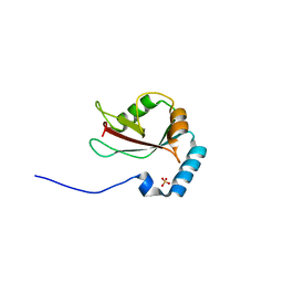
2KBY
 | | The Tetramerization Domain of Human p73 | | 分子名称: | Tumor protein p73 | | 著者 | Coutandin, D, Ikeya, T, Loehr, F, Guntert, P, Ou, H.D, Doetsch, V. | | 登録日 | 2008-12-12 | | 公開日 | 2009-09-29 | | 最終更新日 | 2022-03-16 | | 実験手法 | SOLUTION NMR | | 主引用文献 | Conformational stability and activity of p73 require a second helix in the tetramerization domain.
Cell Death Differ., 16, 2009
|
|
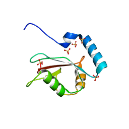
2KX7
 | | Solution structure of the E.coli RcsD-ABL domain (residues 688-795) | | 分子名称: | Sensor-like histidine kinase yojN | | 著者 | Rogov, V.V, Schmoee, K, Rogova, N.Y, Loehr, F, Bernhard, F, Doetsch, V. | | 登録日 | 2010-04-27 | | 公開日 | 2011-04-06 | | 最終更新日 | 2024-05-01 | | 実験手法 | SOLUTION NMR | | 主引用文献 | Structural Insights into Rcs Phosphotransfer: The Newly Identified RcsD-ABL Domain Enhances Interaction with the Response Regulator RcsB.
Structure, 19, 2011
|
|


