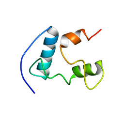
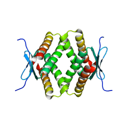
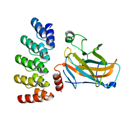
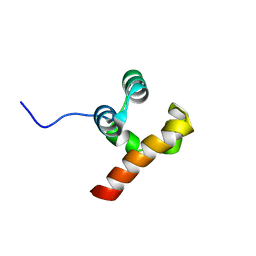
2KR6
 
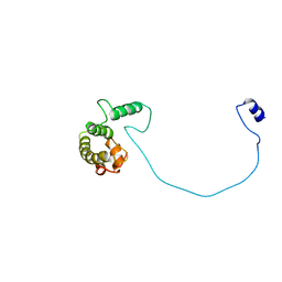 | |
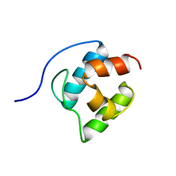
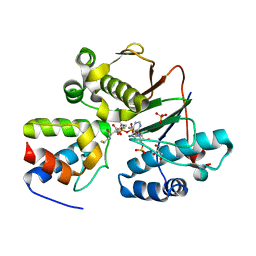
5HKH
 | | Crystal structure of Ufm1 in complex with UBA5 | | 分子名称: | ASP-ASN-GLU-TRP-GLY-ILE-GLU-LEU-VAL, Ubiquitin-fold modifier 1 | | 著者 | Huber, J, Doetsch, V, Rogov, V.V, Akutsu, M. | | 登録日 | 2016-01-14 | | 公開日 | 2016-03-09 | | 最終更新日 | 2024-01-10 | | 実験手法 | X-RAY DIFFRACTION (2.55 Å) | | 主引用文献 | Structural and Functional Analysis of a Novel Interaction Motif within UFM1-activating Enzyme 5 (UBA5) Required for Binding to Ubiquitin-like Proteins and Ufmylation.
J.Biol.Chem., 291, 2016
|
|
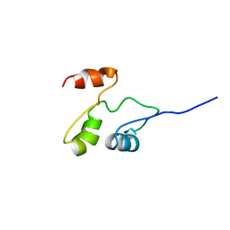
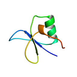
1ZGW
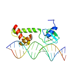 | | NMR structure of E. Coli Ada protein in complex with DNA | | 分子名称: | 5'-D(*GP*CP*AP*AP*AP*TP*TP*AP*AP*AP*GP*CP*GP*CP*AP*AP*GP*A)-3', 5'-D(*TP*CP*TP*TP*GP*CP*GP*CP*TP*TP*TP*AP*AP*TP*TP*TP*GP*C)-3', Ada polyprotein, ... | | 著者 | He, C, Hus, J.C, Sun, L.J, Zhou, P, Norman, D.P, Doetsch, V, Wei, H, Gross, J.D, Lane, W.S, Wagner, G, Verdine, G.L. | | 登録日 | 2005-04-22 | | 公開日 | 2005-10-18 | | 最終更新日 | 2022-03-02 | | 実験手法 | SOLUTION NMR | | 主引用文献 | A Methylation-Dependent Electrostatic Switch Controls DNA Repair and Transcriptional Activation by E. coli Ada.
Mol.Cell, 20, 2005
|
|
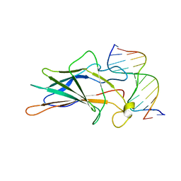
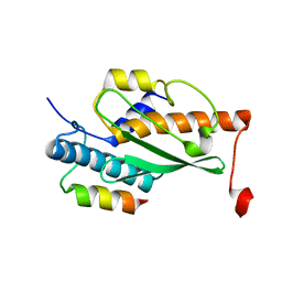
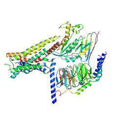
1SR2
 | |
2GDX
 | | Solution structure of the B. brevis TycC3-PCP in H-state | | 分子名称: | Tyrocidine synthetase III | | 著者 | Koglin, A, Loehr, F, Rogov, V.V, Marahiel, M.A, Bernhard, F, Doetsch, V. | | 登録日 | 2006-03-17 | | 公開日 | 2006-08-01 | | 最終更新日 | 2021-11-10 | | 実験手法 | SOLUTION NMR | | 主引用文献 | Conformational switches modulate protein interactions in peptide antibiotic synthetases
Science, 312, 2006
|
|
2GDW
 | | Solution structure of the B. brevis TycC3-PCP in A/H-state | | 分子名称: | Tyrocidine synthetase III | | 著者 | Koglin, A, Loehr, F, Rogov, V.V, Marahiel, M.A, Bernhard, F, Doetsch, V. | | 登録日 | 2006-03-17 | | 公開日 | 2006-08-01 | | 最終更新日 | 2021-11-10 | | 実験手法 | SOLUTION NMR | | 主引用文献 | Conformational switches modulate protein interactions in peptide antibiotic synthetases
Science, 312, 2006
|
|
2GDY
 | | Solution structure of the B. brevis TycC3-PCP in A-state | | 分子名称: | Tyrocidine synthetase III | | 著者 | Koglin, A, Loehr, F, Rogov, V.V, Marahiel, M.A, Bernhard, F, Doetsch, V. | | 登録日 | 2006-03-17 | | 公開日 | 2006-08-01 | | 最終更新日 | 2021-11-10 | | 実験手法 | SOLUTION NMR | | 主引用文献 | Conformational switches modulate protein interactions in peptide antibiotic synthetases
Science, 312, 2006
|
|
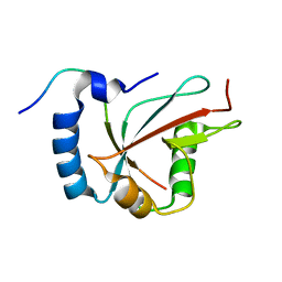
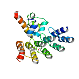
2RP5
 | |
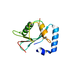
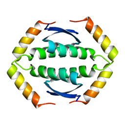
2RP4
 | |
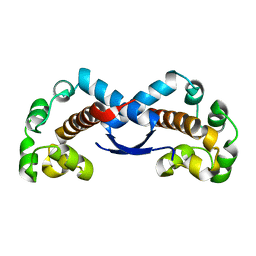
1A66
 | | SOLUTION NMR STRUCTURE OF THE CORE NFATC1/DNA COMPLEX, 18 STRUCTURES | | 分子名称: | CORE NFATC1, DNA (5'-D(*CP*AP*AP*TP*TP*TP*TP*CP*CP*TP*CP*G)-3'), DNA (5'-D(*CP*GP*AP*GP*GP*AP*AP*AP*AP*TP*TP*G)-3') | | 著者 | Zhou, P, Sun, L.J, Doetsch, V, Wagner, G, Verdine, G.L. | | 登録日 | 1998-03-06 | | 公開日 | 1998-06-17 | | 最終更新日 | 2021-11-03 | | 実験手法 | SOLUTION NMR | | 主引用文献 | Solution structure of the core NFATC1/DNA complex.
Cell(Cambridge,Mass.), 92, 1998
|
|
6H8C
 | | Structure of the human GABARAPL2 protein in complex with the UBA5 LIR motif | | 分子名称: | Gamma-aminobutyric acid receptor-associated protein-like 2, Ubiquitin-like modifier-activating enzyme 5 | | 著者 | Huber, J, Loehr, F, Gruber, J, Akutsu, M, Guentert, P, Doetsch, V, Rogov, V.V. | | 登録日 | 2018-08-02 | | 公開日 | 2019-05-01 | | 最終更新日 | 2020-01-29 | | 実験手法 | SOLUTION NMR | | 主引用文献 | An atypical LIR motif within UBA5 (ubiquitin like modifier activating enzyme 5) interacts with GABARAP proteins and mediates membrane localization of UBA5.
Autophagy, 16, 2020
|
|
4XC2
 | | Crystal structure of GABARAP in complex with KBTBD6 LIR peptide | | 分子名称: | GABA(A) receptor-associated protein, Kelch repeat and BTB domain-containing protein 6 | | 著者 | Huber, J, Genau, H.M, Baschieri, F, Doetsch, V, Farhan, H, Rogov, V.V, Behrends, C, Akutsu, M. | | 登録日 | 2014-12-17 | | 公開日 | 2015-03-04 | | 最終更新日 | 2024-01-10 | | 実験手法 | X-RAY DIFFRACTION (1.9 Å) | | 主引用文献 | CUL3-KBTBD6/KBTBD7 Ubiquitin Ligase Cooperates with GABARAP Proteins to Spatially Restrict TIAM1-RAC1 Signaling.
Mol.Cell, 57, 2015
|
|
7Z7E
 | | Crystal structure of p63 DNA binding domain in complex with inhibitory DARPin G4 | | 分子名称: | DARPIN, Isoform 4 of Tumor protein 63, ZINC ION | | 著者 | Strubel, A, Gebel, J, Chaikuad, A, Muenick, P, Doetsch, V. | | 登録日 | 2022-03-15 | | 公開日 | 2022-06-29 | | 最終更新日 | 2024-01-31 | | 実験手法 | X-RAY DIFFRACTION (1.8 Å) | | 主引用文献 | Designed Ankyrin Repeat Proteins as a tool box for analyzing p63.
Cell Death Differ., 29, 2022
|
|
4MRT
 | | Structure of the Phosphopantetheine Transferase Sfp in Complex with Coenzyme A and a Peptidyl Carrier Protein | | 分子名称: | 4'-phosphopantetheinyl transferase sfp, COENZYME A, GLYCEROL, ... | | 著者 | Tufar, P, Rahighi, S, Kraas, F.I, Kirchner, D.K, Loehr, F, Henrich, E, Koepke, J, Dikic, I, Guentert, P, Marahiel, M.A, Doetsch, V. | | 登録日 | 2013-09-17 | | 公開日 | 2014-04-23 | | 最終更新日 | 2024-02-28 | | 実験手法 | X-RAY DIFFRACTION (2 Å) | | 主引用文献 | Crystal Structure of a PCP/Sfp Complex Reveals the Structural Basis for Carrier Protein Posttranslational Modification.
Chem.Biol., 21, 2014
|
|
1GYF
 | | GYF DOMAIN FROM HUMAN CD2BP2 PROTEIN | | 分子名称: | PROTEIN (CYTOPLASMIC DOMAIN BINDING PROTEIN (CD2BP2)) | | 著者 | Freund, C, Doetsch, V, Nishizawa, K, Reinherz, E.L, Wagner, G. | | 登録日 | 1999-04-30 | | 公開日 | 2000-01-05 | | 最終更新日 | 2023-12-27 | | 実験手法 | SOLUTION NMR | | 主引用文献 | The GYF domain is a novel structural fold that is involved in lymphoid signaling through proline-rich sequences.
Nat.Struct.Biol., 6, 1999
|
|
7OVC
 | | Structure of the human UFC1 protein in complex with the UBA5 C-terminal UFC1-binding motif. | | 分子名称: | Ubiquitin-fold modifier-conjugating enzyme 1, Ubiquitin-like modifier-activating enzyme 5 | | 著者 | Wesch, W, Loehr, F, Rogova, N, Doetsch, V, Rogov, V.V. | | 登録日 | 2021-06-14 | | 公開日 | 2021-08-04 | | 最終更新日 | 2023-06-14 | | 実験手法 | SOLUTION NMR | | 主引用文献 | A Concerted Action of UBA5 C-Terminal Unstructured Regions Is Important for Transfer of Activated UFM1 to UFC1.
Int J Mol Sci, 22, 2021
|
|
5N2O
 | | Structure Of P63 SAM Domain L514F Mutant Causative Of AEC Syndrome | | 分子名称: | Tumor protein 63 | | 著者 | Rinnenthal, J, Wuerz, J.M, Osterburg, C, Guentert, P, Doetsch, V. | | 登録日 | 2017-02-08 | | 公開日 | 2018-02-07 | | 最終更新日 | 2023-06-14 | | 実験手法 | SOLUTION NMR | | 主引用文献 | Protein aggregation of the p63 transcription factor underlies severe skin fragility in AEC syndrome.
Proc. Natl. Acad. Sci. U.S.A., 115, 2018
|
|
8P9C
 | | Crystal structure of p63-p73 heterotetramer (tetramerisation domain) in complex with darpin 1810 F11 | | 分子名称: | 1,2-ETHANEDIOL, Darpin 1810 F11, Tumor protein 63, ... | | 著者 | Chaikuad, A, Strubel, A, Doetsch, V, Knapp, S, Structural Genomics Consortium (SGC) | | 登録日 | 2023-06-05 | | 公開日 | 2023-11-08 | | 最終更新日 | 2023-11-15 | | 実験手法 | X-RAY DIFFRACTION (1.76 Å) | | 主引用文献 | DARPins detect the formation of hetero-tetramers of p63 and p73 in epithelial tissues and in squamous cell carcinoma.
Cell Death Dis, 14, 2023
|
|
8P9E
 | | Crystal structure of wild type p63-p73 heterotetramer (tetramerisation domain) in complex with darpin 1810 F11 | | 分子名称: | Darpin 1810 F11, GLYCEROL, Isoform 2 of Tumor protein 63, ... | | 著者 | Chaikuad, A, Strubel, A, Doetsch, V, Knapp, S, Structural Genomics Consortium (SGC) | | 登録日 | 2023-06-05 | | 公開日 | 2023-11-08 | | 最終更新日 | 2023-11-15 | | 実験手法 | X-RAY DIFFRACTION (2.25 Å) | | 主引用文献 | DARPins detect the formation of hetero-tetramers of p63 and p73 in epithelial tissues and in squamous cell carcinoma.
Cell Death Dis, 14, 2023
|
|
8P9D
 | | Crystal structure of p63-p73 heterotetramer (tetramerisation domain) in complex with darpin 1810 A2 | | 分子名称: | Darpin 1810 A2, Tumor protein 63, Tumor protein p73 | | 著者 | Chaikuad, A, Strubel, A, Doetsch, V, Knapp, S, Structural Genomics Consortium (SGC) | | 登録日 | 2023-06-05 | | 公開日 | 2023-11-08 | | 最終更新日 | 2023-11-15 | | 実験手法 | X-RAY DIFFRACTION (2.7 Å) | | 主引用文献 | DARPins detect the formation of hetero-tetramers of p63 and p73 in epithelial tissues and in squamous cell carcinoma.
Cell Death Dis, 14, 2023
|
|
8POK
 | | Cryo-EM structure of cell-free synthesized human histamine H2 receptor coupled to heterotrimeric Gs protein in lipid environment | | 分子名称: | Guanine nucleotide-binding protein G(I)/G(S)/G(O) subunit gamma-2, Guanine nucleotide-binding protein G(I)/G(S)/G(T) subunit beta-1, HISTAMINE, ... | | 著者 | Schnelle, K, Koeck, Z, Persechino, M, Umbach, S, Schihada, H, Januliene, D, Parey, K, Pockes, S, Kolb, P, Doetsch, V, Moeller, A, Hilger, D, Bernhard, F. | | 登録日 | 2023-07-05 | | 公開日 | 2024-03-06 | | 最終更新日 | 2024-03-13 | | 実験手法 | ELECTRON MICROSCOPY (3.4 Å) | | 主引用文献 | Cryo-EM structure of cell-free synthesized human histamine 2 receptor/G s complex in nanodisc environment.
Nat Commun, 15, 2024
|
|
7Z71
 | | Crystal structure of p63 DBD in complex with darpin C14 | | 分子名称: | Darpin C14, Isoform 4 of Tumor protein 63, ZINC ION | | 著者 | Chaikuad, A, Strubel, A, Doetsch, V, Knapp, S, Structural Genomics Consortium (SGC) | | 登録日 | 2022-03-14 | | 公開日 | 2022-07-06 | | 最終更新日 | 2024-01-31 | | 実験手法 | X-RAY DIFFRACTION (1.85 Å) | | 主引用文献 | Designed Ankyrin Repeat Proteins as a tool box for analyzing p63.
Cell Death Differ., 29, 2022
|
|
7Z72
 | | Crystal structure of p63 SAM in complex with darpin A5 | | 分子名称: | DI(HYDROXYETHYL)ETHER, Darpin A5, Isoform 9 of Tumor protein 63 | | 著者 | Chaikuad, A, Strubel, A, Doetsch, V, Knapp, S, Structural Genomics Consortium (SGC) | | 登録日 | 2022-03-14 | | 公開日 | 2022-07-06 | | 最終更新日 | 2024-01-31 | | 実験手法 | X-RAY DIFFRACTION (1.8 Å) | | 主引用文献 | Designed Ankyrin Repeat Proteins as a tool box for analyzing p63.
Cell Death Differ., 29, 2022
|
|
7Z73
 | | Crystal structure of p63 tetramerization domain in complex with darpin 8F1 | | 分子名称: | Darpin 8F1, Isoform 2 of Tumor protein 63 | | 著者 | Chaikuad, A, Strubel, A, Doetsch, V, Knapp, S, Structural Genomics Consortium (SGC) | | 登録日 | 2022-03-14 | | 公開日 | 2022-07-06 | | 最終更新日 | 2024-01-31 | | 実験手法 | X-RAY DIFFRACTION (2.27 Å) | | 主引用文献 | Designed Ankyrin Repeat Proteins as a tool box for analyzing p63.
Cell Death Differ., 29, 2022
|
|
2KBY
 | | The Tetramerization Domain of Human p73 | | 分子名称: | Tumor protein p73 | | 著者 | Coutandin, D, Ikeya, T, Loehr, F, Guntert, P, Ou, H.D, Doetsch, V. | | 登録日 | 2008-12-12 | | 公開日 | 2009-09-29 | | 最終更新日 | 2022-03-16 | | 実験手法 | SOLUTION NMR | | 主引用文献 | Conformational stability and activity of p73 require a second helix in the tetramerization domain.
Cell Death Differ., 16, 2009
|
|


