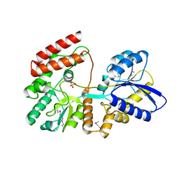
5BU9
 
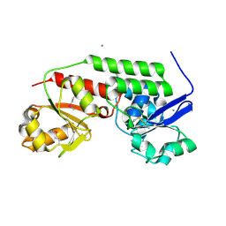
 | | Crystal structure of Beta-N-acetylhexosaminidase from Beutenbergia cavernae DSM 12333 | | 分子名称: | Beta-N-acetylhexosaminidase, GLYCEROL | | 著者 | Chang, C, Tan, K, Li, H, Endres, M, Joachimiak, A, Midwest Center for Structural Genomics (MCSG) | | 登録日 | 2015-06-03 | | 公開日 | 2015-06-17 | | 実験手法 | X-RAY DIFFRACTION (2.255 Å) | | 主引用文献 | Crystal structure of Beta-N-acetylhexosaminidase from Beutenbergia cavernae DSM 12333
To Be Published
|
|
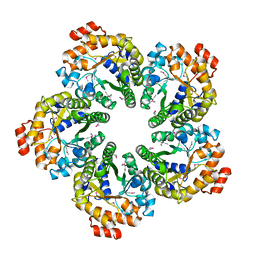
5DA8
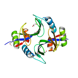
 | | Crystal structure of chaperonin GroEL from | | 分子名称: | 60 kDa chaperonin, CALCIUM ION, MAGNESIUM ION, ... | | 著者 | Chang, C, Marshall, N, Feldmann, B, Joachimiak, A, Midwest Center for Structural Genomics (MCSG) | | 登録日 | 2015-08-19 | | 公開日 | 2015-10-14 | | 最終更新日 | 2023-09-27 | | 実験手法 | X-RAY DIFFRACTION (3 Å) | | 主引用文献 | Crystal structure of chaperonin GroEL from
To Be Published
|
|
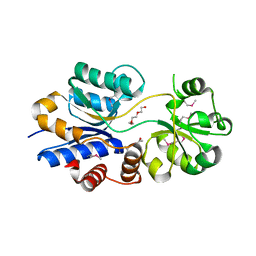
4KVH
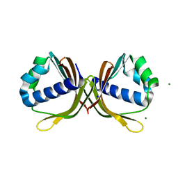
 | | Crystal structure of ketosteroid isomerase fold protein Hmuk_0747 | | 分子名称: | BROMIDE ION, CACODYLATE ION, FORMIC ACID, ... | | 著者 | Chang, C, Holowicki, J, Bearden, J, Joachimiak, A, Midwest Center for Structural Genomics (MCSG) | | 登録日 | 2013-05-22 | | 公開日 | 2013-06-12 | | 最終更新日 | 2024-02-28 | | 実験手法 | X-RAY DIFFRACTION (1.61 Å) | | 主引用文献 | Crystal structure of ketosteroid isomerase fold protein Hmuk_0747
To be Published
|
|
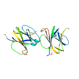
1IHR
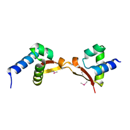
 | | Crystal structure of the dimeric C-terminal domain of TonB | | 分子名称: | BROMIDE ION, TonB protein | | 著者 | Chang, C, Mooser, A, Pluckthun, A, Wlodawer, A. | | 登録日 | 2001-04-20 | | 公開日 | 2001-08-01 | | 最終更新日 | 2024-02-07 | | 実験手法 | X-RAY DIFFRACTION (1.55 Å) | | 主引用文献 | Crystal structure of the dimeric C-terminal domain of TonB reveals a novel fold.
J.Biol.Chem., 276, 2001
|
|
4LMI
 | |
4MDY
 | |
5DUK
 | |
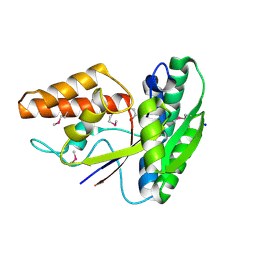
5E2C
 | | Crystal structure of N-terminal domain of cytoplasmic peptidase PepQ from Mycobacterium tuberculosis H37Rv | | 分子名称: | Xaa-Pro dipeptidase | | 著者 | Chang, C, Endres, L, Endres, M, SACCHETTINI, J, JOACHIMIAK, A, Midwest Center for Structural Genomics (MCSG), Structures of Mtb Proteins Conferring Susceptibility to Known Mtb Inhibitors (MTBI) | | 登録日 | 2015-09-30 | | 公開日 | 2015-10-14 | | 実験手法 | X-RAY DIFFRACTION (1.7 Å) | | 主引用文献 | Crystal structure of N-terminal domain of cytoplasmic peptidase PepQ from Mycobacterium tuberculosis H37Rv
To Be Published
|
|
4LQB
 | | Crystal structure of uncharacterized protein Kfla3161 | | 分子名称: | CITRIC ACID, GLYCEROL, Uncharacterized protein | | 著者 | Chang, C, Chhor, G, Endres, M, Joachimiak, A, Midwest Center for Structural Genomics (MCSG) | | 登録日 | 2013-07-17 | | 公開日 | 2013-07-31 | | 最終更新日 | 2017-11-15 | | 実験手法 | X-RAY DIFFRACTION (1.72 Å) | | 主引用文献 | Crystal structure of uncharacterized protein Kfla3161
To be Published
|
|
4MLZ
 | | Crystal structure of periplasmic binding protein from Jonesia denitrificans | | 分子名称: | CALCIUM ION, POTASSIUM ION, Periplasmic binding protein | | 著者 | Chang, C, Chhor, G, Endres, M, Joachimiak, A, Midwest Center for Structural Genomics (MCSG) | | 登録日 | 2013-09-06 | | 公開日 | 2013-09-18 | | 最終更新日 | 2017-11-15 | | 実験手法 | X-RAY DIFFRACTION (1.72 Å) | | 主引用文献 | Crystal structure of periplasmic binding protein from Jonesia denitrificans
To be Published
|
|
4MJD
 | | Crystal structure of ketosteroid isomerase fold protein Hmuk_0747 | | 分子名称: | Ketosteroid isomerase fold protein Hmuk_0747, MAGNESIUM ION, SODIUM ION | | 著者 | Chang, C, Holowicki, J, Bearden, J, Joachimiak, A, Midwest Center for Structural Genomics (MCSG) | | 登録日 | 2013-09-03 | | 公開日 | 2013-09-18 | | 最終更新日 | 2023-09-20 | | 実験手法 | X-RAY DIFFRACTION (1.28 Å) | | 主引用文献 | Crystal structure of ketosteroid isomerase fold protein Hmuk_0747
To be Published
|
|
4MOZ
 | |
4MQD
 | | Crystal structure of ComJ, inhibitor of the DNA degrading activity of NucA, from Bacillus subtilis | | 分子名称: | DNA-entry nuclease inhibitor | | 著者 | Chang, C, Mack, J, Clancy, S, Joachimiak, A, Midwest Center for Structural Genomics (MCSG) | | 登録日 | 2013-09-16 | | 公開日 | 2013-10-09 | | 最終更新日 | 2017-11-15 | | 実験手法 | X-RAY DIFFRACTION (2.16 Å) | | 主引用文献 | Crystal structure of ComJ, inhibitor of the DNA degrading activity of NucA, from Bacillus subtilis
To be Published
|
|
3O6P
 | | Crystal structure of peptide ABC transporter, peptide-binding protein | | 分子名称: | Peptide ABC transporter, peptide-binding protein, SODIUM ION | | 著者 | Chang, C, Bigelow, L, Bearden, J, Joachimiak, A, Midwest Center for Structural Genomics (MCSG) | | 登録日 | 2010-07-29 | | 公開日 | 2010-09-22 | | 最終更新日 | 2017-11-08 | | 実験手法 | X-RAY DIFFRACTION (1.65 Å) | | 主引用文献 | Crystal structure of peptide ABC transporter, peptide-binding protein
To be Published
|
|
3O66
 | | Crystal structure of glycine betaine/carnitine/choline ABC transporter | | 分子名称: | ACETATE ION, Glycine betaine/carnitine/choline ABC transporter, TRIETHYLENE GLYCOL | | 著者 | Chang, C, Bigelow, L, Carroll, J, Joachimiak, A, Midwest Center for Structural Genomics (MCSG) | | 登録日 | 2010-07-28 | | 公開日 | 2010-08-18 | | 最終更新日 | 2017-11-08 | | 実験手法 | X-RAY DIFFRACTION (1.86 Å) | | 主引用文献 | Crystal structure of glycine betaine/carnitine/choline ABC transporter
To be Published
|
|
3OCR
 | | Crystal structure of aldolase II superfamily protein from Pseudomonas syringae | | 分子名称: | Class II aldolase/adducin domain protein, SULFATE ION | | 著者 | Chang, C, Kagan, O, Savchenko, A, Edwards, A, Joachimiak, A, Midwest Center for Structural Genomics (MCSG) | | 登録日 | 2010-08-10 | | 公開日 | 2010-08-25 | | 最終更新日 | 2017-11-08 | | 実験手法 | X-RAY DIFFRACTION (1.95 Å) | | 主引用文献 | Crystal structure of aldolase II superfamily protein from Pseudomonas syringae
To be Published
|
|
3OIO
 | | Crystal structure of transcriptional regulator (AraC-type DNA-binding domain-containing proteins) from Chromobacterium violaceum | | 分子名称: | CHLORIDE ION, SULFATE ION, Transcriptional regulator (AraC-type DNA-binding domain-containing proteins) | | 著者 | Chang, C, Mack, J, Feldman, B, Joachimiak, A, Midwest Center for Structural Genomics (MCSG) | | 登録日 | 2010-08-19 | | 公開日 | 2010-09-08 | | 最終更新日 | 2019-12-18 | | 実験手法 | X-RAY DIFFRACTION (1.65 Å) | | 主引用文献 | Crystal structure of transcriptional regulator (AraC-type DNA-binding domain-containing proteins) from Chromobacterium violaceum
To be Published
|
|
3OIZ
 | | Crystal structure of antisigma-factor antagonist, STAS domain from Rhodobacter sphaeroides | | 分子名称: | Antisigma-factor antagonist, STAS | | 著者 | Chang, C, Marshall, N, Freeman, L, Joachimiak, A, Midwest Center for Structural Genomics (MCSG) | | 登録日 | 2010-08-20 | | 公開日 | 2010-09-08 | | 最終更新日 | 2011-07-13 | | 実験手法 | X-RAY DIFFRACTION (1.65 Å) | | 主引用文献 | Crystal structure of antisigma-factor antagonist, STAS domain from Rhodobacter sphaeroides
To be Published
|
|
3OMB
 | | Crystal structure of extracellular solute-binding protein from Bifidobacterium longum subsp. infantis | | 分子名称: | Extracellular solute-binding protein, family 1, MAGNESIUM ION | | 著者 | Chang, C, Xu, X, Chin, S, Cui, H, Dong, A, Savchenko, A, Edwards, A, Joachimiak, A, Midwest Center for Structural Genomics (MCSG) | | 登録日 | 2010-08-26 | | 公開日 | 2010-09-08 | | 最終更新日 | 2011-07-13 | | 実験手法 | X-RAY DIFFRACTION (2.1 Å) | | 主引用文献 | Crystal structure of extracellular solute-binding protein from Bifidobacterium longum subsp. infantis
To be Published
|
|
3OMD
 | | Crystal structure of unknown function protein from Leptospirillum rubarum | | 分子名称: | Uncharacterized protein | | 著者 | Chang, C, Xu, X, Cui, H, Chen, Z, Savchenko, A, Edwards, A, Joachimiak, A, Midwest Center for Structural Genomics (MCSG) | | 登録日 | 2010-08-26 | | 公開日 | 2010-09-15 | | 最終更新日 | 2011-07-13 | | 実験手法 | X-RAY DIFFRACTION (1.5 Å) | | 主引用文献 | Crystal structure of unknown function protein from Leptospirillum rubarum
To be Published
|
|
3OKX
 | | Crystal structure of YaeB-like protein from Rhodopseudomonas palustris | | 分子名称: | S-ADENOSYLMETHIONINE, YaeB-like protein RPA0152 | | 著者 | Chang, C, Evdokimova, E, Liu, F, Savchenko, A, Edwards, A, Joachimiak, A, Midwest Center for Structural Genomics (MCSG) | | 登録日 | 2010-08-25 | | 公開日 | 2010-09-08 | | 最終更新日 | 2011-07-13 | | 実験手法 | X-RAY DIFFRACTION (1.8 Å) | | 主引用文献 | Crystal structure of YaeB-like protein from Rhodopseudomonas palustris
To be Published
|
|
3P01
 | |
4O2I
 | | The crystal structure of non-LEE encoded type III effector C from Citrobacter rodentium | | 分子名称: | Non-LEE encoded type III effector C, ZINC ION | | 著者 | Chang, C, Xu, X, Cui, H, Savchenko, A, Adkins, J, Joachimiak, A, Midwest Center for Structural Genomics (MCSG), Program for the Characterization of Secreted Effector Proteins (PCSEP) | | 登録日 | 2013-12-17 | | 公開日 | 2014-01-15 | | 最終更新日 | 2014-05-07 | | 実験手法 | X-RAY DIFFRACTION (2.2 Å) | | 主引用文献 | The crystal structure of non-LEE encoded type III effector C from Citrobacter rodentium
To be Published
|
|
4OVX
 | | Crystal structure of Xylose isomerase domain protein from Planctomyces limnophilus DSM 3776 | | 分子名称: | 1,2-ETHANEDIOL, Xylose isomerase domain protein TIM barrel | | 著者 | Chang, C, Bigelow, L, Endres, M, Joachimiak, A, Midwest Center for Structural Genomics (MCSG) | | 登録日 | 2014-01-22 | | 公開日 | 2014-02-12 | | 最終更新日 | 2023-12-27 | | 実験手法 | X-RAY DIFFRACTION (2.253 Å) | | 主引用文献 | Crystal structure of Xylose isomerase domain protein from Planctomyces limnophilus DSM 3776
To be published
|
|
4OVJ
 | |


