Loading
PDBj
MenuPDBj@FacebookPDBj@TwitterPDBj@YouTubewwPDB FoundationwwPDB
RCSB PDBPDBeBMRBAdv. SearchSearch help
Search by PDB author
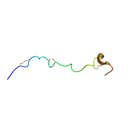
2JM2
DownloadVisualize
BU of 2jm2 by Molmil
Structure of the N-terminal subdomain of insulin-like growth factor (IGF) binding protein-6 and its interactions with IGFs
Descriptor: Insulin-like growth factor-binding protein 6
Authors:Chandrashekaran, I.R, Yao, S, Wang, C.C, Bansal, P.S, Alewood, P.F, Forbes, B.E, Wallace, J.C, Bach, L.A, Norton, R.S.
Deposit date:2006-09-18
Release date:2007-03-27
Last modified:2023-12-20
Method:SOLUTION NMR
Cite:The N-Terminal Subdomain of Insulin-like Growth Factor (IGF) Binding Protein 6. Structure and Interaction with IGFs
Biochemistry, 46, 2007
2N34
DownloadVisualize
BU of 2n34 by Molmil
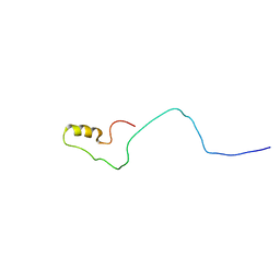
NMR assignments and solution structure of the JAK interaction region of SOCS5
Descriptor: Suppressor of cytokine signaling 5
Authors:Chandrashekaran, I.R, Mohanty, B, Linossi, E.M, Nicholson, S.E, Babon, J, Norton, R.S, Dagley, L.F, Leung, E.W.W, Murphy, J.M.
Deposit date:2015-05-21
Release date:2015-07-29
Last modified:2024-05-15
Method:SOLUTION NMR
Cite:Structure and Functional Characterization of the Conserved JAK Interaction Region in the Intrinsically Disordered N-Terminus of SOCS5.
Biochemistry, 54, 2015
7N3K
DownloadVisualize
BU of 7n3k by Molmil
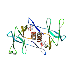
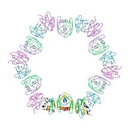
Oridonin-bound SARS-CoV-2 Nsp9
Descriptor: (1beta,6beta,7beta,8alpha,9beta,10alpha,13alpha,14R,16beta)-1,6,7,14-tetrahydroxy-7,20-epoxykauran-15-one, Non-structural protein 9, SULFATE ION
Authors:Littler, D.R, Gully, B.S, Rossjohn, J.
Deposit date:2021-06-01
Release date:2021-10-27
Last modified:2023-10-18
Method:X-RAY DIFFRACTION (3 Å)
Cite:A natural product compound inhibits coronaviral replication in vitro by binding to the conserved Nsp9 SARS-CoV-2 protein.
J.Biol.Chem., 297, 2021
4R1B
DownloadVisualize
BU of 4r1b by Molmil
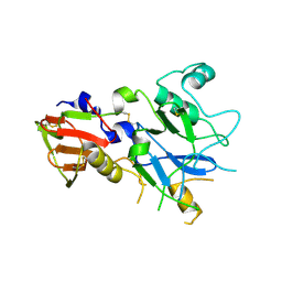
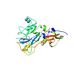
Crystal Structure of 3D7 strain Plasmodium falciparum AMA1
Descriptor: Apical membrane antigen 1, AMA1
Authors:Lim, S.S, Norton, R.S, McGowan, S.
Deposit date:2014-08-04
Release date:2015-01-14
Last modified:2023-09-20
Method:X-RAY DIFFRACTION (1.6 Å)
Cite:Structure and Dynamics of Apical Membrane Antigen 1 from Plasmodium falciparum FVO.
Biochemistry, 53, 2014
4R1C
DownloadVisualize
BU of 4r1c by Molmil
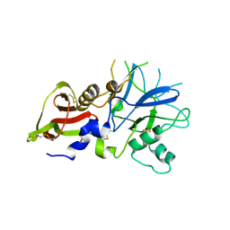
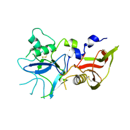
Crystal Structure of 3D7 strain Plasmodium falciparum AMA1
Descriptor: Apical membrane antigen 1, AMA1
Authors:Lim, S.S, Norton, R.S, McGowan, S.
Deposit date:2014-08-04
Release date:2015-01-14
Last modified:2023-09-20
Method:X-RAY DIFFRACTION (2 Å)
Cite:Structure and Dynamics of Apical Membrane Antigen 1 from Plasmodium falciparum FVO.
Biochemistry, 53, 2014
4R19
DownloadVisualize
BU of 4r19 by Molmil
Crystal Structure of 3D7 strain Plasmodium falciparum AMA1
Descriptor: Apical membrane antigen 1, AMA1
Authors:Lim, S.S, Norton, R.S, McGowan, S.
Deposit date:2014-08-04
Release date:2015-01-14
Last modified:2023-09-20
Method:X-RAY DIFFRACTION (1.8 Å)
Cite:Structure and Dynamics of Apical Membrane Antigen 1 from Plasmodium falciparum FVO.
Biochemistry, 53, 2014
4R1A
DownloadVisualize
BU of 4r1a by Molmil
Crystal Structure of FVO strain Plasmodium falciparum AMA1
Descriptor: Apical membrane antigen 1
Authors:Lim, S.S, Norton, R.S, McGowan, S.
Deposit date:2014-08-04
Release date:2015-01-14
Last modified:2023-09-20
Method:X-RAY DIFFRACTION (2 Å)
Cite:Structure and Dynamics of Apical Membrane Antigen 1 from Plasmodium falciparum FVO.
Biochemistry, 53, 2014
6DO7
DownloadVisualize
BU of 6do7 by Molmil
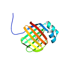
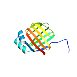
NMR solution structure of wild type hFABP1 with GW7647
Descriptor: Fatty acid-binding protein, liver
Authors:Scanlon, M.J, Mohanty, B, Doak, B.C, Patil, R.
Deposit date:2018-06-09
Release date:2019-01-02
Last modified:2024-05-01
Method:SOLUTION NMR
Cite:A ligand-induced structural change in fatty acid-binding protein 1 is associated with potentiation of peroxisome proliferator-activated receptor alpha agonists.
J. Biol. Chem., 294, 2019
6DRG
DownloadVisualize
BU of 6drg by Molmil
NMR solution structure of wild type hFABP1 with GW7647
Descriptor: 2-[(4-{2-[(4-cyclohexylbutyl)(cyclohexylcarbamoyl)amino]ethyl}phenyl)sulfanyl]-2-methylpropanoic acid, Fatty acid-binding protein, liver
Authors:Scanlon, M.J, Mohanty, B, Doak, B.C, Patil, R.
Deposit date:2018-06-11
Release date:2018-12-26
Last modified:2024-05-01
Method:SOLUTION NMR
Cite:A ligand-induced structural change in fatty acid-binding protein 1 is associated with potentiation of peroxisome proliferator-activated receptor alpha agonists.
J. Biol. Chem., 294, 2019
6DO6
DownloadVisualize
BU of 6do6 by Molmil
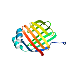
NMR solution structure of wild type apo hFABP1 at 308 K
Descriptor: Fatty acid-binding protein, liver
Authors:Scanlon, M.J, Mohanty, B, Doak, B.C, Patil, R.
Deposit date:2018-06-09
Release date:2018-12-26
Last modified:2024-05-01
Method:SOLUTION NMR
Cite:A ligand-induced structural change in fatty acid-binding protein 1 is associated with potentiation of peroxisome proliferator-activated receptor alpha agonists.
J. Biol. Chem., 294, 2019
8D12
DownloadVisualize
BU of 8d12 by Molmil
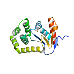
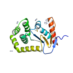
Crystal Structure of EcDsbA in a complex with 1-methyl-1H-pyrazol-4-amine
Descriptor: 1-methyl-1H-pyrazol-4-amine, COPPER (II) ION, Thiol:disulfide interchange protein DsbA
Authors:Whitehouse, R.L, Ilyichova, O.V, Taylor, A.J.
Deposit date:2022-05-26
Release date:2022-12-07
Last modified:2023-10-25
Method:X-RAY DIFFRACTION (1.6 Å)
Cite:Fragment screening libraries for the identification of protein hot spots and their minimal binding pharmacophores.
Rsc Med Chem, 14, 2023
8CZM
DownloadVisualize
BU of 8czm by Molmil
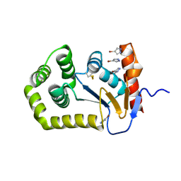
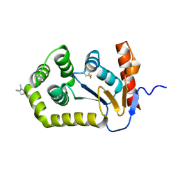
Crystal Structure of EcDsbA in a complex with 4-bromo-1H-pyrazole
Descriptor: 4-bromo-1H-pyrazole, TRIETHYLENE GLYCOL, Thiol:disulfide interchange protein DsbA
Authors:Whitehouse, R.L, Ilyichova, O.V, Taylor, A.J.
Deposit date:2022-05-25
Release date:2022-12-07
Last modified:2023-10-25
Method:X-RAY DIFFRACTION (1.8 Å)
Cite:Fragment screening libraries for the identification of protein hot spots and their minimal binding pharmacophores.
Rsc Med Chem, 14, 2023
8DG0
DownloadVisualize
BU of 8dg0 by Molmil
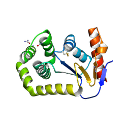
Crystal Structure of EcDsbA in a complex with Urea
Descriptor: COPPER (II) ION, Thiol:disulfide interchange protein DsbA, UREA
Authors:Whitehouse, R.L, Ilyichova, O.V, Taylor, A.J.
Deposit date:2022-06-23
Release date:2022-12-07
Last modified:2023-10-25
Method:X-RAY DIFFRACTION (2.5 Å)
Cite:Fragment screening libraries for the identification of protein hot spots and their minimal binding pharmacophores.
Rsc Med Chem, 14, 2023
8CXD
DownloadVisualize
BU of 8cxd by Molmil
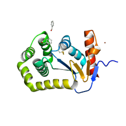
Crystal Structure of EcDsbA in a complex with phenylmethanol
Descriptor: COPPER (II) ION, Thiol:disulfide interchange protein DsbA, phenylmethanol
Authors:Whitehouse, R.L, Ilyichova, O.V, Taylor, A.J.
Deposit date:2022-05-20
Release date:2022-12-07
Last modified:2023-10-25
Method:X-RAY DIFFRACTION (1.8 Å)
Cite:Fragment screening libraries for the identification of protein hot spots and their minimal binding pharmacophores.
Rsc Med Chem, 14, 2023
8CZN
DownloadVisualize
BU of 8czn by Molmil
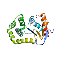
Crystal Structure of EcDsbA in a complex with 1H-pyrrole-3-carboxylic acid
Descriptor: 1H-pyrrole-3-carboxylic acid, COPPER (II) ION, Thiol:disulfide interchange protein DsbA
Authors:Whitehouse, R.L, Ilyichova, O.V, Taylor, A.J.
Deposit date:2022-05-25
Release date:2022-12-07
Last modified:2023-10-25
Method:X-RAY DIFFRACTION (1.7 Å)
Cite:Fragment screening libraries for the identification of protein hot spots and their minimal binding pharmacophores.
Rsc Med Chem, 14, 2023
8DG1
DownloadVisualize
BU of 8dg1 by Molmil
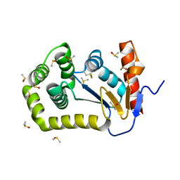
Crystal Structure of EcDsbA in a complex with DMSO
Descriptor: COPPER (II) ION, DIMETHYL SULFOXIDE, Thiol:disulfide interchange protein DsbA
Authors:Whitehouse, R.L, Ilyichova, O.V, Taylor, A.J.
Deposit date:2022-06-23
Release date:2022-12-07
Last modified:2023-10-25
Method:X-RAY DIFFRACTION (1.95 Å)
Cite:Fragment screening libraries for the identification of protein hot spots and their minimal binding pharmacophores.
Rsc Med Chem, 14, 2023
8CXE
DownloadVisualize
BU of 8cxe by Molmil
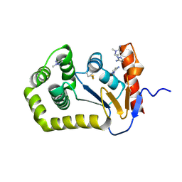
Crystal Structure of EcDsbA in a complex with 1H-imidazole
Descriptor: IMIDAZOLE, Thiol:disulfide interchange protein DsbA
Authors:Whitehouse, R.L, Ilyichova, O.V, Taylor, A.J.
Deposit date:2022-05-20
Release date:2022-12-07
Last modified:2023-10-25
Method:X-RAY DIFFRACTION (1.47 Å)
Cite:Fragment screening libraries for the identification of protein hot spots and their minimal binding pharmacophores.
Rsc Med Chem, 14, 2023
8D10
DownloadVisualize
BU of 8d10 by Molmil
Crystal Structure of EcDsbA in a complex with (1-methyl-1H-pyrazol-5-yl)methanamine
Descriptor: 1-(1-methyl-1H-pyrazol-5-yl)methanamine, COPPER (II) ION, Thiol:disulfide interchange protein DsbA
Authors:Whitehouse, R.L, Ilyichova, O.V, Taylor, A.J.
Deposit date:2022-05-26
Release date:2022-12-07
Last modified:2023-10-25
Method:X-RAY DIFFRACTION (1.6 Å)
Cite:Fragment screening libraries for the identification of protein hot spots and their minimal binding pharmacophores.
Rsc Med Chem, 14, 2023
8DG2
DownloadVisualize
BU of 8dg2 by Molmil
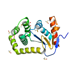
Crystal Structure of EcDsbA in a complex with DMSO
Descriptor: COPPER (II) ION, DIMETHYL SULFOXIDE, GLYCEROL, ...
Authors:Whitehouse, R.L, Ilyichova, O.V, Taylor, A.J.
Deposit date:2022-06-23
Release date:2022-12-07
Last modified:2023-10-25
Method:X-RAY DIFFRACTION (1.95 Å)
Cite:Fragment screening libraries for the identification of protein hot spots and their minimal binding pharmacophores.
Rsc Med Chem, 14, 2023
8D11
DownloadVisualize
BU of 8d11 by Molmil
Crystal Structure of EcDsbA in a complex with 1-methyl-1H-pyrazol-5-amine
Descriptor: 1-methyl-1H-pyrazol-5-amine, COPPER (II) ION, Thiol:disulfide interchange protein DsbA
Authors:Whitehouse, R.L, Ilyichova, O.V, Taylor, A.J.
Deposit date:2022-05-26
Release date:2022-12-07
Last modified:2023-10-25
Method:X-RAY DIFFRACTION (1.85 Å)
Cite:Fragment screening libraries for the identification of protein hot spots and their minimal binding pharmacophores.
Rsc Med Chem, 14, 2023
8DQU
DownloadVisualize
BU of 8dqu by Molmil
Nanobody bound SARS-CoV-2 Nsp9
Descriptor: Nanobody, Non-structural protein 9
Authors:Littler, D.R, Gully, B.S, Rossjohn, J.
Deposit date:2022-07-20
Release date:2023-02-08
Last modified:2023-10-25
Method:X-RAY DIFFRACTION (2.45003176 Å)
Cite:Inside-out: Antibody-binding reveals potential folding hinge-points within the SARS-CoV-2 replication co-factor nsp9.
Plos One, 18, 2023

221371

PDB entries from 2024-06-19

PDB statisticsPDBj update infoContact PDBjnumon