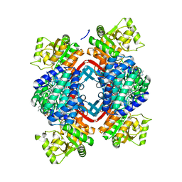
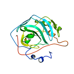
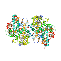
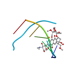
5IY2
 
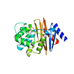 | | Structure of apo OXA-143 carbapenemase | | 分子名称: | Beta-lactamase OXA-143, GLYCEROL | | 著者 | Smith, C.A, Vakulenko, S.B. | | 登録日 | 2016-03-23 | | 公開日 | 2017-08-09 | | 最終更新日 | 2019-12-11 | | 実験手法 | X-RAY DIFFRACTION (1.15 Å) | | 主引用文献 | The role of conserved surface hydrophobic residues in the carbapenemase activity of the class D beta-lactamases.
Acta Crystallogr D Struct Biol, 73, 2017
|
|
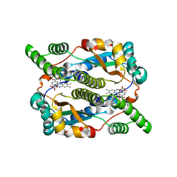
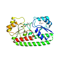
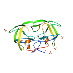
5J7N
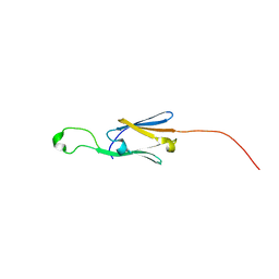 | | Crystal structure of a small heat-shock protein from Xylella fastidiosa reveals a distinct high order structure | | 分子名称: | Low molecular weight heat shock protein | | 著者 | Fonseca, E.M.B, Scorsato, V, dos Santos, C.A, Tomazini Jr, A, Aparicio, R, Polikarpov, I. | | 登録日 | 2016-04-06 | | 公開日 | 2017-04-12 | | 最終更新日 | 2023-09-27 | | 実験手法 | X-RAY DIFFRACTION (2.9 Å) | | 主引用文献 | Crystal structure of a small heat-shock protein from Xylella fastidiosa reveals a distinct high-order structure.
Acta Crystallogr F Struct Biol Commun, 73, 2017
|
|
5JCO
 | | Structure and dynamics of single-isoform recombinant neuronal human tubulin | | 分子名称: | GUANOSINE-5'-TRIPHOSPHATE, MAGNESIUM ION, PHOSPHOMETHYLPHOSPHONIC ACID GUANYLATE ESTER, ... | | 著者 | Vemu, A, Atherton, J, Spector, J.O, Szyk, A, Moores, C.A, Roll-Mecak, A. | | 登録日 | 2016-04-15 | | 公開日 | 2016-05-04 | | 最終更新日 | 2024-03-06 | | 実験手法 | ELECTRON MICROSCOPY (4 Å) | | 主引用文献 | Structure and Dynamics of Single-isoform Recombinant Neuronal Human Tubulin.
J.Biol.Chem., 291, 2016
|
|
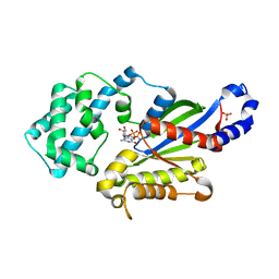
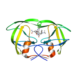
5J8D
 | | Structure of nitroreductase from E. cloacae complexed with nicotinic acid adenine dinucleotide | | 分子名称: | FLAVIN MONONUCLEOTIDE, NICOTINIC ACID ADENINE DINUCLEOTIDE, Oxygen-insensitive NAD(P)H nitroreductase | | 著者 | Haynes, C.A, Koder, R.L, Miller, A.F, Rodgers, D.W. | | 登録日 | 2016-04-07 | | 公開日 | 2017-05-17 | | 最終更新日 | 2023-09-27 | | 実験手法 | X-RAY DIFFRACTION (1.85 Å) | | 主引用文献 | Mechanism-Informed Refinement Reveals Altered Substrate-Binding Mode for Catalytically Competent Nitroreductase.
Structure, 25, 2017
|
|
5JFL
 | |
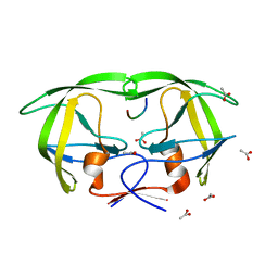
5J8G
 | | Structure of nitroreductase from E. cloacae complexed with para-nitrobenzoic acid | | 分子名称: | 4-NITROBENZOIC ACID, FLAVIN MONONUCLEOTIDE, Oxygen-insensitive NAD(P)H nitroreductase | | 著者 | Haynes, C.A, Koder, R.L, Miller, A.-F, Rodgers, D.W. | | 登録日 | 2016-04-07 | | 公開日 | 2017-05-17 | | 最終更新日 | 2024-03-06 | | 実験手法 | X-RAY DIFFRACTION (1.9 Å) | | 主引用文献 | Mechanism-Informed Refinement Reveals Altered Substrate-Binding Mode for Catalytically Competent Nitroreductase.
Structure, 25, 2017
|
|
1ZSC
 | |
1ZSB
 | | CARBONIC ANHYDRASE II MUTANT E117Q, TRANSITION STATE ANALOGUE ACETAZOLAMIDE | | 分子名称: | 5-ACETAMIDO-1,3,4-THIADIAZOLE-2-SULFONAMIDE, CARBONIC ANHYDRASE II, ZINC ION | | 著者 | Lesburg, C.A, Christianson, D.W. | | 登録日 | 1996-01-09 | | 公開日 | 1996-07-11 | | 最終更新日 | 2024-02-14 | | 実験手法 | X-RAY DIFFRACTION (2 Å) | | 主引用文献 | Reversal of the hydrogen bond to zinc ligand histidine-119 dramatically diminishes catalysis and enhances metal equilibration kinetics in carbonic anhydrase II.
Biochemistry, 35, 1996
|
|
1ZSA
 | |
116D
 | |
117D
 | |
1A1R
 | | HCV NS3 PROTEASE DOMAIN:NS4A PEPTIDE COMPLEX | | 分子名称: | NS3 PROTEIN, NS4A PROTEIN, ZINC ION | | 著者 | Kim, J.L, Morgenstern, K.A, Lin, C, Fox, T, Dwyer, M.D, Landro, J.A, Chambers, S.P, Markland, W, Lepre, C.A, O'Malley, E.T, Harbeson, S.L, Rice, C.M, Murcko, M.A, Caron, P.R, Thomson, J.A. | | 登録日 | 1997-12-15 | | 公開日 | 1998-06-17 | | 最終更新日 | 2024-02-07 | | 実験手法 | X-RAY DIFFRACTION (2.5 Å) | | 主引用文献 | Crystal structure of the hepatitis C virus NS3 protease domain complexed with a synthetic NS4A cofactor peptide.
Cell(Cambridge,Mass.), 87, 1996
|
|
1AIO
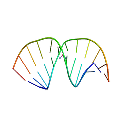 | | CRYSTAL STRUCTURE OF A DOUBLE-STRANDED DNA CONTAINING THE MAJOR ADDUCT OF THE ANTICANCER DRUG CISPLATIN | | 分子名称: | Cisplatin, DNA (5'-D(*CP*CP*(BRU)P*CP*TP*[PT(NH3)2(GP*GP)]*TP*CP*TP*CP*C)-3'), DNA (5'-D(*GP*GP*AP*GP*AP*CP*CP*AP*GP*AP*GP*G)-3') | | 著者 | Takahara, P.M, Rosenzweig, A.C, Frederick, C.A, Lippard, S.J. | | 登録日 | 1997-04-23 | | 公開日 | 1997-04-24 | | 最終更新日 | 2024-02-07 | | 実験手法 | X-RAY DIFFRACTION (2.6 Å) | | 主引用文献 | Crystal structure of double-stranded DNA containing the major adduct of the anticancer drug cisplatin.
Nature, 377, 1995
|
|
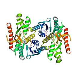
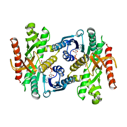
3UMS
 | | Crystal structure of the G202A mutant of human G-alpha-i1 | | 分子名称: | CHLORIDE ION, GUANOSINE-5'-DIPHOSPHATE, Guanine nucleotide-binding protein G(i) subunit alpha-1, ... | | 著者 | Lambert, N.A, Johnston, C.A, Cappell, S.D, Kuravi, S, Kimple, A.J, Willard, F.S, Siderovski, D.P. | | 登録日 | 2011-11-14 | | 公開日 | 2012-02-08 | | 最終更新日 | 2023-09-13 | | 実験手法 | X-RAY DIFFRACTION (2.343 Å) | | 主引用文献 | Correction for Regulators of G-protein Signaling accelerate GPCR signaling kinetics and govern sensitivity solely by accelerating GTPase activity
Proc.Natl.Acad.Sci.USA, 2012
|
|
1BDM
 | |
1BGO
 | | CRYSTAL STRUCTURE OF CYSTEINE PROTEASE HUMAN CATHEPSIN K IN COMPLEX WITH A COVALENT PEPTIDOMIMETIC INHIBITOR | | 分子名称: | 1-[2-(3-BIPHENYL)-4-METHYLVALERYL)]AMINO-3-(2-PYRIDYLSULFONYL)AMINO-2-PROPANONE, CATHEPSIN K | | 著者 | Desjarlais, R.L, Yamashita, D.S, Oh, H.-J, Bondinell, W.E, Uzinskas, I.N, Erhard, K.F, Allen, A.C, Haltiwanger, R.C, Zhao, B, Smith, W.W, Abdel-Meguid, S.S, D'Alessio, K, Janson, C.A, Mcqueney, M.S, Tomaszek, T.A, Levy, M.A, Veber, D.F. | | 登録日 | 1998-05-29 | | 公開日 | 1999-06-08 | | 最終更新日 | 2023-08-02 | | 実験手法 | X-RAY DIFFRACTION (2.3 Å) | | 主引用文献 | Use of X-Ray Co-Crystal Structures and Molecular Modeling to Design Potent and Selective Non-Peptide Inhibitors of Cathepsin K
J.Am.Chem.Soc., 120, 1998
|
|
1LCF
 | | CRYSTAL STRUCTURE OF COPPER-AND OXALATE-SUBSTITUTED HUMAN LACTOFERRIN AT 2.0 ANGSTROMS RESOLUTION | | 分子名称: | 2-acetamido-2-deoxy-beta-D-glucopyranose, CARBONATE ION, COPPER (II) ION, ... | | 著者 | Smith, C.A, Anderson, B.F, Baker, H.M, Baker, E.N. | | 登録日 | 1994-01-11 | | 公開日 | 1994-08-31 | | 最終更新日 | 2020-07-29 | | 実験手法 | X-RAY DIFFRACTION (2 Å) | | 主引用文献 | Structure of copper- and oxalate-substituted human lactoferrin at 2.0 A resolution.
Acta Crystallogr.,Sect.D, 50, 1994
|
|
1BMD
 | |
1BKA
 | | OXALATE-SUBSTITUTED DIFERRIC LACTOFERRIN | | 分子名称: | FE (III) ION, LACTOFERRIN, OXALATE ION | | 著者 | Baker, H.M, Smith, C.A, Baker, E.N. | | 登録日 | 1996-04-15 | | 公開日 | 1996-11-08 | | 最終更新日 | 2011-07-13 | | 実験手法 | X-RAY DIFFRACTION (2.4 Å) | | 主引用文献 | Anion binding by transferrins: importance of second-shell effects revealed by the crystal structure of oxalate-substituted diferric lactoferrin.
Biochemistry, 35, 1996
|
|
1D2V
 | | CRYSTAL STRUCTURE OF BROMIDE-BOUND HUMAN MYELOPEROXIDASE ISOFORM C AT PH 5.5 | | 分子名称: | 2-acetamido-2-deoxy-beta-D-glucopyranose, ACETATE ION, BROMIDE ION, ... | | 著者 | Fiedler, T.J, Davey, C.A, Fenna, R.E. | | 登録日 | 1999-09-28 | | 公開日 | 2000-04-24 | | 最終更新日 | 2020-07-29 | | 実験手法 | X-RAY DIFFRACTION (1.75 Å) | | 主引用文献 | X-ray crystal structure and characterization of halide-binding sites of human myeloperoxidase at 1.8 A resolution.
J.Biol.Chem., 275, 2000
|
|
3ZK7
 | | CRYSTAL STRUCTURE OF PNEUMOCOCCAL SURFACE ANTIGEN PSAA IN THE METAL-FREE, OPEN STATE | | 分子名称: | 2-AMINO-2-HYDROXYMETHYL-PROPANE-1,3-DIOL, MANGANESE ABC TRANSPORTER SUBSTRATE-BINDING LIPOPROTEIN | | 著者 | Counago, R.M, Ween, M.P, Bajaj, M, Zuegg, J, Cooper, M.A, McEwan, A.G, Paton, J.C, Kobe, B, McDevitt, C.A. | | 登録日 | 2013-01-22 | | 公開日 | 2013-11-06 | | 最終更新日 | 2023-12-20 | | 実験手法 | X-RAY DIFFRACTION (1.69 Å) | | 主引用文献 | Imperfect coordination chemistry facilitates metal ion release in the Psa permease.
Nat. Chem. Biol., 10, 2014
|
|
1MTB
 | | Viability of a drug-resistant HIV-1 protease mutant: structural insights for better antiviral therapy | | 分子名称: | (2S)-2-amino-3-phenylpropane-1,1-diol, 2-METHYL-DECAHYDRO-ISOQUINOLINE-3-CARBOXYLIC ACID, ASPARAGINE, ... | | 著者 | Prabu-Jeyabalan, M, Nalivaika, E.A, King, N.M, Schiffer, C.A. | | 登録日 | 2002-09-20 | | 公開日 | 2003-01-07 | | 最終更新日 | 2024-03-13 | | 実験手法 | X-RAY DIFFRACTION (2.5 Å) | | 主引用文献 | Viability of drug-resistant human immunodeficiency virus type 1 protease variant: structural insights for better antiviral therapy
J.Virol., 77, 2003
|
|
1MT7
 | | Viability of a drug-resistant HIV-1 protease mutant: structural insights for better antiviral therapy | | 分子名称: | ACETATE ION, PROTEASE RETROPEPSIN, Substrate analogue | | 著者 | Prabu-Jeyabalan, M, Nalivaika, E.A, King, N.M, Schiffer, C.A. | | 登録日 | 2002-09-20 | | 公開日 | 2003-01-07 | | 最終更新日 | 2024-02-14 | | 実験手法 | X-RAY DIFFRACTION (1.9 Å) | | 主引用文献 | Viability of drug-resistant human immunodeficiency virus type 1 protease variant: structural insights for better antiviral therapy
J.Virol., 77, 2003
|
|
1D17
 | | DNA-NOGALAMYCIN INTERACTIONS | | 分子名称: | DNA (5'-D(*(5CM)P*GP*TP*AP*(5CM)P*G)-3'), NOGALAMYCIN | | 著者 | Egli, M, Williams, L.D, Frederick, C.A, Rich, A. | | 登録日 | 1990-08-08 | | 公開日 | 1991-07-15 | | 最終更新日 | 2024-02-07 | | 実験手法 | X-RAY DIFFRACTION (2 Å) | | 主引用文献 | DNA-nogalamycin interactions.
Biochemistry, 30, 1991
|
|
1MT9
 | | Viability of a drug-resistant HIV-1 protease mutant: structural insights for better antiviral therapy | | 分子名称: | PHOSPHATE ION, PROTEASE RETROPEPSIN, p1-p6 Gag substrate decapeptide | | 著者 | Prabu-Jeyabalan, M, Nalivaika, E.A, King, N.M, Schiffer, C.A. | | 登録日 | 2002-09-20 | | 公開日 | 2003-01-07 | | 最終更新日 | 2024-02-14 | | 実験手法 | X-RAY DIFFRACTION (2 Å) | | 主引用文献 | Viability of drug-resistant human immunodeficiency virus type 1 protease variant: structural insights for better antiviral therapy
J.Virol., 77, 2003
|
|


