1SXJ
 
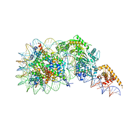
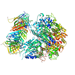 | | Crystal Structure of the Eukaryotic Clamp Loader (Replication Factor C, RFC) Bound to the DNA Sliding Clamp (Proliferating Cell Nuclear Antigen, PCNA) | | 分子名称: | ADENOSINE-5'-DIPHOSPHATE, Activator 1 37 kDa subunit, Activator 1 40 kDa subunit, ... | | 著者 | Bowman, G.D, O'Donnell, M, Kuriyan, J. | | 登録日 | 2004-03-30 | | 公開日 | 2004-06-22 | | 最終更新日 | 2023-11-15 | | 実験手法 | X-RAY DIFFRACTION (2.85 Å) | | 主引用文献 | Structural analysis of a eukaryotic sliding DNA clamp-clamp loader complex.
Nature, 429, 2004
|
|
1F7S
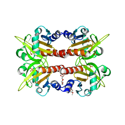
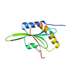 | | CRYSTAL STRUCTURE OF ADF1 FROM ARABIDOPSIS THALIANA | | 分子名称: | ACTIN DEPOLYMERIZING FACTOR (ADF), LAURYL DIMETHYLAMINE-N-OXIDE | | 著者 | Bowman, G.D, Nodelman, I.M, Lindberg, U, Chua, N.H, Schutt, C.E. | | 登録日 | 2000-06-27 | | 公開日 | 2000-11-15 | | 最終更新日 | 2024-02-07 | | 実験手法 | X-RAY DIFFRACTION (2 Å) | | 主引用文献 | A comparative structural analysis of the ADF/cofilin family.
Proteins, 41, 2000
|
|
1G1J
 | |
1G1I
 | |
5J70
 | |
4Y6C
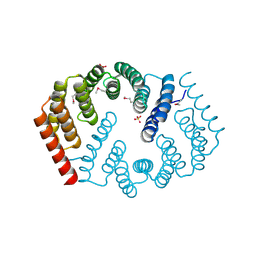 | | Q17M crystal structure of Podosopora anserina putative kinesin light chain nearly identical TPR-like repeats | | 分子名称: | ANSERINA PUTATIVE KINESIN LIGHT chain, SULFATE ION | | 著者 | Marold, J.D, Kavran, J.M, Bowman, G.D, Barrick, D. | | 登録日 | 2015-02-12 | | 公開日 | 2015-10-07 | | 最終更新日 | 2017-11-01 | | 実験手法 | X-RAY DIFFRACTION (1.772 Å) | | 主引用文献 | A Naturally Occurring Repeat Protein with High Internal Sequence Identity Defines a New Class of TPR-like Proteins.
Structure, 23, 2015
|
|
4Y6W
 | |
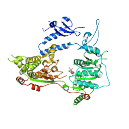
3TED
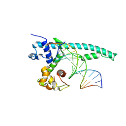
 | | Crystal structure of the Chd1 DNA-binding domain in complex with a DNA duplex | | 分子名称: | 5'-D(*CP*CP*AP*TP*AP*TP*AP*TP*AP*TP*GP*C)-3', 5'-D(*GP*CP*AP*TP*AP*TP*AP*TP*AP*TP*GP*G)-3', Chromo domain-containing protein 1 | | 著者 | Sharma, A, Jenkins, K.R, Heroux, A, Bowman, G.D. | | 登録日 | 2011-08-12 | | 公開日 | 2011-11-02 | | 最終更新日 | 2023-09-13 | | 実験手法 | X-RAY DIFFRACTION (2 Å) | | 主引用文献 | DNA-binding domain of Chd1 in complex with a DNA duplex
J.Biol.Chem., 2011
|
|
7SWY
 | | 2.6 A structure of a 40-601[TA-rich+1]-40 nucleosome | | 分子名称: | DNA Guide Strand, DNA Tracking Strand, Histone H2A, ... | | 著者 | Nodelman, I.M, Bowman, G.D, Armache, J.-P. | | 登録日 | 2021-11-21 | | 公開日 | 2022-03-02 | | 最終更新日 | 2023-03-29 | | 実験手法 | ELECTRON MICROSCOPY (2.6 Å) | | 主引用文献 | Nucleosome recognition and DNA distortion by the Chd1 remodeler in a nucleotide-free state.
Nat.Struct.Mol.Biol., 29, 2022
|
|
7TN2
 | | Composite model of a Chd1-nucleosome complex in the nucleotide-free state derived from 2.3A and 2.7A Cryo-EM maps | | 分子名称: | Chromo domain-containing protein 1, DNA Lagging Strand, DNA Tracking Strand, ... | | 著者 | Nodelman, I.M, Bowman, G.D, Armache, J.-P. | | 登録日 | 2022-01-20 | | 公開日 | 2022-03-02 | | 最終更新日 | 2024-02-21 | | 実験手法 | ELECTRON MICROSCOPY (2.3 Å) | | 主引用文献 | Nucleosome recognition and DNA distortion by the Chd1 remodeler in a nucleotide-free state.
Nat.Struct.Mol.Biol., 29, 2022
|
|
3MWY
 | |
1D1J
 | | CRYSTAL STRUCTURE OF HUMAN PROFILIN II | | 分子名称: | 1-(2-METHOXY-ETHOXY)-2-{2-[2-(2-METHOXY-ETHOXY]-ETHOXY}-ETHANE, 1-METHOXY-2-[2-(2-METHOXY-ETHOXY]-ETHANE, PROFILIN II, ... | | 著者 | Nodelman, I.M, Bowman, G.D, Lindberg, U, Schutt, C.E. | | 登録日 | 1999-09-17 | | 公開日 | 2000-12-22 | | 最終更新日 | 2024-02-07 | | 実験手法 | X-RAY DIFFRACTION (2.2 Å) | | 主引用文献 | X-ray structure determination of human profilin II: A comparative structural analysis of human profilins.
J.Mol.Biol., 294, 1999
|
|
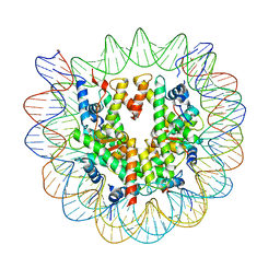
3K4X
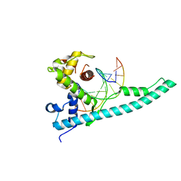
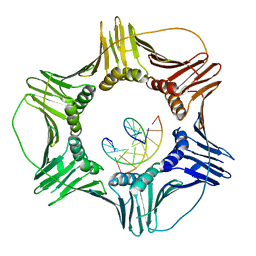 | | Eukaryotic Sliding Clamp PCNA Bound to DNA | | 分子名称: | DNA (5'-D(*CP*CP*CP*AP*TP*CP*GP*TP*AP*T)-3'), DNA (5'-D(*TP*TP*TP*TP*AP*TP*AP*CP*GP*AP*TP*GP*GP*G)-3'), Proliferating cell nuclear antigen | | 著者 | McNally, R, Kuriyan, J. | | 登録日 | 2009-10-06 | | 公開日 | 2010-02-16 | | 最終更新日 | 2023-09-06 | | 実験手法 | X-RAY DIFFRACTION (2.98 Å) | | 主引用文献 | Analysis of the role of PCNA-DNA contacts during clamp loading.
Bmc Struct.Biol., 10, 2010
|
|


