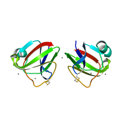
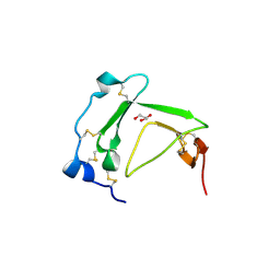
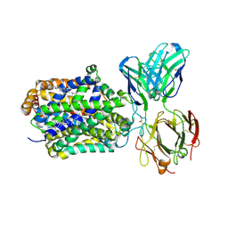
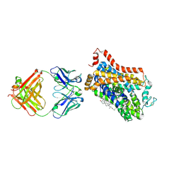
3CW3
 
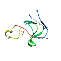 | | Crystal structure of AIM1g1 | | 分子名称: | Absent in melanoma 1 protein, GLYCEROL | | 著者 | Aravind, P, Sankaranarayanan, R, Sharma, Y. | | 登録日 | 2008-04-21 | | 公開日 | 2008-06-17 | | 最終更新日 | 2024-02-21 | | 実験手法 | X-RAY DIFFRACTION (1.88 Å) | | 主引用文献 | Exploring the limits of sequence and structure in a variant betagamma-crystallin domain of the protein absent in melanoma-1 (AIM1).
J.Mol.Biol., 381, 2008
|
|
3ENT
 | |
3ENU
 | |
3HZ2
 | |
3HZB
 | |
3I9H
 | |
3IAJ
 | |
4FD9
 | |
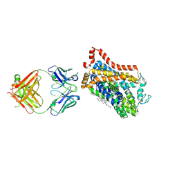
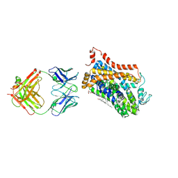
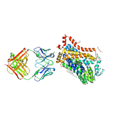
3BT4
 | | Crystal Structure Analysis of AmFPI-1, fungal protease inhibitor from Antheraea mylitta | | 分子名称: | Fungal protease inhibitor-1, GLYCEROL | | 著者 | Roy, S, Aravind, P, Madhurantakam, C, Ghosh, A.K, Sankarananarayanan, R, Das, A.K. | | 登録日 | 2007-12-27 | | 公開日 | 2008-12-30 | | 最終更新日 | 2017-10-25 | | 実験手法 | X-RAY DIFFRACTION (2.1 Å) | | 主引用文献 | Crystal structure of a fungal protease inhibitor from Antheraea mylitta
J.Struct.Biol., 166, 2009
|
|
4XP4
 | | X-ray structure of Drosophila dopamine transporter in complex with cocaine | | 分子名称: | 1-ETHOXY-2-(2-ETHOXYETHOXY)ETHANE, ANTIBODY FRAGMENT HEAVY CHAIN-PROTEIN, 9D5-HEAVY CHAIN, ... | | 著者 | Aravind, P, Wang, K, Gouaux, E. | | 登録日 | 2015-01-16 | | 公開日 | 2015-05-06 | | 最終更新日 | 2020-07-29 | | 実験手法 | X-RAY DIFFRACTION (2.8 Å) | | 主引用文献 | Neurotransmitter and psychostimulant recognition by the dopamine transporter.
Nature, 521, 2015
|
|
4XNU
 | | X-ray structure of Drosophila dopamine transporter in complex with nisoxetine | | 分子名称: | CHLORIDE ION, CHOLESTEROL, Integral membrane protein-dopamine transporter, ... | | 著者 | Aravind, P, Wang, K, Gouaux, E. | | 登録日 | 2015-01-16 | | 公開日 | 2015-05-13 | | 最終更新日 | 2023-09-27 | | 実験手法 | X-RAY DIFFRACTION (2.98 Å) | | 主引用文献 | X-ray structures of Drosophila dopamine transporter in complex with nisoxetine and reboxetine.
Nat.Struct.Mol.Biol., 22, 2015
|
|
4XPB
 | | X-ray structure of Drosophila dopamine transporter with subsiteB mutations (D121G/S426M) bound to cocaine | | 分子名称: | Antibody fragment heavy chain-protein, 9D5-heavy chain, Antibody fragment light chain-protein, ... | | 著者 | Aravind, P, Wang, K, Gouaux, E. | | 登録日 | 2015-01-16 | | 公開日 | 2015-05-13 | | 最終更新日 | 2020-07-29 | | 実験手法 | X-RAY DIFFRACTION (3.05 Å) | | 主引用文献 | Neurotransmitter and psychostimulant recognition by the dopamine transporter.
Nature, 521, 2015
|
|
4XPT
 | | X-ray structure of Drosophila dopamine transporter with subsiteB mutations D121G/S426M and EL2 deletion of 162-201 in complex with substrate analogue 3,4 dichlorophen ethylamine | | 分子名称: | 2-(3,4-dichlorophenyl)ethanamine, 2-acetamido-2-deoxy-beta-D-glucopyranose, CHLORIDE ION, ... | | 著者 | Aravind, P, Wang, K, Gouaux, E. | | 登録日 | 2015-01-17 | | 公開日 | 2015-05-06 | | 最終更新日 | 2023-09-27 | | 実験手法 | X-RAY DIFFRACTION (3.36 Å) | | 主引用文献 | Neurotransmitter and psychostimulant recognition by the dopamine transporter.
Nature, 521, 2015
|
|
4XNX
 | | X-ray structure of Drosophila dopamine transporter in complex with reboxetine | | 分子名称: | (2R)-2-[(R)-(2-ethoxyphenoxy)(phenyl)methyl]morpholine, Antibody fragment heavy chain, CHLORIDE ION, ... | | 著者 | Aravind, P, Wang, K, Gouaux, E. | | 登録日 | 2015-01-16 | | 公開日 | 2015-05-13 | | 最終更新日 | 2023-09-27 | | 実験手法 | X-RAY DIFFRACTION (3 Å) | | 主引用文献 | X-ray structures of Drosophila dopamine transporter in complex with nisoxetine and reboxetine.
Nat.Struct.Mol.Biol., 22, 2015
|
|
4XP6
 | | X-ray structure of Drosophila dopamine transporter bound to psychostimulant methamphetamine | | 分子名称: | (2S)-N-methyl-1-phenylpropan-2-amine, 2-acetamido-2-deoxy-beta-D-glucopyranose, Antibody fragment heavy chain-protein, ... | | 著者 | Aravind, P, Wang, K, Gouaux, E. | | 登録日 | 2015-01-16 | | 公開日 | 2015-05-13 | | 最終更新日 | 2023-09-27 | | 実験手法 | X-RAY DIFFRACTION (3.1 Å) | | 主引用文献 | Neurotransmitter and psychostimulant recognition by the dopamine transporter.
Nature, 521, 2015
|
|
4XP9
 | | X-ray structure of Drosophila dopamine transporter bound to psychostimulant D-amphetamine | | 分子名称: | (2S)-1-phenylpropan-2-amine, 1-ETHOXY-2-(2-ETHOXYETHOXY)ETHANE, 2-acetamido-2-deoxy-beta-D-glucopyranose, ... | | 著者 | Aravind, P, Wang, K, Gouaux, E. | | 登録日 | 2015-01-16 | | 公開日 | 2015-05-06 | | 最終更新日 | 2023-09-27 | | 実験手法 | X-RAY DIFFRACTION (2.8 Å) | | 主引用文献 | Neurotransmitter and psychostimulant recognition by the dopamine transporter.
Nature, 521, 2015
|
|
4XPA
 | | X-ray structure of Drosophila dopamine transporter bound to 3,4dichlorophenethylamine | | 分子名称: | 2-(3,4-dichlorophenyl)ethanamine, Antibody fragment heavy chain-protein, 9D5-heavy chain, ... | | 著者 | Aravind, P, Wang, K, Gouaux, E. | | 登録日 | 2015-01-16 | | 公開日 | 2015-05-20 | | 最終更新日 | 2023-09-27 | | 実験手法 | X-RAY DIFFRACTION (2.95 Å) | | 主引用文献 | Neurotransmitter and psychostimulant recognition by the dopamine transporter.
Nature, 521, 2015
|
|


