4WMA
 
 | |
4WM0
 | |
4WN2
 | |
4WNH
 | |
4WLZ
 | |
4WMI
 | |
4WLM
 | |
4WLG
 | |
4WMB
 | |
4WMK
 | |
1FWQ
 | |
1SRM
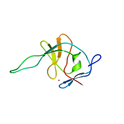
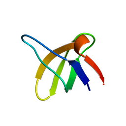
 | | 1H AND 15N ASSIGNMENTS AND SECONDARY STRUCTURE OF THE SRC SH3 DOMAIN | | 分子名称: | SRC TYROSINE KINASE SH3 DOMAIN | | 著者 | Yu, H, Rosen, M.K, Shin, T.B, Seidel-Dugan, C, Brugge, J.S, Schreiber, S.L. | | 登録日 | 1994-03-07 | | 公開日 | 1994-05-31 | | 最終更新日 | 2017-11-29 | | 実験手法 | SOLUTION NMR | | 主引用文献 | 1H and 15N assignments and secondary structure of the Src SH3 domain.
FEBS Lett., 324, 1993
|
|
1SRL
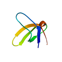
 | | 1H AND 15N ASSIGNMENTS AND SECONDARY STRUCTURE OF THE SRC SH3 DOMAIN | | 分子名称: | SRC TYROSINE KINASE SH3 DOMAIN | | 著者 | Yu, H, Rosen, M.K, Shin, T.B, Seidel-Dugan, C, Brugge, J.S, Schreiber, S.L. | | 登録日 | 1994-03-07 | | 公開日 | 1994-05-31 | | 最終更新日 | 2017-11-29 | | 実験手法 | SOLUTION NMR | | 主引用文献 | 1H and 15N assignments and secondary structure of the Src SH3 domain.
FEBS Lett., 324, 1993
|
|
1OMB
 | |
1OMA
 | |
5BKG
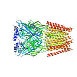
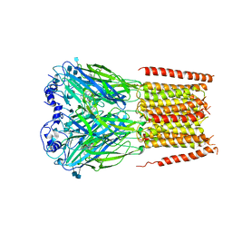
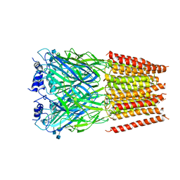 | | Cyro-EM structure of human Glycine Receptor alpha2-beta heteromer, glycine bound, (semi)open state | | 分子名称: | 2-acetamido-2-deoxy-beta-D-glucopyranose, GLYCINE, Glycine receptor subunit alpha-2, ... | | 著者 | Yu, H, Wang, W. | | 登録日 | 2021-03-19 | | 公開日 | 2021-09-08 | | 実験手法 | ELECTRON MICROSCOPY (3.8 Å) | | 主引用文献 | Characterization of the subunit composition and structure of adult human glycine receptors
Neuron, 109, 2021
|
|
5BKF
 | | Cyro-EM structure of human Glycine Receptor alpha2-beta heteromer, Glycine bound, desensitized state | | 分子名称: | 2-acetamido-2-deoxy-beta-D-glucopyranose, GLYCINE, Glycine receptor subunit alpha-2, ... | | 著者 | Yu, H, Wang, W. | | 登録日 | 2021-03-19 | | 公開日 | 2021-09-08 | | 実験手法 | ELECTRON MICROSCOPY (3.6 Å) | | 主引用文献 | Characterization of the subunit composition and structure of adult human glycine receptors
Neuron, 109, 2021
|
|
8I4N
 | |
8I4Q
 | |
7L31
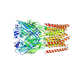 | | Cyro-EM structure of human Glycine Receptor alpha2-beta heteromer, strychnine bound state, 3.8 Angstrom | | 分子名称: | 2-acetamido-2-deoxy-beta-D-glucopyranose, Glycine receptor subunit alpha-2, Glycine receptor subunit beta,Green fluorescent protein, ... | | 著者 | Yu, H, Wang, W. | | 登録日 | 2020-12-17 | | 公開日 | 2021-09-08 | | 実験手法 | ELECTRON MICROSCOPY (3.8 Å) | | 主引用文献 | Characterization of the subunit composition and structure of adult human glycine receptors
Neuron, 109, 2021
|
|
7KUY
 | |
7CC9
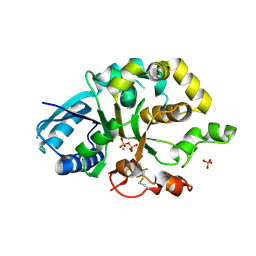
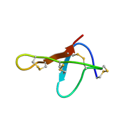
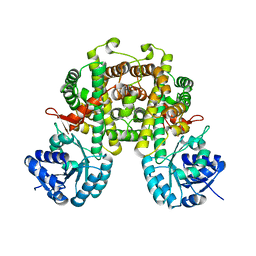
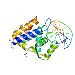 | | Sulfur binding domain of SprMcrA complexed with phosphorothioated DNA | | 分子名称: | ACETATE ION, DNA (5'-D(*GP*GP*CP*GP*GS*CP*CP*C)-3'), DNA (5'-D(*GP*GP*GP*CP*CP*GP*CP*C)-3'), ... | | 著者 | Yu, H, Zhao, G, Gan, J, Liu, G, Wu, G, He, X. | | 登録日 | 2020-06-16 | | 公開日 | 2020-07-08 | | 最終更新日 | 2023-11-29 | | 実験手法 | X-RAY DIFFRACTION (2.063 Å) | | 主引用文献 | DNA backbone interactions impact the sequence specificity of DNA sulfur-binding domains: revelations from structural analyses.
Nucleic Acids Res., 48, 2020
|
|
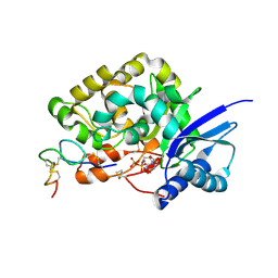
7CCJ
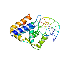 | | Sulfur binding domain of SprMcrA complexed with phosphorothioated DNA | | 分子名称: | DNA (5'-D(*GP*GP*AP*TP*CP*AP*TP*C)-3'), HNHc domain-containing protein | | 著者 | Yu, H, Zhao, G, Gan, J, Liu, G, Wu, G, He, X. | | 登録日 | 2020-06-17 | | 公開日 | 2020-07-08 | | 最終更新日 | 2023-11-29 | | 実験手法 | X-RAY DIFFRACTION (3.3 Å) | | 主引用文献 | DNA backbone interactions impact the sequence specificity of DNA sulfur-binding domains: revelations from structural analyses.
Nucleic Acids Res., 48, 2020
|
|
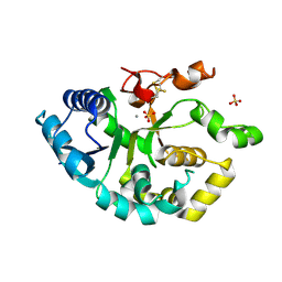
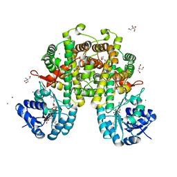
7CCD
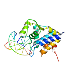 | | Sulfur binding domain of SprMcrA complexed with phosphorothioated DNA | | 分子名称: | DNA (5'-D(*CP*AP*CP*GP*TP*TP*CP*GP*CP*C)-3'), DNA (5'-D(*GP*GP*CP*GP*AS*AP*CP*GP*TP*G)-3'), HNHc domain-containing protein | | 著者 | Yu, H, Li, J, Liu, G, Zhao, G, Wang, Y, Hu, W, Deng, Z, Gan, J, Zhao, Y, He, X. | | 登録日 | 2020-06-16 | | 公開日 | 2020-07-08 | | 最終更新日 | 2023-11-29 | | 実験手法 | X-RAY DIFFRACTION (2.42 Å) | | 主引用文献 | DNA backbone interactions impact the sequence specificity of DNA sulfur-binding domains: revelations from structural analyses.
Nucleic Acids Res., 48, 2020
|
|
7EDA
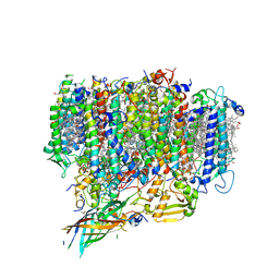 | | Structure of monomeric photosystem II | | 分子名称: | (3R)-beta,beta-caroten-3-ol, 1,2-DI-O-ACYL-3-O-[6-DEOXY-6-SULFO-ALPHA-D-GLUCOPYRANOSYL]-SN-GLYCEROL, 1,2-DIPALMITOYL-PHOSPHATIDYL-GLYCEROLE, ... | | 著者 | Yu, H, Hamaguchi, T, Nakajima, Y, Kato, K, kawakami, K, Akita, F, Yonekura, K, Shen, J.R. | | 登録日 | 2021-03-15 | | 公開日 | 2021-07-07 | | 最終更新日 | 2021-08-04 | | 実験手法 | ELECTRON MICROSCOPY (2.78 Å) | | 主引用文献 | Cryo-EM structure of monomeric photosystem II at 2.78 angstrom resolution reveals factors important for the formation of dimer.
Biochim Biophys Acta Bioenerg, 1862, 2021
|
|


