3QS7
 
 | |
3QS9
 | |
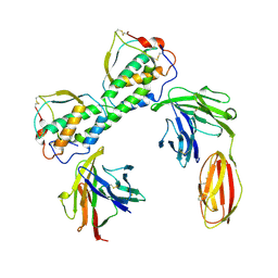
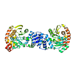
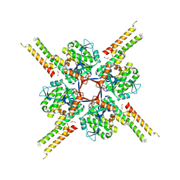
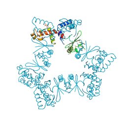
4EC2
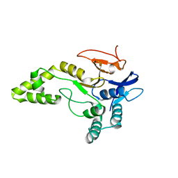
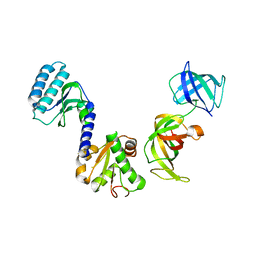
 | | Crystal structure of trimeric frataxin from the yeast Saccharomyces cerevisiae, complexed with ferrous | | 分子名称: | FE (II) ION, Frataxin homolog, mitochondrial | | 著者 | Soderberg, C.A.G, Rajan, S, Gakh, O, Isaya, G, Al-Karadaghi, S. | | 登録日 | 2012-03-26 | | 公開日 | 2013-01-30 | | 最終更新日 | 2023-09-13 | | 実験手法 | X-RAY DIFFRACTION (3.002 Å) | | 主引用文献 | The molecular basis of iron-induced oligomerization of frataxin and the role of the ferroxidation reaction in oligomerization.
J.Biol.Chem., 288, 2013
|
|
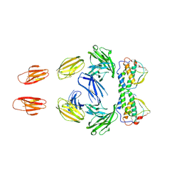
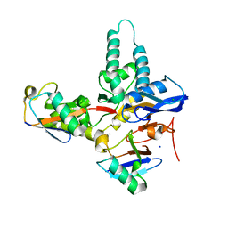
2HAK
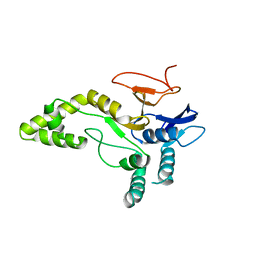
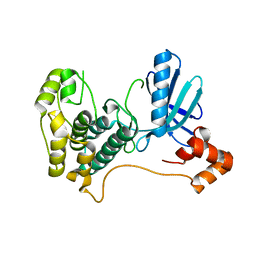 | | Catalytic and ubiqutin-associated domains of MARK1/PAR-1 | | 分子名称: | Serine/threonine-protein kinase MARK1 | | 著者 | Marx, A, Nugoor, C, Mueller, J, Panneerselvam, S, Mandelkow, E.-M, Mandelkow, E. | | 登録日 | 2006-06-13 | | 公開日 | 2006-07-11 | | 最終更新日 | 2023-10-25 | | 実験手法 | X-RAY DIFFRACTION (2.6 Å) | | 主引用文献 | Structural variations in the catalytic and ubiquitin-associated domains of microtubule-associated protein/microtubule affinity regulating kinase (MARK) 1 and MARK2
J.Biol.Chem., 281, 2006
|
|
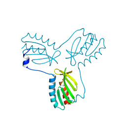
2GVQ
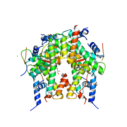
 | | Anthranilate phosphoribosyl-transferase (TRPD) from S. solfataricus in complex with anthranilate | | 分子名称: | 2-AMINOBENZOIC ACID, Anthranilate phosphoribosyltransferase | | 著者 | Marino, M, Deuss, M, Sterner, R, Mayans, O. | | 登録日 | 2006-05-03 | | 公開日 | 2006-05-23 | | 最終更新日 | 2023-11-15 | | 実験手法 | X-RAY DIFFRACTION (2.43 Å) | | 主引用文献 | Structural and mutational analysis of substrate complexation by anthranilate phosphoribosyltransferase from Sulfolobus solfataricus.
J.Biol.Chem., 281, 2006
|
|
2RIK
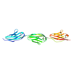 | | I-band fragment I67-I69 from titin | | 分子名称: | Titin | | 著者 | Marino, M, von Castelmur, E, Labeit, D, Labeit, S, Mayans, O. | | 登録日 | 2007-10-11 | | 公開日 | 2008-01-22 | | 最終更新日 | 2023-10-25 | | 実験手法 | X-RAY DIFFRACTION (1.6 Å) | | 主引用文献 | A regular pattern of Ig super-motifs defines segmental flexibility as the elastic mechanism of the titin chain
Proc.Natl.Acad.Sci.Usa, 105, 2008
|
|
2RJM
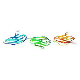 | | 3Ig structure of titin domains I67-I69 E-to-A mutated variant | | 分子名称: | Titin | | 著者 | von Castelmur, E, Marino, M, Labeit, D, Labeit, S, Mayans, O. | | 登録日 | 2007-10-15 | | 公開日 | 2008-01-22 | | 最終更新日 | 2023-10-25 | | 実験手法 | X-RAY DIFFRACTION (2 Å) | | 主引用文献 | A regular pattern of Ig super-motifs defines segmental flexibility as the elastic mechanism of the titin chain
Proc.Natl.Acad.Sci.Usa, 105, 2008
|
|
6SNK
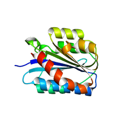 | | Crystal structure of the Collagen VI alpha3 N2 domain | | 分子名称: | Collagen alpha-3(VI) chain | | 著者 | Gebauer, J.M, Degefa, H.S, Paulsson, M, Wagener, R, Baumann, U. | | 登録日 | 2019-08-26 | | 公開日 | 2020-07-29 | | 最終更新日 | 2024-01-24 | | 実験手法 | X-RAY DIFFRACTION (2.2 Å) | | 主引用文献 | Structure of a collagen VI alpha 3 chain VWA domain array: adaptability and functional implications of myopathy causing mutations.
J.Biol.Chem., 295, 2020
|
|
6T69
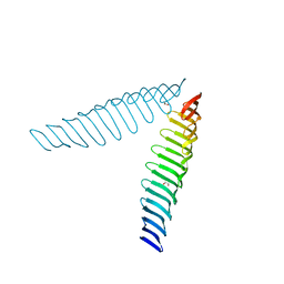 | | Crystal structure of Toxoplasma gondii Morn1(V shape) | | 分子名称: | 1,2-ETHANEDIOL, GLYCEROL, Membrane occupation and recognition nexus protein MORN1, ... | | 著者 | Grishkovskaya, I, Kostan, J, Sajko, S, Morriswood, B, Djinovic-Carugo, K. | | 登録日 | 2019-10-18 | | 公開日 | 2020-11-18 | | 最終更新日 | 2024-01-24 | | 実験手法 | X-RAY DIFFRACTION (2.5 Å) | | 主引用文献 | Structures of three MORN repeat proteins and a re-evaluation of the proposed lipid-binding properties of MORN repeats.
Plos One, 15, 2020
|
|
6T4D
 | | Crystal structure of Plasmodium falciparum Morn1 | | 分子名称: | Morn1, ZINC ION | | 著者 | Grishkovskaya, I, Kostan, J, Sajko, S, Morriswood, B, Djinovic-Carugo, K. | | 登録日 | 2019-10-13 | | 公開日 | 2020-11-18 | | 最終更新日 | 2024-05-15 | | 実験手法 | X-RAY DIFFRACTION (2.14 Å) | | 主引用文献 | Structures of three MORN repeat proteins and a re-evaluation of the proposed lipid-binding properties of MORN repeats.
Plos One, 15, 2020
|
|
6TLB
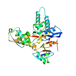
 | | Plasmodium falciparum lipocalin (PF3D7_0925900) | | 分子名称: | GLYCEROL, SODIUM ION, Serine/threonine protein kinase | | 著者 | Burda, P.C, Crosskey, T.D, Lauk, K, Wilmanns, M, Gilberger, T.W. | | 登録日 | 2019-12-02 | | 公開日 | 2020-06-24 | | 最終更新日 | 2024-01-24 | | 実験手法 | SOLUTION SCATTERING (2.85 Å), X-RAY DIFFRACTION | | 主引用文献 | Structure-Based Identification and Functional Characterization of a Lipocalin in the Malaria Parasite Plasmodium falciparum.
Cell Rep, 31, 2020
|
|
6T5A
 | |
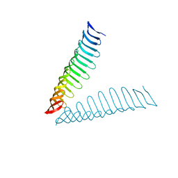
6T6Q
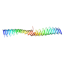 | | Crystal structure of Toxoplasma gondii Morn1 (extended conformation). | | 分子名称: | Membrane occupation and recognition nexus protein MORN1 | | 著者 | Grishkovskaya, I, Kostan, J, Sajko, S, Morriswood, B, Djinovic-Carugo, K. | | 登録日 | 2019-10-18 | | 公開日 | 2020-11-18 | | 最終更新日 | 2024-01-24 | | 実験手法 | X-RAY DIFFRACTION (2.902 Å) | | 主引用文献 | Structures of three MORN repeat proteins and a re-evaluation of the proposed lipid-binding properties of MORN repeats.
Plos One, 15, 2020
|
|
6T68
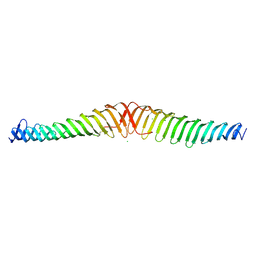 | | Crystal structure of Trypanosoma brucei Morn1 | | 分子名称: | CHLORIDE ION, MORN repeat-containing protein 1 | | 著者 | Grishkovskaya, I, Kostan, J, Sajko, S, Morriswood, B, Djinovic-Carugo, K. | | 登録日 | 2019-10-17 | | 公開日 | 2020-11-18 | | 最終更新日 | 2024-01-24 | | 実験手法 | X-RAY DIFFRACTION (2.54 Å) | | 主引用文献 | Structures of three MORN repeat proteins and a re-evaluation of the proposed lipid-binding properties of MORN repeats.
Plos One, 15, 2020
|
|
6T4R
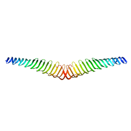 | | Crystal structure of Trypanosoma brucei Morn1 | | 分子名称: | MORN repeat-containing protein 1 | | 著者 | Grishkovskaya, I, Kostan, J, Sajko, S, Morriswood, B, Djinovic-Carugo, K. | | 登録日 | 2019-10-14 | | 公開日 | 2020-11-18 | | 最終更新日 | 2024-01-24 | | 実験手法 | X-RAY DIFFRACTION (2.352 Å) | | 主引用文献 | Structures of three MORN repeat proteins and a re-evaluation of the proposed lipid-binding properties of MORN repeats.
Plos One, 15, 2020
|
|
6TUV
 | |
6XWG
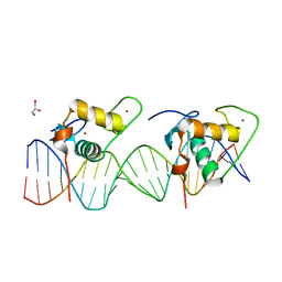 | | Crystal Structure of the Human RXR/RAR DNA-Binding Domain Heterodimer Bound to the Human RARb2 DR5 Response Element | | 分子名称: | CHLORIDE ION, GLYCEROL, RARb2 DR5 Response Element, ... | | 著者 | McEwen, A.G, Poussin-Courmontagne, P, Peluso-Iltis, C, Rochel, N. | | 登録日 | 2020-01-23 | | 公開日 | 2020-09-09 | | 最終更新日 | 2024-01-24 | | 実験手法 | X-RAY DIFFRACTION (2.4 Å) | | 主引用文献 | Structural basis for DNA recognition and allosteric control of the retinoic acid receptors RAR-RXR.
Nucleic Acids Res., 48, 2020
|
|
6XWH
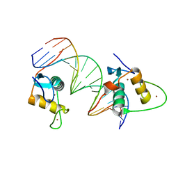 | | Crystal Structure of the Human RXR DNA-Binding Domain Homodimer Bound to the Human Hoxb13 DR0 Response Element | | 分子名称: | Hoxb13 DR0 Response Element, 3'-5' strand, 5'-3' strand, ... | | 著者 | McEwen, A.G, Poussin-Courmontagne, P, Peluso-Iltis, C, Rochel, N. | | 登録日 | 2020-01-23 | | 公開日 | 2020-09-09 | | 最終更新日 | 2024-01-24 | | 実験手法 | X-RAY DIFFRACTION (2.1 Å) | | 主引用文献 | Structural basis for DNA recognition and allosteric control of the retinoic acid receptors RAR-RXR.
Nucleic Acids Res., 48, 2020
|
|
6XZ5
 | |
6TXB
 | |
6Y6R
 | |
6YJG
 | |
6YN1
 | | Crystal structure of histone chaperone APLF acidic domain bound to the histone H2A-H2B-H3-H4 octamer | | 分子名称: | Aprataxin and PNK-like factor, CHLORIDE ION, GLYCEROL, ... | | 著者 | Corbeski, I, Guo, X, Van Ingen, H, Sixma, T.K. | | 登録日 | 2020-04-10 | | 公開日 | 2021-11-17 | | 最終更新日 | 2024-01-24 | | 実験手法 | X-RAY DIFFRACTION (2.35 Å) | | 主引用文献 | Chaperoning of the histone octamer by the acidic domain of DNA repair factor APLF.
Sci Adv, 8, 2022
|
|
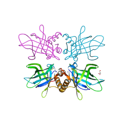
3E1Y
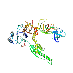 | | Crystal structure of human eRF1/eRF3 complex | | 分子名称: | ADENOSINE-5'-TRIPHOSPHATE, Eukaryotic peptide chain release factor GTP-binding subunit ERF3A, Eukaryotic peptide chain release factor subunit 1 | | 著者 | Cheng, Z, Lim, M, Kong, C, Song, H. | | 登録日 | 2008-08-05 | | 公開日 | 2009-05-19 | | 最終更新日 | 2023-11-01 | | 実験手法 | X-RAY DIFFRACTION (3.8 Å) | | 主引用文献 | Structural insights into eRF3 and stop codon recognition by eRF1
Genes Dev., 23, 2009
|
|
3E20
 | | Crystal structure of S.pombe eRF1/eRF3 complex | | 分子名称: | Eukaryotic peptide chain release factor GTP-binding subunit, Eukaryotic peptide chain release factor subunit 1 | | 著者 | Cheng, Z, Lim, M, Kong, C, Song, H. | | 登録日 | 2008-08-05 | | 公開日 | 2009-05-19 | | 最終更新日 | 2023-11-01 | | 実験手法 | X-RAY DIFFRACTION (3.5 Å) | | 主引用文献 | Structural insights into eRF3 and stop codon recognition by eRF1
Genes Dev., 23, 2009
|
|


