1YRD
 
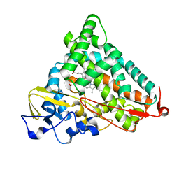 | | X-ray crystal structure of PERDEUTERATED Cytochrome P450cam | | 分子名称: | CAMPHOR, Cytochrome P450-cam, POTASSIUM ION, ... | | 著者 | Meilleur, F, Dauvergne, M.-T, Schlichting, I, Myles, D.A.A. | | 登録日 | 2005-02-03 | | 公開日 | 2005-02-15 | | 最終更新日 | 2023-10-25 | | 実験手法 | X-RAY DIFFRACTION (1.7 Å) | | 主引用文献 | Production and X-ray crystallographic analysis of fully deuterated cytochrome P450cam.
Acta Crystallogr.,Sect.D, 61, 2005
|
|
1YRC
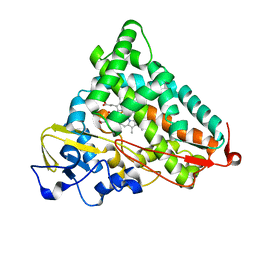 | | X-ray Crystal Structure of hydrogenated Cytochrome P450cam | | 分子名称: | CAMPHOR, Cytochrome P450-cam, POTASSIUM ION, ... | | 著者 | Meilleur, F, Dauvergne, M.-T, Schlichting, I, Myles, D.A.A. | | 登録日 | 2005-02-03 | | 公開日 | 2005-02-15 | | 最終更新日 | 2023-10-25 | | 実験手法 | X-RAY DIFFRACTION (1.4 Å) | | 主引用文献 | Production and X-ray crystallographic analysis of fully deuterated cytochrome P450cam.
Acta Crystallogr.,Sect.D, 61, 2005
|
|
8RBR
 | |
8RBN
 | |
8DYK
 | |
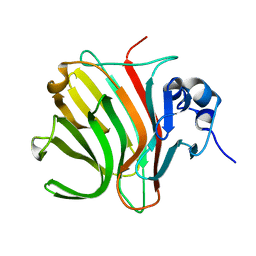
8E1W
 | | Neutron crystal structure of Panus similis AA9A at room temperature | | 分子名称: | 2-acetamido-2-deoxy-beta-D-glucopyranose, CHLORIDE ION, COPPER (II) ION, ... | | 著者 | Meilleur, F, Tandrup, T, Lo Leggio, L. | | 登録日 | 2022-08-11 | | 公開日 | 2023-01-11 | | 最終更新日 | 2024-04-03 | | 実験手法 | NEUTRON DIFFRACTION (2.1 Å), X-RAY DIFFRACTION | | 主引用文献 | Joint X-ray/neutron structure of Lentinus similis AA9_A at room temperature.
Acta Crystallogr.,Sect.F, 79, 2023
|
|
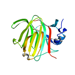
4K9F
 | | Neutron structure of Perdeuterated Rubredoxin refined against 1.75 resolution data collected on the new IMAGINE instrument at HFIR, ORNL | | 分子名称: | FE (III) ION, Rubredoxin | | 著者 | Munshi, P, Meilleur, F, Myles, D. | | 登録日 | 2013-04-19 | | 公開日 | 2013-12-04 | | 最終更新日 | 2023-09-20 | | 実験手法 | NEUTRON DIFFRACTION (1.75 Å) | | 主引用文献 | The IMAGINE instrument: first neutron protein structure and new capabilities for neutron macromolecular crystallography.
Acta Crystallogr.,Sect.D, 69, 2013
|
|
4RSG
 | | Neutron crystal structure of Ras bound to the GTP analogue GppNHp | | 分子名称: | GTPase HRas, MAGNESIUM ION, PHOSPHOAMINOPHOSPHONIC ACID-GUANYLATE ESTER | | 著者 | Knihtila, R.R, Holzapfel, G, Weiss, K.L, Meilleur, F, Mattos, C. | | 登録日 | 2014-11-07 | | 公開日 | 2015-11-04 | | 最終更新日 | 2024-02-28 | | 実験手法 | NEUTRON DIFFRACTION (1.907 Å) | | 主引用文献 | Neutron Crystal Structure of RAS GTPase Puts in Question the Protonation State of the GTP gamma-Phosphate.
J.Biol.Chem., 290, 2015
|
|
5KWF
 | | Joint X-ray Neutron Structure of Cholesterol Oxidase | | 分子名称: | Cholesterol oxidase, FLAVIN-ADENINE DINUCLEOTIDE | | 著者 | Golden, E, Vrielink, A, Meilleur, F, Blakeley, M. | | 登録日 | 2016-07-18 | | 公開日 | 2017-02-01 | | 最終更新日 | 2024-03-06 | | 実験手法 | NEUTRON DIFFRACTION (1.499 Å), X-RAY DIFFRACTION | | 主引用文献 | An extended N-H bond, driven by a conserved second-order interaction, orients the flavin N5 orbital in cholesterol oxidase.
Sci Rep, 7, 2017
|
|
2QXW
 | | Perdeuterated alr2 in complex with idd594 | | 分子名称: | Aldose reductase, CITRIC ACID, IDD594, ... | | 著者 | Blakeley, M.P, Ruiz, F, Cachau, R, Hazemann, I, Meilleur, F, Mitschler, A, Ginell, S, Afonine, P, Ventura, O, Cousido-Siah, A, Joachimiak, A, Myles, D, Podjarny, A. | | 登録日 | 2007-08-13 | | 公開日 | 2008-01-22 | | 最終更新日 | 2023-08-30 | | 実験手法 | X-RAY DIFFRACTION (0.8 Å) | | 主引用文献 | Quantum model of catalysis based on a mobile proton revealed by subatomic x-ray and neutron diffraction studies of h-aldose reductase.
Proc.Natl.Acad.Sci.Usa, 105, 2008
|
|
2R24
 | | Human Aldose Reductase structure | | 分子名称: | Aldose reductase, IDD594, NADP NICOTINAMIDE-ADENINE-DINUCLEOTIDE PHOSPHATE | | 著者 | Blakeley, M.P, Ruiz, F, Cachau, R, Hazemann, I, Meilleur, F, Mitschler, A, Ginell, S, Afonine, P, Ventura, O.N, Cousido-Siah, A, Haertlein, M, Joachimiak, A, Myles, D, Podjarny, A. | | 登録日 | 2007-08-24 | | 公開日 | 2008-12-23 | | 最終更新日 | 2024-02-21 | | 実験手法 | NEUTRON DIFFRACTION (1.752 Å), X-RAY DIFFRACTION | | 主引用文献 | Quantum model of catalysis based on mobile proton revealed by subatomic X-Ray and neutron diffraction studies of h-Aldose Reductase
Proc.Natl.Acad.Sci.USA, 105, 2008
|
|
2PLL
 | | Crystal structure of perdeuterated human arginase I | | 分子名称: | 2(S)-AMINO-6-BORONOHEXANOIC ACID, MANGANESE (II) ION, arginase-1 | | 著者 | Di Costanzo, L, Moulin, M, Haertlein, M, Meilleur, F, Christianson, D.W. | | 登録日 | 2007-04-19 | | 公開日 | 2007-08-14 | | 最終更新日 | 2023-08-30 | | 実験手法 | X-RAY DIFFRACTION (1.9 Å) | | 主引用文献 | Expression, purification, assay, and crystal structure of perdeuterated human arginase I
Arch.Biochem.Biophys., 465, 2007
|
|
3RZ6
 | | Neutron structure of perdeuterated rubredoxin using 40 hours 1st pass data | | 分子名称: | FE (III) ION, Rubredoxin | | 著者 | Munshi, P, Chung, C.-L, Weiss, K.L, Blakeley, M.P, Myles, D.A.A, Meilleur, F. | | 登録日 | 2011-05-11 | | 公開日 | 2011-12-28 | | 最終更新日 | 2023-09-13 | | 実験手法 | NEUTRON DIFFRACTION (1.75 Å) | | 主引用文献 | Rapid visualization of hydrogen positions in protein neutron crystallographic structures.
Acta Crystallogr.,Sect.D, 68, 2012
|
|
3RZT
 | | Neutron structure of perdeuterated rubredoxin using rapid (14 hours) data | | 分子名称: | FE (III) ION, Rubredoxin | | 著者 | Munshi, P, Chung, C.-L, Weiss, K.L, Blakeley, M.P, Myles, D.A.A, Meilleur, F. | | 登録日 | 2011-05-12 | | 公開日 | 2011-12-28 | | 最終更新日 | 2023-09-13 | | 実験手法 | NEUTRON DIFFRACTION (1.7504 Å) | | 主引用文献 | Rapid visualization of hydrogen positions in protein neutron crystallographic structures.
Acta Crystallogr.,Sect.D, 68, 2012
|
|
3RYG
 | | 128 hours neutron structure of perdeuterated rubredoxin | | 分子名称: | FE (III) ION, Rubredoxin | | 著者 | Munshi, P, Chung, C.-L, Weiss, K.L, Blakeley, M.P, Myles, D.A.A, Meilleur, F. | | 登録日 | 2011-05-11 | | 公開日 | 2011-12-28 | | 最終更新日 | 2023-09-13 | | 実験手法 | NEUTRON DIFFRACTION (1.75 Å) | | 主引用文献 | Rapid visualization of hydrogen positions in protein neutron crystallographic structures.
Acta Crystallogr.,Sect.D, 68, 2012
|
|
7T5D
 | |
7T5E
 | |
7T5C
 | |
3SS2
 | | Neutron structure of perdeuterated rubredoxin using 48 hours 3rd pass data | | 分子名称: | FE (III) ION, Rubredoxin | | 著者 | Munshi, P, Chung, C.-L, Blakeley, M.P, Weiss, K.L, Myles, D.A.A, Meilleur, F. | | 登録日 | 2011-07-07 | | 公開日 | 2011-12-28 | | 最終更新日 | 2023-09-13 | | 実験手法 | NEUTRON DIFFRACTION (1.75 Å) | | 主引用文献 | Rapid visualization of hydrogen positions in protein neutron crystallographic structures.
Acta Crystallogr.,Sect.D, 68, 2012
|
|
4LNC
 | | Neutron structure of the cyclic glucose bound Xylose Isomerase E186Q mutant | | 分子名称: | MAGNESIUM ION, MANGANESE (II) ION, Xylose isomerase, ... | | 著者 | Munshi, P, Meilleur, F, Myles, D. | | 登録日 | 2013-07-11 | | 公開日 | 2014-02-12 | | 最終更新日 | 2024-02-28 | | 実験手法 | NEUTRON DIFFRACTION (2.19 Å) | | 主引用文献 | Neutron structure of the cyclic glucose-bound xylose isomerase E186Q mutant.
Acta Crystallogr.,Sect.D, 70, 2014
|
|
4P1H
 | | Crystal structure of wild type Hypocrea jecorina Cel7a in a monoclinic crystal form | | 分子名称: | 2-acetamido-2-deoxy-beta-D-glucopyranose, BENZAMIDINE, Exoglucanase 1, ... | | 著者 | Bodenheimer, A.B, Cuneo, M.J, Swartz, P.D, Myles, D.A, Meilleur, F. | | 登録日 | 2014-02-26 | | 公開日 | 2015-03-04 | | 最終更新日 | 2023-09-27 | | 実験手法 | X-RAY DIFFRACTION (1.5 Å) | | 主引用文献 | Crystal structure of wild type Hypocrea jecorina Cel7a in a monoclinic crystal form
to be published
|
|
4P1J
 | | Crystal structure of wild type Hypocrea jecorina Cel7a in a hexagonal crystal form | | 分子名称: | 2-acetamido-2-deoxy-beta-D-glucopyranose, Exoglucanase 1, SAMARIUM (III) ION, ... | | 著者 | Bodenheimer, A.B, Cuneo, M.J, Swartz, P.D, Myles, D.A, Meilleur, F. | | 登録日 | 2014-02-26 | | 公開日 | 2015-03-25 | | 最終更新日 | 2023-12-27 | | 実験手法 | X-RAY DIFFRACTION (2.62 Å) | | 主引用文献 | Crystal structure of wild type Hypocrea jecorina Cel7a in a hexagonal crystal form
To Be Published
|
|
3KYX
 | |
7JOR
 | | Neutron structure of ferric Dehaloperoxidase B | | 分子名称: | 2-(2-METHOXYETHOXY)ETHANOL, Dehaloperoxidase B, PROTOPORPHYRIN IX CONTAINING FE | | 著者 | Carey, L.M, Ghiladi, R.A, Meilleur, F, Myles, D.A.A. | | 登録日 | 2020-08-07 | | 公開日 | 2021-09-08 | | 最終更新日 | 2024-04-03 | | 実験手法 | NEUTRON DIFFRACTION (2.05 Å) | | 主引用文献 | Complementarity of neutron, XFEL and synchrotron crystallography for defining the structures of metalloenzymes at room temperature.
Iucrj, 9, 2022
|
|
7KCU
 | | Joint neutron/X-ray structure of Oxyferrous Dehaloperoxidase B | | 分子名称: | Dehaloperoxidase B, OXYGEN MOLECULE, PROTOPORPHYRIN IX CONTAINING FE, ... | | 著者 | Carey, L.M, Ghiladi, R.A, Meilleur, F, Myles, D. | | 登録日 | 2020-10-07 | | 公開日 | 2021-10-13 | | 最終更新日 | 2023-10-25 | | 実験手法 | NEUTRON DIFFRACTION (2.2 Å), X-RAY DIFFRACTION | | 主引用文献 | Complementarity of neutron, XFEL and synchrotron crystallography for defining the structures of metalloenzymes at room temperature.
Iucrj, 9, 2022
|
|


